What Causes Jowls: Real Anatomy and Risk Factors 2026
What causes jowls is a combination of three distinct anatomical processes: the progressive laxity of specific facial retaining ligaments that normally anchor soft tissue above the jawline, the age-related loss of volume in the facial fat compartments that support overlying skin, and the decline in dermal collagen and elastin that reduces the skin’s ability to resist gravitational forces. Understanding which of these processes is most active in a given person determines why jowls form when they do and what, if anything, can slow or address the change.
The anatomy involved is more specific than most health content acknowledges. According to studies published in the journal Plastic and Reconstructive Surgery, the lower face contains a system of named retaining ligaments, including the mandibular ligament and the masseteric cutaneous ligament, that physically hold facial soft tissue in position against the pull of gravity. When these structures weaken, tissue descends below the mandibular border in a predictable pattern that produces the characteristic jowl silhouette.
This article covers every documented cause of jowls, from the primary anatomical mechanisms to the hormonal, genetic, environmental, and lifestyle factors that accelerate or delay onset. It also identifies when seeing a board-certified dermatologist or plastic surgeon adds genuine clinical value beyond what self-assessment can provide.
What Causes Jowls
Jowls are caused by the downward displacement of lower facial soft tissue past the mandibular border, driven by retaining ligament laxity, fat compartment volume loss, and dermal collagen decline working simultaneously or in varying combinations.
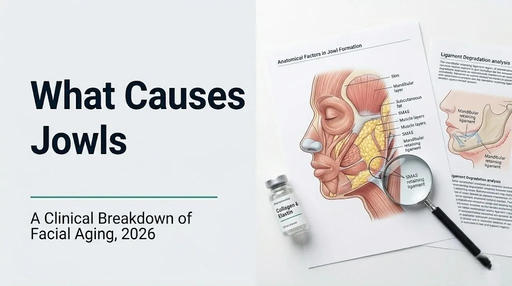
The lower face contains layers of tissue stacked from bone outward: the craniofacial skeleton, the periosteum covering the bone surface, named muscles of facial expression, a connective tissue layer called the superficial musculoaponeurotic system (SMAS), named fat compartments, subcutaneous tissue, and finally the dermis and epidermis. Jowls form when the connections between these layers weaken, allowing the entire soft tissue stack to shift downward and forward relative to the mandibular border.
This is not a simple “skin loosening” process. Three distinct failure modes contribute. First, the named retaining ligaments that tether soft tissue to the underlying bony and fascial framework lengthen and weaken over time. Second, the fat compartments that give the lower cheek its volume shrink, removing the structural support that holds overlying skin in its youthful position. Third, the skin itself loses the collagen and elastin that allow it to rebound and conform to the underlying structure.
Primary causes of jowls, ranked by clinical frequency:
- Age-related retaining ligament laxity (most common primary driver, affects virtually everyone with sufficient time)
- Dermal collagen and elastin loss from intrinsic aging and photoaging (universal contributor, rate varies)
- Facial fat compartment volume loss (becomes more prominent in the fifth decade and beyond)
- Genetic predisposition to early ligament laxity or thin dermis (variable, can cause jowls before age 40)
- Postmenopausal estrogen decline accelerating fibroblast-driven collagen synthesis reduction (women after menopause)
- Significant weight fluctuation causing fat compartment deflation and stretch-then-loose skin (can occur at any age)
The National Institute on Aging documents that skin structural protein loss is a universal feature of biological aging, though the rate at which it produces visible facial changes varies widely between individuals based on genetic, hormonal, and environmental factors.
What Are Jowls and Which Facial Structures Are Involved
Jowls refer to the soft tissue bulge or sagging that develops below the mandible (lower jaw bone), disrupting the clean jawline definition that characterizes a younger facial profile.
Anatomically, the lower face is organized around the mandible, the U-shaped lower jaw bone that provides the foundational skeletal support for the overlying soft tissue. Above the mandible, the masseter muscle (the large chewing muscle) and the depressor anguli oris muscle (which pulls the corners of the mouth downward) are surrounded by named fat compartments. The buccal fat pad, a discrete encapsulated fat structure occupying the cheek space between the buccinator and masseter muscles, provides volume to the midface and contributes to the contour of the lower face. Immediately below and adjacent to this sits the jowl fat compartment, a separate anatomical fat pocket.
Holding all of this tissue in position against gravity is the named retaining ligament system. The mandibular ligament (also called McGregor’s patch in surgical literature) runs from the periosteum of the mandible directly through the subcutaneous fat to the overlying dermis, acting like a biological anchor point. The masseteric cutaneous ligament performs a similar function along the anterior border of the masseter muscle.
Think of this retaining ligament system like the suspension cables on a drawbridge. As long as the cables are taut and anchored at both ends, the bridge deck stays level. When the cables lengthen or the anchor points weaken, the deck sags between the fixed points. Jowls form in exactly the same way: the soft tissue between ligament anchor points descends when the cables go slack.
Key anatomical structures involved in jowl formation:
- Mandible (lower jaw bone): skeletal foundation; undergoes resorption with age
- Mandibular ligament (McGregor’s patch): primary tethering structure at the mandibular border
- Masseteric cutaneous ligament: secondary retaining structure along the masseter
- Superficial musculoaponeurotic system (SMAS): fibromuscular layer connecting facial muscle to overlying fat and skin
- Buccal fat pad: volume contributor in the midface and lower cheek
- Jowl fat compartment: discrete fat pocket whose deflation contributes to tissue ptosis
- Platysma muscle: broad superficial neck muscle whose anterior fibers connect to the lower face SMAS
How Collagen and Elastin Loss Causes Jowls
Collagen and elastin loss causes jowls by reducing the dermis’s structural integrity and recoil capacity, allowing skin to stretch and descend under gravitational force without returning to its original position.
Type I collagen is the primary structural protein of the dermis, forming the dense fibrous scaffold that gives skin its firmness and resistance to mechanical deformation. Type III collagen contributes flexibility to this scaffold. Together they are synthesized by dermal fibroblasts, the connective tissue cells embedded throughout the dermis. Research published in the British Journal of Dermatology found that dermal collagen content declines by approximately 1% per year from early adulthood onward in women, with the rate accelerating after menopause. This cumulative loss represents a substantial reduction in the dermis’s load-bearing capacity over decades.
Elastin fibers are the second critical structural component. Where collagen provides tensile strength, elastin provides elastic recoil, the ability of the skin to spring back after being stretched or displaced. Elastin synthesis essentially stops after adolescence, meaning the fibers present in adult skin are the ones formed during development. As these fibers fragment and lose cross-linking integrity over decades, the skin progressively loses its ability to rebound.
The degradation mechanism is well-characterized. Matrix metalloproteinase-1 (MMP-1), an enzyme produced by aging and UV-stressed fibroblasts, cleaves the triple-helix structure of type I collagen, breaking it into fragments that are then cleared by macrophages. A 2009 study published in JAMA Dermatology demonstrated that even a single dose of UV radiation significantly increases MMP-1 expression in human skin within hours of exposure, meaning that years of cumulative UV exposure produces proportionally greater collagen loss.
Advanced glycation end-products (AGEs) represent a third collagen-disrupting mechanism. Glucose molecules bind non-enzymatically to the amino groups of collagen fibers in a process called glycation. This abnormal cross-linking makes the collagen scaffold stiffer and more brittle rather than maintaining the organized, flexible structure needed for youthful skin function. People with chronically elevated blood glucose, including those with type 2 diabetes mellitus or prediabetes, experience accelerated AGE accumulation in the dermis and may develop skin aging changes, including jowl formation, earlier than age-matched controls without metabolic dysfunction.
Facial Retaining Ligament Laxity and Jowl Formation
Facial retaining ligament laxity is the primary anatomical driver of jowl formation because it allows the entire lower facial soft tissue mass to descend below the mandibular border, regardless of skin quality.
Cadaveric anatomical studies published in Plastic and Reconstructive Surgery by researchers including Furnas (1989) and Mendelson (2013) established the detailed anatomy of the facial retaining ligament system. These structures are fibrous condensations that run perpendicularly from the periosteum of the facial bones or the deep fascia, through the facial soft tissue layers, to anchor in the overlying dermis. They function as fixed tethering points that define the boundaries between facial compartments and prevent the gravitational descent of soft tissue.
The mandibular ligament, arising from the periosteum of the mandible at its anteromedial surface, is the key structure. Its attachment at the skin surface corresponds to the prejowl area. When this ligament maintains its tension, it holds the lower cheek tissue above the mandibular border. As the ligament elongates with age, the tissue anterior and posterior to its attachment point descends, but the attachment point itself remains relatively fixed. The result is that tissue bulges on either side of the mandibular ligament attachment, producing the jowl prominence with the prejowl sulcus (a subtle depression) visible at the ligament’s skin attachment point.
The masseteric cutaneous ligament, running from the anterior border of the masseter muscle through the overlying fat to the lateral cheek skin, provides a secondary anchor along the lateral face. Its weakening allows the cheek fat to descend toward the jowl region, adding tissue volume to the descending jowl mass.
This ligament laxity mechanism explains a clinical observation that puzzles many people: jowls tend to form in a specific location rather than producing uniform skin loosening across the entire face. The position of the mandibular ligament attachment point determines exactly where the tissue bulge appears. This precision is why surgical procedures targeting jowls must address the SMAS and retaining ligament system, not simply tighten the overlying skin.
Postmenopausal women show accelerated ligament laxity alongside accelerated collagen loss, because estrogen receptors are present in facial ligament tissue as well as in dermal fibroblasts, and declining estradiol reduces the maintenance of connective tissue integrity throughout the face.
Buccal Fat Pad Volume Loss and Jowl Appearance
Buccal fat pad volume loss contributes to jowl formation by removing the structural support that holds lower cheek skin in its youthful position, causing overlying tissue to descend and pile up at the mandibular border.
The buccal fat pad (Bichat’s fat pad) is a discrete, encapsulated structure occupying the buccal space between the buccinator muscle and the masseter muscle. It consists of a central body and three extensions (buccal, pterygoid, and temporal extensions). Its primary function in adult faces is structural: it provides volume to the midface and lower cheek, supporting the overlying subcutaneous fat and skin from beneath.
Age-related fat compartment changes in the face do not occur uniformly. Research published in Aesthetic Surgery Journal using magnetic resonance imaging (MRI) and computed tomography (CT) imaging of aging faces documented that the buccal fat pad undergoes gradual volume reduction from approximately the fourth decade onward, with the rate accelerating in the fifth and sixth decades. This deflation is clinically distinct from the retaining ligament descent mechanism: deflation produces a hollowed or flattened cheek appearance with tissue draping rather than the rounded bulge of a ligament descent jowl.
The jowl fat compartment, a separate anatomical fat pocket located anterior to the masseter and inferior to the jowl region, can paradoxically increase in volume with age while adjacent compartments decrease. This differential volume change, deflation above and potential retention or thickening below the mandibular border, contributes to the visual contrast that makes jowls more apparent.
For people who experience primarily fat-compartment-driven jowl changes, the clinical picture differs from ligament-descent jowls. A board-certified plastic surgeon or board-certified facial plastic surgeon assessing jowls will evaluate whether the primary change is tissue descent, volume loss, or a combination, because these mechanisms respond differently to different intervention approaches.
People who undergo significant intentional weight loss through dietary restriction or bariatric surgery may experience disproportionate facial fat compartment deflation relative to body fat loss, producing facial aging changes including jowl formation at younger ages than would otherwise occur.
Key Takeaway: The reason jowls form in a specific, predictable location rather than producing generalized facial drooping is that the mandibular retaining ligament creates a fixed point, and tissue descends on either side of it. The same anatomical precision that causes jowls also explains why targeting those specific ligament structures surgically produces more predictable results than skin-only tightening.
The Platysma Muscle Role in Jowl Development
The platysma muscle contributes to jowl formation by pulling the lower facial soft tissue and skin downward when its superior fibers lose their connection to the overlying SMAS and lower face anatomy.
The platysma is a broad, thin sheet of muscle arising from the fascia overlying the pectoralis major and deltoid muscles of the chest and shoulder. It travels superiorly across the neck and inserts into the lower face region, with its anterior fibers interdigitating with the depressor anguli oris and orbicularis oris muscles at the corners of the mouth. In younger anatomy, the platysma’s superior margin is closely integrated with the lower face SMAS, providing a functional connection that helps maintain lower face position.
With age, two platysma-related changes contribute to the jowl picture. First, the anterior platysma fibers on each side of the neck separate in the midline, creating platysmal banding, the vertical cord-like lines visible in the neck. This separation reduces the integrated tension that the platysma normally provides to the lower face structure. Second, the lateral platysma loses its attachment to the anterior SMAS of the lower face as SMAS integrity declines, removing a secondary supporting element.
The relationship between platysmal changes and jowl formation is why surgical procedures addressing jowls often include a procedure called platysmaplasty, which tightens and re-anchors the platysma as part of a lower face and neck lift. Simply tightening skin over a lax platysma produces an unnatural result because the underlying muscular anatomy has not been addressed.
In men, the platysma tends to be slightly thicker and more strongly anchored to the overlying lower face tissue, contributing to the clinical observation that male jowls develop later and have a somewhat different appearance than female jowls. This sex-based anatomical difference, rather than skin thickness alone, is part of why jowl formation timing differs between men and women.
How Aging Causes Jowls to Form and Worsen
Aging causes jowls through the simultaneous progression of all three mechanisms: retaining ligament laxity, fat compartment deflation, and skin collagen loss, along with a fourth factor that most health content ignores entirely: resorption of the underlying facial skeleton.
The craniofacial skeleton is not static after adulthood. Research published in Plastic and Reconstructive Surgery using three-dimensional CT imaging of skulls across age groups documented progressive resorption of the mandibular and maxillary bones with aging. The mandibular angle becomes less defined, the mandibular border loses bone height, and the anterior projection of the chin region diminishes. This skeletal recession means the underlying structural platform for all of the overlying soft tissue is physically shrinking over time.
Think of it like a tent collapsing as its center pole shortens. The tent fabric does not change, but without full pole height, the fabric cannot stay taut. As mandibular bone volume decreases, the soft tissue drapes over a smaller skeletal frame, producing the appearance of excess tissue at the jawline.
This skeletal resorption happens gradually across decades, typically becoming clinically noticeable between the fifth and seventh decades. According to the National Institute on Aging, bone density and bone volume decline across the entire skeleton with aging due to an imbalance between osteoclast (bone-resorbing) and osteoblast (bone-forming) activity. The face is not exempt from this process, and the clinical impact on lower face contour is documented in facial aging research literature.
The progression of jowls without intervention follows a predictable timeline in most people. Initial subtle changes in jawline definition begin in the late 30s to early 40s, with the mandibular border becoming less sharp. By the late 40s to mid-50s, the soft tissue bulge at the jowl region is typically visible at rest. By the 60s and 70s, the jowl prominence increases as all contributing mechanisms continue to progress simultaneously.
Quick Tip:
- Sun protection applied to the lower face, jaw, and neck daily reduces the rate of UV-driven MMP-1 collagen degradation at a skin level
- Maintaining a stable body weight reduces the volume fluctuation in facial fat compartments that accelerates deflation-driven ptosis
- People with a family history of early, pronounced jowls should know their risk is partly structural and seek a consultation with a board-certified dermatologist for a baseline skin assessment in their early 40s
Sun Damage and Photoaging as a Cause of Jowls
Sun damage causes jowls by directly activating collagen-degrading enzymes in the dermis through ultraviolet radiation, accelerating the structural protein loss that reduces skin’s ability to resist the gravitational and mechanical forces acting on the lower face.
Ultraviolet A (UVA) radiation, which penetrates to the dermal level and is present year-round regardless of cloud cover, is the primary driver of photoaging in the skin. UVA photons generate reactive oxygen species within dermal fibroblasts and keratinocytes. These reactive oxygen species activate the transcription of matrix metalloproteinase genes, particularly MMP-1 (collagenase) and MMP-3 (stromelysin), through the AP-1 transcription factor pathway. The result is upregulated production of collagen-degrading enzymes in direct proportion to cumulative UV exposure over a lifetime.
Research published in JAMA Dermatology by Krutmann and colleagues documented that people with high cumulative UV exposure show dermal collagen fragmentation patterns on histological examination that are distinctly different from age-matched unexposed controls. The collagen architecture in photoaged skin shows disorganized, fragmented fibers rather than the organized parallel bundle pattern seen in sun-protected skin of similar chronological age. This disorganization directly reduces the skin’s load-bearing capacity.
The clinical result of photoaging in the lower face is that people with high cumulative sun exposure, particularly those with Fitzpatrick skin phototypes I and II (lighter skin with less melanin to absorb UV), develop visible jowl changes earlier than sun-protected individuals of the same age. A 2013 twin study published in Plastic and Reconstructive Surgery examined identical twins with different sun exposure histories and found that the more sun-exposed twin showed consistently greater facial aging signs, including lower face ptosis and jowl prominence.
Photoaging at the lower face specifically: The lower face and jaw receive significant UV exposure from reflective surfaces such as water, sand, and snow. People who spend significant time outdoors without sunscreen applied to the lower face, jaw, and neck accumulate UV-driven collagen degradation in exactly the anatomical region where jowls form. The American Academy of Dermatology recommends broad-spectrum sunscreen with SPF 30 or higher applied to all exposed skin, including the neck and jawline, as the primary evidence-supported measure for reducing cumulative UV-driven skin aging.
Genetics and Why Some People Get Jowls Earlier
Genetics causes early jowl formation by determining the baseline density and architecture of facial retaining ligaments, the thickness and collagen composition of the dermis, the distribution and volume of facial fat compartments, and the strength and geometry of the underlying craniofacial skeleton.
These inherited structural characteristics explain the well-known clinical observation that jowl development runs in families. A person whose parents developed visible jowls in their late 30s is substantially more likely to follow the same timeline than a person whose family maintains a defined jawline into their 60s. This familial pattern reflects inheritance of multiple structural traits that collectively determine how quickly the three primary jowl mechanisms progress.
Genetic variation in collagen gene expression is one documented contributor. Studies in dermatology literature have identified that individuals vary meaningfully in their baseline rates of collagen I and III synthesis and in their sensitivity of MMP-1 upregulation to UV exposure. People at the lower end of baseline collagen synthesis rates, combined with higher MMP-1 UV-sensitivity, will experience faster structural collagen decline.
Facial ligament laxity also has a genetic component. Connective tissue disorder presentations that include ligamentous hyperlaxity across the body, such as hypermobile Ehlers-Danlos syndrome (hEDS), are associated with earlier and more pronounced facial soft tissue ptosis including jowl formation, because the retaining ligament structures share the same underlying fibrillar collagen composition affected by the disorder. A person with documented generalized joint hyperlaxity who notices early facial aging changes should mention this history to a board-certified dermatologist or plastic surgeon during any evaluation, as it is clinically relevant to the likely progression and to the surgical planning if intervention is pursued.
Ethnic facial anatomy also influences jowl formation patterns. Research published in the Aesthetic Surgery Journal documented that individuals with a naturally stronger, wider mandible and denser facial retaining ligament attachments (structural features that are more common in certain ethnic populations) tend to show jowl formation later and with less severity than individuals with a narrower mandible and thinner ligament structures. These are population-level observations with wide individual variation.
Key Takeaway: Genetics sets the starting point and the rate of progression for jowl formation, but UV exposure and weight fluctuation are the two most modifiable accelerating factors, and acting on both consistently from early adulthood produces a measurable difference in the rate at which collagen loss and tissue descent progress.
Weight Changes and Jowl Formation
Weight changes cause jowls by producing expansion and contraction cycles in the facial fat compartments that stretch the overlying retaining ligaments and skin beyond their elastic recovery capacity, leaving the tissue permanently displaced after the fat volume that had stretched them decreases.
Facial fat compartments respond to body fat gain and loss in the same way as other adipose depots, though individual variation in facial fat distribution means some people store and lose fat in the face more readily than others. When body weight increases, the buccal fat pad and jowl fat compartment expand, placing tension on the overlying retaining ligaments and stretching the dermis. If this expanded state persists for months to years, the ligaments and skin adapt by elongating.
When body weight subsequently decreases, the fat compartment volume contracts. The retaining ligaments and skin, which had permanently elongated, do not return to their pre-expansion dimensions. The result is skin and ligament laxity disproportionate to the current fat compartment volume, producing ptosis in the same pattern as age-related jowl formation.
This mechanism is clinically distinct from pure aging-related jowls, though the two are often present simultaneously in older adults who have had weight fluctuations over their lifetime. Clinicians generally observe that a single episode of large-magnitude weight loss, greater than approximately 15% to 20% of body weight, produces more pronounced facial fat compartment changes than gradual, smaller fluctuations.
Bariatric surgery patients represent a population at particularly high risk for post-weight-loss facial aging changes including jowls, because the magnitude of weight loss is typically large and occurs over a shorter time period than dietary weight loss alone. Research published in Plastic and Reconstructive Surgery documented that bariatric surgery patients frequently report facial aging as one of the post-surgical changes that most affects their quality of life, particularly when the surgery is performed after age 40 when baseline collagen reserves are already declining.
People considering significant intentional weight loss should discuss the potential facial tissue effects with their primary care physician as part of their overall planning, particularly if they are over 40 and have a family history of early jowl formation.
Why Women Develop Jowls Faster After Menopause
Women develop jowls faster after menopause because the decline in circulating estradiol (17-beta-estradiol) directly reduces dermal fibroblast activity and type I collagen synthesis, accelerating the structural protein loss that underlies jowl formation.
Estrogen receptors are present in dermal fibroblasts, specifically estrogen receptor alpha (ERalpha) and estrogen receptor beta (ERbeta). When estradiol binds to these receptors, it upregulates the transcription of type I procollagen genes, increasing the rate at which fibroblasts produce new collagen to replace degraded fibers. This estrogen-driven collagen maintenance is a significant contributor to the observation that premenopausal women often have better skin structural integrity than age-matched postmenopausal women.
Research published in the British Journal of Dermatology found that skin collagen content declines by approximately 2% per year in the years immediately following menopause, compared to the approximately 1% per year baseline decline seen in younger premenopausal women. This doubling of the collagen loss rate in the early postmenopausal period directly accelerates jowl-formation mechanisms in the lower face. The same research documented that the most rapid phase of postmenopausal collagen loss occurs in the first five years after menopause onset.
Estrogen also has a direct effect on the retaining ligaments of the face. As noted in the retaining ligament section, these structures contain connective tissue fibers whose integrity depends in part on estrogen-mediated collagen maintenance. Postmenopausal estrogen decline therefore weakens the ligament system and the dermal collagen simultaneously, explaining why many women report noticing visible jowl changes in the first five to seven years after menopause even if they had maintained good skin care habits prior.
Women who experience surgical menopause, meaning sudden complete estrogen withdrawal following bilateral oophorectomy (surgical removal of both ovaries), may notice accelerated facial aging changes including jowl formation more rapidly than women experiencing natural gradual menopause. Any postmenopausal woman noticing rapid facial tissue changes should discuss the hormonal timeline with their obstetrician-gynecologist or primary care physician, as this history is clinically relevant context for any dermatological or cosmetic evaluation.
Jowls in Men: Causes and Differences from Women
Jowls in men develop through the same three primary anatomical mechanisms as in women, but they typically appear later, progress more slowly, and produce a somewhat different visual appearance due to sex-based differences in skin thickness, fat compartment distribution, and facial muscle anatomy.
Men have, on average, a dermis that is approximately 20% to 25% thicker than women’s dermis at matched anatomical sites, according to ultrasound-based dermal thickness studies published in Dermatologic Surgery. This greater baseline dermal collagen mass means that even as men experience the same 1% per year collagen decline, they begin from a higher structural baseline and therefore reach clinically visible skin laxity thresholds later than women of the same age.
The facial fat compartment distribution also differs between sexes. Men tend to have more subcutaneous fat in the lower face and neck region relative to women, and the fat is distributed differently around the mandibular border. This additional fat volume in the lower face can initially delay the visual appearance of jowl formation because the fat provides structural support, but once the retaining ligaments weaken and the tissue descends, this same fat contributes to the volume of the jowl bulge.
Testosterone has some role in maintaining collagen synthesis in men, though the mechanism is less directly studied than the estrogen-collagen pathway in women. Men do not experience the equivalent of menopause in terms of a rapid hormonal withdrawal. Instead, testosterone levels decline gradually, by approximately 1% per year from the early 30s onward according to the National Institute on Aging. This gradual decline produces a proportionally slower rate of hormone-driven collagen loss compared to the accelerated postmenopausal decline in women.
Male jowls also tend to appear heavier and more pronounced when they do form, partly because of the greater overall facial soft tissue mass. Men who notice early jowl formation and are concerned about it should seek evaluation by a board-certified plastic surgeon or board-certified facial plastic surgeon, as the surgical anatomy relevant to jowl correction differs between sexes and requires provider experience with male-specific facial anatomy.
Key Takeaway: Postmenopausal women experience jowl formation at roughly double the collagen loss rate of premenopausal women, making the five years after menopause onset the highest-risk period for visible lower face changes, and also the period when evidence-supported protective measures such as daily broad-spectrum sun protection have the greatest incremental benefit.
Premature Jowl Causes Before Age 40
Premature jowl formation before age 40 is caused by accelerated versions of the standard mechanisms, typically driven by genetic ligament laxity, high cumulative sun exposure, significant weight fluctuation, connective tissue disorders, or a combination of these factors.
The most common reason a person in their late 20s or 30s develops jowls is inherited structural characteristics: a naturally weaker mandibular retaining ligament, a thinner dermis with lower baseline collagen density, or a facial fat distribution pattern that places less supporting fat in the midface and more in the lower face. When these inherited traits are present, even normal rates of age-related collagen decline produce visible tissue descent earlier than would occur in someone with more structurally resilient anatomy.
High cumulative UV exposure before age 40 is the most preventable cause of premature jowl formation. Someone who spent significant time outdoors without sun protection through childhood and young adulthood may have already accumulated decades of UV-driven MMP-1 collagen degradation by their mid-30s. The American Academy of Dermatology notes that the majority of lifetime UV damage accumulates in the first two decades of life for people who grow up in sunny climates, meaning that skin aging manifestations of this damage can appear well before middle age.
People with hypermobile Ehlers-Danlos syndrome (hEDS) or other heritable connective tissue disorders affecting fibrillar collagen integrity develop premature facial ligament laxity as part of their broader connective tissue presentation. Any person under 40 with generalized joint hypermobility who is developing visible jowls should mention the connective tissue history to a board-certified dermatologist during any evaluation, as it changes both the expected progression trajectory and the approach to any intervention.
Risk factors specifically associated with jowl formation before age 40:
- Family history of early jowl formation (one or both parents with jowls before 45)
- High cumulative UV exposure with minimal sun protection in childhood and young adulthood
- Weight fluctuations greater than 20 to 30 pounds on two or more occasions before age 35
- Fitzpatrick skin phototype I or II (high UV sensitivity, less natural photoprotection)
- Documented connective tissue disorder affecting collagen integrity
- Chronic corticosteroid medication use (systemic or high-potency topical), which suppresses collagen synthesis via glucocorticoid receptor signaling in fibroblasts
- Smoking history (nicotine and combustion products impair dermal blood flow and accelerate oxidative damage to collagen fibers)
Lifestyle Factors That Worsen Jowl Formation
Several lifestyle factors accelerate jowl formation by independently increasing the rate of collagen degradation, fat compartment deflation, or retaining ligament stress, and each operates through a specific and named physiological mechanism.
Smoking is the lifestyle factor with the most well-documented direct effect on facial skin aging. Nicotine causes vasoconstriction in the dermal microvasculature, reducing blood flow and oxygen delivery to fibroblasts and impeding collagen synthesis. Combustion products in cigarette smoke generate reactive oxygen species that directly activate MMP-1 production. Research published in the British Journal of Dermatology documented that smokers show greater dermal collagen fragmentation and more pronounced facial aging changes at matched ages compared to non-smokers, independent of sun exposure history.
Chronic psychological stress activates the hypothalamic-pituitary-adrenal (HPA) axis, producing elevated circulating cortisol from the adrenal cortex. Cortisol binds to glucocorticoid receptors in dermal fibroblasts and directly suppresses the transcription of type I procollagen genes while simultaneously increasing MMP activity. The result is a dual effect: slower new collagen production and faster existing collagen degradation. People with chronic, unmanaged stress levels, particularly those with documented elevated cortisol, may notice accelerated skin aging changes relative to their chronological age.
Poor sleep quality is increasingly recognized in dermatology research as a factor that impairs the nocturnal dermal repair cycle. Growth hormone, which stimulates fibroblast activity and collagen synthesis, is secreted in highest concentration during deep sleep stages. Chronic sleep restriction reduces total growth hormone output, limiting the dermal repair capacity that partially offsets daily collagen degradation.
Sleeping position adds a mechanical component. Consistent side sleeping or stomach sleeping presses the lower face into a pillow, creating repetitive mechanical compression and stretching of the lower face skin and fat compartments over years. A 2016 study published in Aesthetic Surgery Journal examined the relationship between preferred sleeping position and facial aging asymmetry, finding that side sleepers showed asymmetric facial aging changes consistent with mechanical compression on the preferred sleeping side, including earlier skin laxity at the lower face.
Additional lifestyle factors that worsen jowl formation:
- High dietary glycemic load (promotes AGE formation and collagen glycation)
- Alcohol consumption (impairs hepatic protein synthesis including collagen precursor production, and causes dehydration affecting dermal fluid balance)
- Rapid yo-yo weight cycling (stretches then deflates facial fat compartments repeatedly)
- Consistently dehydrated skin without barrier maintenance (reduces the dermal hydration that maintains turgor and skin position)
When to See a Provider About Jowls and What to Expect
Seeing a provider about jowls is appropriate when the degree of change is affecting quality of life, when early evidence-based intervention options could slow progression most effectively, or when the rate of change seems disproportionate to age and suggests an underlying cause worth evaluating.
Jowl formation is not a medical emergency and carries no health risk on its own. However, it can be the visible surface expression of underlying physiological changes worth knowing about, including the accelerated collagen loss of the early postmenopausal period, a connective tissue disorder, or the skin aging effects of a modifiable lifestyle factor. A board-certified dermatologist is the appropriate first provider for most people, offering clinical skin quality assessment, discussion of evidence-based topical and procedural options for slowing collagen loss, and referral to a board-certified plastic surgeon or board-certified facial plastic surgeon if structural anatomical evaluation and discussion of surgical or nonsurgical intervention is desired.
To prepare for a dermatology or plastic surgery consultation about jowls:
- Bring dated photographs showing the lower face over the past five to ten years to give the provider a documented timeline of change
- Note when you first observed the change and whether it progressed quickly or gradually
- List all medications currently used, including any corticosteroids (oral, inhaled, or topical), as these affect collagen synthesis and are clinically relevant
- Describe your sun exposure history honestly, including years in high-UV climates and typical sun protection practices
- Disclose family history of early facial aging or any diagnosed connective tissue disorder
- Bring a list of any supplements currently used, as some (including blood-thinning supplements) are relevant to any procedural planning
| Provider Type | What They Assess | When to See Them |
|---|---|---|
| Board-certified dermatologist | Skin quality, collagen status, photoaging, topical options, energy-based devices | First evaluation; skin health baseline; nonsurgical options |
| Board-certified plastic surgeon | SMAS anatomy, retaining ligament assessment, fat compartment volume, surgical planning | When surgical or significant procedural options are being considered |
| Board-certified facial plastic surgeon | Lower face and neck surgical anatomy specialist | When surgery targeting specifically the face and neck is under consideration |
| Primary care physician | Underlying hormonal or metabolic contributors (menopause, cortisol, glycemic status) | When the rate of change seems disproportionate or systemic cause is suspected |
No emergency medical evaluation is needed for jowl formation itself. The only circumstance that would prompt urgent medical evaluation is new or rapidly progressive swelling, firmness, or asymmetric tissue change in the lower face or jaw that develops acutely rather than gradually, as these presentations could indicate a lymph node abnormality or other structural change requiring clinical evaluation by a primary care physician or, depending on the presentation, a maxillofacial surgeon.
Frequently Asked Questions About What Causes Jowls
What is the main cause of jowls?
The primary cause of jowls is progressive laxity of the facial retaining ligaments, particularly the mandibular ligament and masseteric cutaneous ligament, which allow lower facial soft tissue to descend below the mandibular border with advancing age.
This ligament laxity works in combination with dermal collagen and elastin loss and facial fat compartment volume changes, producing the characteristic soft tissue bulge at the jawline.
The relative contribution of each mechanism varies between individuals based on genetics, hormone status, sun exposure history, and weight history.
At what age do jowls typically start forming?
Most people begin to notice subtle jowl changes in their late 30s to early 40s, when retaining ligament laxity and cumulative collagen loss first become visible at the mandibular border.
Visible jowl prominence at rest typically becomes more apparent in the late 40s to 50s, with progression continuing through the sixth and seventh decades as all contributing mechanisms advance simultaneously.
People with significant sun damage history, genetic predisposition, or postmenopausal estrogen decline may experience earlier onset, sometimes in the mid-30s.
Can you get jowls if you are thin?
Yes, thin people can develop jowls, because jowls are primarily driven by retaining ligament laxity and dermal collagen loss rather than fat accumulation.
In some thin-faced individuals, reduced facial fat compartment volume actually makes jowl formation more apparent at an earlier stage because there is less supporting volume to compensate for tissue descent.
Genetics, UV exposure history, and hormonal status are more predictive of jowl onset timing than body weight alone for thin individuals.
Do jowls get worse if left untreated?
Jowls do worsen over time without any intervention because the underlying anatomical processes of retaining ligament laxity, collagen loss, and fat compartment deflation all continue to progress with aging.
The rate of worsening varies between individuals based on sun protection habits, weight stability, hormonal status, and genetic factors, but no natural reversal of these anatomical changes occurs spontaneously.
Evidence-based measures such as consistent broad-spectrum sun protection can slow the rate of UV-driven collagen loss, but they cannot reverse established ligament laxity or restore lost fat compartment volume.
Why do some people get jowls earlier than others?
Genetic differences in facial retaining ligament density, baseline dermal collagen thickness, facial fat compartment architecture, and facial skeleton geometry create wide variation in the age at which jowls become visible.
People with inherited connective tissue characteristics that include thinner retaining ligaments or lower baseline collagen production develop jowls earlier, often in their 30s, while others with structurally denser anatomy may not develop visible jowls until their 60s.
High cumulative UV exposure from early life is the most preventable factor that causes early jowl onset by accelerating MMP-1-driven collagen degradation beginning in young adulthood.
Can losing weight cause jowls?
Significant weight loss can cause or worsen jowls by deflating the facial fat compartments that structurally support lower face skin, leaving the retaining ligaments and skin with less underlying volume to maintain their position.
Weight loss greater than approximately 15% to 20% of body weight, particularly when it occurs over a short time period as with bariatric surgery or rapid dietary restriction, produces the most pronounced facial fat compartment changes.
People who lose substantial weight after age 40 face the highest risk of weight-loss-associated jowl formation because baseline collagen reserves are already declining, reducing the skin’s ability to contract toward the reduced underlying volume.
The most actionable thing this article establishes is that jowl formation is an anatomically specific process driven by named structures in a predictable sequence, not a vague consequence of “aging skin.” That specificity matters practically: it means the cause of your jowls can be assessed, the rate of progression can be influenced by specific modifiable behaviors, and the right provider can give you a genuinely informed picture of your anatomy and your options.
Start with sun protection applied daily to the lower face, jaw, and neck if you are not already doing this. Maintain weight stability to reduce fat compartment fluctuation. If you want a clinical assessment of where your lower face anatomy stands and what intervention options, nonsurgical or surgical, would actually address your specific anatomical picture, schedule an evaluation with a board-certified dermatologist as your first step.
You now have the anatomical language and the mechanistic understanding to ask specific, informed questions in that appointment and to understand the answers you receive.