Hand Swelling Causes: Why Your Hands Swell and When to Act
Hand swelling causes range from minor positional fluid retention to joint infection, blood clots, and heart failure, and the specific cause determines not just the treatment but whether the situation requires emergency evaluation within the next hour. The most diagnostically useful question is not which condition caused your hand swelling but whether the swelling is in one hand or both, because that single observation narrows the differential diagnosis more effectively than any other feature.
According to the National Institute of Arthritis and Musculoskeletal and Skin Diseases (NIAMS), musculoskeletal conditions affecting the hands are among the most prevalent causes of disability in adults in the United States, with inflammatory arthritis, gout, and hand injuries collectively affecting tens of millions of people. Hand swelling is frequently the first visible sign of these conditions, making accurate understanding of the cause practically important for anyone experiencing it.
This article covers the four physiological mechanisms that produce hand swelling, the specific anatomical and biochemical pathways behind every major cause, the unilateral-versus-bilateral distinction that determines the diagnostic direction, which presentations are self-limiting and which are emergencies, and the specific type of physician to contact for each clinical pattern.
Hand Swelling Causes: The Four Mechanistic Categories
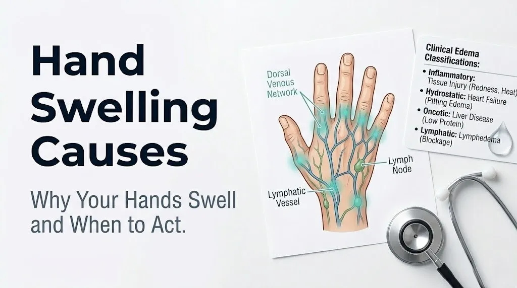
Hand swelling causes fall into four physiological categories based on the mechanism that drives fluid into the tissue, and identifying which category applies is the first step in understanding why your hands are swollen.
Think of the hand’s tissue spaces like a sponge. Fluid flows into the sponge from the tiny blood vessels called capillaries and flows back out through the lymphatic system. When this in-flow and out-flow balance is disrupted by any mechanism, the sponge fills with excess fluid. That excess fluid is what produces the visible swelling, tightness, and puffiness most people experience.
The four mechanisms, governed by what physiologists call Starling forces, are:
| Mechanism | Core Disruption | Example Causes | Typical Presentation |
|---|---|---|---|
| Inflammatory edema | Increased capillary permeability from mediators | Arthritis, infection, gout, allergy | Warm, red, painful; often unilateral or asymmetric |
| Hydrostatic edema | Elevated capillary pressure from venous backup | Heart failure, venous insufficiency, positional stasis | Cool, non-painful, bilateral, worse with dependency |
| Oncotic edema | Low plasma albumin, reduced fluid reabsorption | Liver disease, kidney disease, malnutrition | Bilateral, pitting, gradual onset, no warmth |
| Lymphatic edema | Impaired lymphatic drainage of interstitial fluid | Lymph node removal, fibrosis, infection, cancer | Non-pitting, progressive, often unilateral, no redness |
According to the National Heart Lung and Blood Institute (NHLBI), understanding whether edema is pitting (leaves an indentation when pressed) or non-pitting helps distinguish lymphatic from other forms of edema at the bedside, with non-pitting edema characteristic of lymphedema and hypothyroidism.
Older adults commonly present with overlapping mechanisms: mild heart failure reducing cardiac output, concurrent polypharmacy with edema-producing medications, and reduced lymphatic reserve from age-related lymphatic vessel degeneration may all contribute simultaneously, making the clinical picture harder to categorize cleanly.
What Causes Hands to Swell: Unilateral Versus Bilateral Hand Swelling
Whether one hand or both hands are swollen is the single most diagnostically useful clinical observation in evaluating hand swelling causes.
Unilateral hand swelling (one hand swollen) points strongly toward a local cause: direct trauma or fracture, a localized infection such as cellulitis or an infected tendon sheath, a single-joint inflammatory condition such as gout or septic arthritis, a blood clot in the veins draining that arm, or complex regional pain syndrome (CRPS) following injury or surgery. The local cause produces a local effect, which is why only one extremity is involved.
Bilateral hand swelling (both hands swollen) shifts the diagnostic probability toward systemic causes: heart failure producing elevated venous pressure throughout the body, chronic kidney disease reducing the kidneys’ ability to excrete sodium and water, liver disease reducing albumin production and plasma oncotic pressure, hypothyroidism producing glycosaminoglycan deposition in the dermis, or systemic inflammatory conditions such as rheumatoid arthritis and psoriatic arthritis that affect multiple joints symmetrically or simultaneously.
The timing of swelling is the second most important clinical feature:
| Swelling Pattern | Most Likely Cause Categories | Clinical Urgency |
|---|---|---|
| Acute unilateral, appeared within hours, with warmth and redness | Infection, gout, septic arthritis, trauma | Same day to emergency evaluation |
| Acute unilateral, appeared within hours, with arm/hand cord-like pain | Upper extremity DVT | Emergency evaluation |
| Chronic bilateral, worsening over weeks to months | Heart failure, kidney disease, liver disease, hypothyroidism | Within days to one week evaluation |
| Bilateral in morning, improves during day | Inflammatory arthritis (RA, psoriatic) | Within days evaluation |
| Bilateral in a pregnant woman | Gestational edema vs. preeclampsia | Same day if accompanied by headache or elevated BP |
| Unilateral, months after surgery or cancer treatment | Lymphedema | Non-urgent but requires specialist evaluation |
Children with acute unilateral hot swollen hand joints should be evaluated by a pediatric emergency medicine physician on the same day, because septic arthritis in children progresses rapidly and can damage joint cartilage within 24 to 48 hours if untreated.
Inflammatory Arthritis as a Cause of Hand Swelling
Rheumatoid arthritis (RA) and psoriatic arthritis are among the most frequent causes of chronic bilateral hand swelling, acting through an autoimmune-driven inflammatory cascade that targets the synovial membrane lining the joints and tendon sheaths of the hands.
In rheumatoid arthritis, autoantibodies (rheumatoid factor and anti-CCP antibodies) activate complement pathways and trigger macrophage and T-cell infiltration of the synovial membrane. These immune cells release tumor necrosis factor-alpha (TNF-alpha) and interleukin-1 beta (IL-1beta), which stimulate synovial fibroblasts to proliferate, produce vascular endothelial growth factor (VEGF), and release prostaglandins. VEGF increases capillary permeability in the synovium, allowing protein-rich inflammatory fluid to accumulate in the joint space and surrounding tissue. The result is the warm, swollen, tender joints most visible at the metacarpophalangeal (MCP) joints and proximal interphalangeal (PIP) joints in the morning, when overnight inactivity allows fluid to accumulate maximally.
According to the American College of Rheumatology (ACR), rheumatoid arthritis affects approximately 1.3 million adults in the United States, with the highest incidence in women aged 30 to 60. Morning stiffness lasting more than 60 minutes alongside bilateral MCP and PIP joint swelling is one of the cardinal diagnostic features the ACR uses in classification criteria.
Psoriatic arthritis differs from RA in its joint distribution. Swelling often affects the distal interphalangeal (DIP) joints (the finger joints closest to the nail) and can produce dactylitis, the “sausage finger” appearance of diffuse swelling throughout an entire digit from flexor tendon sheath inflammation, rather than isolated joint swelling. Psoriatic arthritis may be seronegative (rheumatoid factor negative), making it diagnostically distinct.
Key clinical features that distinguish RA from psoriatic arthritis in the hands:
- RA: symmetric MCP and PIP joint swelling, morning stiffness over 60 minutes, rheumatoid factor or anti-CCP positive in approximately 70% of cases
- Psoriatic arthritis: DIP joint swelling, dactylitis, nail pitting or onycholysis present in most cases, preceding or concurrent skin psoriasis in approximately 85% of cases
- Both require referral to a board-certified rheumatologist for disease-modifying antirheumatic drug (DMARD) evaluation once diagnosed
Gout and Septic Arthritis Causing Swollen Hands
Gout produces acute, extremely painful, usually unilateral hand or finger swelling through one of the most precisely characterized inflammatory mechanisms in all of clinical medicine. When serum uric acid exceeds its solubility threshold (generally above 6.8 mg/dL), monosodium urate (MSU) crystals precipitate in joint spaces and periarticular tissues, particularly in cooler peripheral joints. In the hand, the wrist, MCP joints, and interphalangeal joints are frequently affected.
The deposited MSU crystals are recognized by NLRP3 inflammasome complexes in resident macrophages and neutrophils. NLRP3 activation triggers caspase-1-mediated cleavage of pro-IL-1beta into its active form, interleukin-1 beta, which is released in large quantities into the joint space and surrounding tissue. IL-1beta drives massive neutrophil recruitment, prostaglandin release, and vascular permeability changes that produce the classic gout flare: exquisitely tender, hot, red, swollen joint that is essentially untouchable within 24 hours of onset.
According to the ACR gout management guidelines, the first metatarsophalangeal joint (big toe) is the most commonly affected site, but wrist and finger involvement occurs in roughly 40% of patients with polyarticular gout. Hand gout is more common in women than men, particularly in postmenopausal women and in people taking thiazide diuretics, because estrogen normally promotes renal uric acid excretion.
Septic arthritis (infectious arthritis from bacteria entering the joint space) is a medical emergency that produces acute, hot, swollen, painful unilateral joint involvement with fever and systemic illness. The hand joints most commonly affected are the wrist and MCP joints. The organisms responsible in most cases are Staphylococcus aureus and Streptococcus pyogenes entering through a skin wound, a hematogenous (bloodstream) route, or direct inoculation from a bite or puncture. Synovial fluid white blood cell counts above 50,000 cells per mm3 with greater than 75% neutrophils on arthrocentesis (joint aspiration) strongly suggests septic arthritis and requires immediate hospitalization with intravenous antibiotics, directed by an emergency medicine physician or infectious disease specialist.
Key Takeaway: Acute hot swollen single hand joint with fever should be treated as septic arthritis until proven otherwise by joint fluid analysis, because delaying arthrocentesis by even 24 hours risks permanent joint destruction from bacterial protease activity on cartilage.
Infection and Cellulitis as Causes of Hand Swelling
Cellulitis of the hand is a bacterial infection of the dermis and subcutaneous tissue that produces progressive unilateral hand swelling through two distinct mechanisms: direct bacterial toxin-mediated tissue damage, and the inflammatory response to bacterial invasion.
Bacteria, most commonly Staphylococcus aureus (including methicillin-resistant S. aureus, MRSA) and Streptococcus pyogenes (Group A streptococcus), enter through a break in the skin. Small wounds, insect bites, abrasions, nail biting, or a hangnail can provide an entry point invisible to the casual observer. Once in the dermis, bacteria release hyaluronidase (which breaks down the hyaluronic acid matrix holding tissue together, allowing bacterial spread) and streptokinase (which dissolves fibrin barriers). The immune response releases histamine, bradykinin, and prostaglandins, producing the characteristic redness, warmth, pain, and swelling that expands outward from the entry point.
The swelling in cellulitis is typically non-pitting and unilateral, extending in an advancing, poorly demarcated front that spreads proximally toward the forearm if untreated. Red streaks extending from the hand up the forearm indicate lymphangitis (bacterial tracking along the lymphatic channels), which requires urgent evaluation by an emergency medicine physician or urgent care with same-day antibiotic initiation.
Flexor tenosynovitis, infection of the flexor tendon sheath, is a surgical emergency specifically involving the hand. The four clinical signs of flexor tenosynovitis described by Kanavel are: uniform fusiform swelling of the entire digit, the digit held in slight flexion, pain with passive extension of the digit, and tenderness along the flexor tendon sheath. This presentation requires immediate evaluation by a hand surgeon, as the tendon sheath forms a closed compartment where bacterial infection rapidly destroys tendon integrity.
According to the Mayo Clinic, animal bites (particularly cat bites) to the hand carry an exceptionally high risk of joint or tendon sheath infection because feline incisor teeth inject bacteria from the oral cavity, especially Pasteurella multocida, directly into deep structures. Any cat bite to the hand warrants same-day evaluation by an emergency medicine physician, regardless of how small the puncture appears.
People with diabetes mellitus are at substantially higher risk for hand infections from cellulitis and flexor tenosynovitis because of impaired neutrophil chemotaxis, reduced opsonization efficiency, and peripheral neuropathy that may delay recognition of the initial injury. Any person with diabetes who develops hand swelling with redness should contact their primary care physician the same day, not wait to see if it improves.
Heart Failure and Kidney Disease Causing Hand Swelling
Heart failure causes bilateral hand swelling through a chain of physiological events that begins in the failing cardiac muscle and ends in fluid accumulation in the peripheral tissues, including the hands and feet.
When the left ventricle cannot pump blood forward efficiently, venous pressure rises backward through the pulmonary circulation and then into the systemic venous system, increasing the hydrostatic pressure in the capillaries of the hands, forearms, and legs. Elevated capillary hydrostatic pressure overwhelms the plasma oncotic pressure that normally keeps fluid in the bloodstream, and net fluid movement shifts into the interstitial space of peripheral tissues.
Simultaneously, the kidneys detect reduced cardiac output as reduced renal perfusion pressure. They respond by activating the renin-angiotensin-aldosterone system (RAAS), increasing aldosterone production from the adrenal cortex. Aldosterone drives sodium and water reabsorption in the renal collecting ducts. The result is systemic fluid retention that amplifies the peripheral edema already produced by elevated capillary pressure. The hormone antidiuretic hormone (ADH), released from the posterior pituitary in response to low cardiac output, contributes further water retention.
The American Heart Association reports that approximately 6.7 million adults in the United States have heart failure, and peripheral edema affecting the hands and feet is one of the cardinal presenting symptoms. Bilateral hand and leg swelling that worsens when lying flat or with exertion and that is accompanied by breathlessness should prompt urgent evaluation by a cardiologist, with a BNP or NT-proBNP blood test and an echocardiogram as key initial investigations.
Chronic kidney disease (CKD) and nephrotic syndrome cause bilateral hand swelling through reduced renal sodium and water excretion (raising systemic fluid volume) and through massive urinary protein losses that reduce serum albumin (lowering plasma oncotic pressure). Both mechanisms together produce bilateral pitting edema that affects the hands, face, and legs in advanced cases.
Older adults with both heart failure and CKD often present with combined hydrostatic and oncotic edema, making the clinical picture more severe than either condition would produce alone. Their primary care physician should coordinate with both a cardiologist and a nephrologist in these overlapping presentations.
Liver Disease and Low Albumin Causing Hand Swelling
Liver disease causes hand and peripheral swelling through a mechanism distinct from cardiac or renal edema, centered on the liver’s failure to produce adequate amounts of serum albumin, the most abundant plasma protein.
Albumin, synthesized exclusively by hepatocytes in the liver, is the primary determinant of plasma oncotic pressure. It accounts for approximately 80% of the colloid osmotic pressure that draws fluid back from the interstitial space into capillaries. Normal serum albumin is 3.5 to 5.0 grams per deciliter. When hepatic cirrhosis, acute liver failure, or severe malnutrition reduces albumin below 2.5 g/dL, the oncotic pulling force in the capillaries falls sharply, and fluid that would normally be reabsorbed into the bloodstream remains in the interstitial tissue.
Liver disease also contributes to fluid accumulation through a second mechanism: portal hypertension. When the portal vein pressure rises because of cirrhotic scarring obstructing blood flow through the liver, splanchnic vasodilation occurs (mediated by increased local nitric oxide production). This reduces effective arterial blood volume, triggering RAAS activation and sodium retention, further expanding extracellular fluid volume.
According to the National Institute of Diabetes and Digestive and Kidney Diseases (NIDDK), approximately 4.5 million adults in the United States have been diagnosed with liver disease, with cirrhosis being one of the leading causes of portal hypertension and hypoalbuminemia-related edema. The peripheral edema from liver disease is characteristically bilateral and pitting and is confirmed with a serum albumin level and liver function tests (LFTs) including alanine aminotransferase (ALT), aspartate aminotransferase (AST), and bilirubin.
People with a history of alcohol use disorder are at substantially higher risk for hepatic cirrhosis-related edema than the general population. A primary care physician who identifies bilateral pitting hand edema alongside signs of liver disease (spider angiomata, jaundice, caput medusae, or palmar erythema) should expedite referral to a gastroenterologist or hepatologist.
Key Takeaway: Bilateral pitting edema in the hands and legs in the presence of known or suspected liver disease, kidney disease, or heart failure reflects a failure of Starling forces in three different ways, and sorting out which mechanism dominates requires specific blood tests ordered by a primary care physician or relevant specialist, not watchful waiting at home.
Hypothyroidism and Lymphedema as Causes of Swollen Hands
Hypothyroidism causes hand swelling through a mechanism that is fundamentally different from edema: it is not excess fluid in the wrong place but rather an accumulation of glycosaminoglycans (hydrophilic polysaccharides including hyaluronic acid and chondroitin sulfate) in the dermis of the skin.
Normally, thyroid hormones (T3 and T4) regulate the metabolic clearance rate of glycosaminoglycans in connective tissue. When thyroid hormone levels fall, glycosaminoglycan clearance slows and these water-binding molecules accumulate in the dermal layer. Because glycosaminoglycans bind water strongly, the dermis becomes thickened and boggy in appearance. This is called myxedema, and unlike standard edema, it is characteristically non-pitting: pressing firmly on the swollen tissue does not leave an indentation, because the glycosaminoglycan-bound water cannot be displaced by pressure the way free interstitial fluid can.
Myxedema from hypothyroidism typically affects the hands, face, and lower legs. The hands may appear swollen and doughy, with periorbital puffiness, macroglossia (enlarged tongue), and hoarseness. A thyroid-stimulating hormone (TSH) blood test is the first-line investigation for non-pitting bilateral hand swelling, particularly when accompanied by fatigue, cold intolerance, constipation, or unexplained weight gain. An elevated TSH confirms hypothyroidism and warrants referral to an endocrinologist for thyroid hormone replacement management.
Lymphedema produces hand swelling through impaired lymphatic drainage of the normal filtrate that continuously leaks from capillaries into the interstitial space. Lymphatic vessels normally collect this filtrate and return it to the venous system at the thoracic duct. When lymphatic vessels are removed (as in axillary lymph node dissection for breast cancer treatment), obstructed (by filariasis or tumor), or congenitally absent, the filtrate accumulates progressively in the upper extremity.
Breast cancer-related lymphedema of the arm and hand is one of the most prevalent forms, affecting an estimated 20 to 30% of women who undergo axillary lymph node dissection, according to research published in the Journal of Clinical Oncology. The swelling is characteristically non-pitting in established lymphedema, unilateral in post-surgical cases, and progressive without intervention. Management by a lymphedema-certified physical therapist using complete decongestive therapy (CDT) is the evidence-based standard approach, per guidelines from the National Lymphedema Network.
Individuals with a history of any cancer treatment involving lymph node removal or radiation to the axillary region who develop ipsilateral arm or hand swelling months to years after treatment should be referred promptly to a lymphedema specialist rather than reassured that the swelling is minor.
Upper Extremity DVT and Blood Clots Causing Hand Swelling
Upper extremity deep vein thrombosis (DVT) is an underrecognized cause of acute unilateral hand and arm swelling that carries a genuine risk of pulmonary embolism and requires emergency evaluation when certain features are present.
The veins that drain the hand and arm include the cephalic and basilic veins superficially, feeding into the deep system via the brachial veins, which join to form the axillary vein, which continues as the subclavian vein before draining into the superior vena cava. A clot forming in any segment of this deep venous system, from the brachial veins proximally, produces obstruction to venous outflow from the hand and arm. Blood backs up upstream from the clot, capillary hydrostatic pressure rises in the hand and forearm, and edema develops rapidly on the affected side only.
Acute upper extremity DVT presents as rapid-onset unilateral arm or hand swelling, often accompanied by a dull, aching heaviness in the arm, prominent superficial veins visible on the hand and forearm (as the body attempts to reroute blood through alternative pathways), and sometimes warmth and discoloration. The presence of a palpable cord along the course of the axillary or brachial vein is a specific finding pointing toward DVT.
Upper extremity DVT occurs in two clinical patterns. Effort-related thrombosis, called Paget-Schroetter syndrome, is caused by repetitive overhead arm movement compressing the subclavian vein between the clavicle and first rib, most often in young athletes (baseball pitchers, swimmers, weightlifters, volleyball players). Catheter-related thrombosis is the more frequent pattern in hospital and clinical settings, occurring in patients with central venous catheters, PICC lines, or implanted ports.
The National Heart Lung and Blood Institute states that upper extremity DVT accounts for approximately 4 to 10% of all DVT cases, with pulmonary embolism occurring in 6 to 20% of upper extremity DVT cases in published cohort studies. This makes it a clinically serious diagnosis, not a benign finding.
A Doppler ultrasound of the upper extremity venous system is the primary diagnostic test. Any person with acute unilateral arm and hand swelling of unexplained onset should have this test ordered urgently, not at a routine appointment, by a primary care physician or emergency medicine physician.
Young male athletes in overhead sports who develop sudden arm heaviness and hand swelling during or after training should be evaluated for Paget-Schroetter syndrome with same-day Doppler ultrasound and referral to a vascular surgeon if confirmed.
Medications That Cause Hand Swelling
Several medication classes cause bilateral hand swelling as a direct pharmacological effect, and this category is frequently overlooked when patients and physicians are searching for the cause of peripheral edema.
Calcium channel blockers (CCBs), including amlodipine, nifedipine, and felodipine, are among the most common medication causes of bilateral hand and ankle swelling. The mechanism is peripheral arteriolar vasodilation: CCBs block L-type voltage-gated calcium channels in vascular smooth muscle, reducing the smooth muscle tone of peripheral arterioles. This vasodilation lowers precapillary resistance, increasing capillary hydrostatic pressure in peripheral tissues and driving net fluid outward into the interstitial space. The edema is typically bilateral, non-pitting, and dose-dependent, appearing within weeks of starting the medication or increasing the dose.
According to a clinical review published in the Annals of Internal Medicine, peripheral edema from calcium channel blockers affects approximately 10 to 30% of patients taking dihydropyridine CCBs at standard doses, making it one of the most frequent medication side effects in hypertension management. Switching to a different antihypertensive class under physician guidance resolves the edema.
Corticosteroids (prednisone, dexamethasone, methylprednisolone) produce bilateral hand and facial swelling through mineralocorticoid receptor activation in the renal collecting ducts, increasing sodium and water reabsorption. The RAAS is not activated in this case; the steroid molecule itself binds to mineralocorticoid receptors alongside aldosterone receptors, producing the same sodium-retaining effect.
Thiazolidinediones (pioglitazone, rosiglitazone, used for type 2 diabetes) cause peripheral edema through a mechanism involving renal sodium retention mediated by increased collecting duct expression of sodium channels. Pregabalin and gabapentin (used for neuropathic pain and epilepsy) produce peripheral edema through unclear mechanisms, possibly involving voltage-gated calcium channel inhibition in vascular tissue similar to CCBs.
Medications commonly associated with bilateral hand and peripheral edema:
- Calcium channel blockers (amlodipine, nifedipine, felodipine): peripheral arteriolar vasodilation increasing capillary pressure
- Corticosteroids (prednisone, methylprednisolone): mineralocorticoid receptor-mediated sodium and water retention
- Thiazolidinediones (pioglitazone): renal sodium channel upregulation
- Pregabalin and gabapentin: mechanism partially unclear, possibly vascular calcium channel-related
- NSAIDs (ibuprofen, naproxen, celecoxib): renal prostaglandin inhibition reducing sodium excretion
- Hormone therapies (estrogen-containing oral contraceptives, HRT): direct sodium retention effect
- Testosterone and anabolic steroids: RAAS activation with aldosterone-mediated retention
Any patient who develops bilateral hand or peripheral edema after starting or changing a medication should contact their prescribing primary care physician or specialist before stopping the medication, as substitution within the same drug class may resolve the edema without compromising the underlying treatment goal.
Key Takeaway: Calcium channel blockers cause bilateral hand and ankle swelling in approximately 10 to 30% of users through peripheral arteriolar vasodilation, not through cardiac or renal pathology, and the edema resolves when the drug is switched under physician supervision, making medication review the first step whenever bilateral hand swelling appears without another clear cause.
Trauma, Fractures, and Overuse Injuries Causing Hand Swelling
Traumatic causes of hand swelling are the most straightforward in terms of mechanism: direct tissue injury triggers an immediate local inflammatory response that produces swelling within minutes to hours of the injury.
When hand tissue is injured, damaged cells and activated mast cells release histamine and bradykinin, which dilate local arterioles and increase capillary permeability. Neutrophils and monocytes are recruited by chemokines including IL-8 (CXCL8) to the injury site. The combination of arteriolar dilation, increased capillary permeability, and protein-rich plasma leaking into the interstitial space produces the classic signs of acute inflammation: swelling, redness, warmth, and pain at the injury site within the first 30 to 60 minutes.
Metacarpal fractures are among the most frequent hand injuries, particularly the fifth metacarpal (the “boxer’s fracture” from a closed-fist punch against a hard surface), which accounts for approximately 40% of all metacarpal fractures. Swelling following a metacarpal fracture is typically dorsal, localized to the fracture site, and accompanied by point tenderness on palpation and loss of normal knuckle contour. An X-ray of the hand is the appropriate initial imaging study; complex or intra-articular fractures may require CT for full characterization.
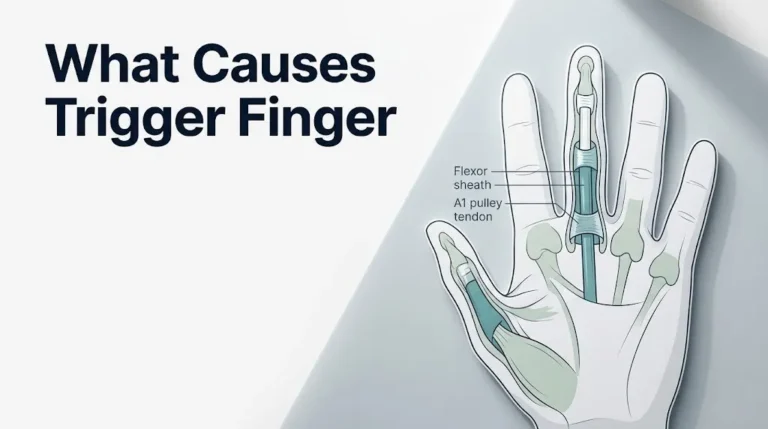
Overuse injuries produce a more gradual, chronic-onset swelling through repeated microtrauma and the cumulative inflammatory response it generates. De Quervain’s tenosynovitis, inflammation of the abductor pollicis longus and extensor pollicis brevis tendons at the radial styloid, produces swelling and tenderness at the base of the thumb. Trigger finger (stenosing tenosynovitis) involves inflammation and thickening of the flexor tendon sheath at the MCP joint level.
According to the American Academy of Orthopaedic Surgeons (AAOS), any hand injury with swelling, deformity, and inability to fully straighten or bend fingers should be evaluated with X-ray imaging to exclude a fracture before any manual reduction or splinting is attempted at home or in a non-specialized setting.
Athletes and manual laborers who develop gradual-onset hand swelling over weeks to months from repetitive use should be evaluated by a primary care physician with referral to an orthopedic hand surgeon or sports medicine physician if the swelling does not resolve with activity modification and appropriate conservative management within 4 to 6 weeks.
Complex Regional Pain Syndrome as a Cause of Hand Swelling
Complex regional pain syndrome (CRPS), formerly called reflex sympathetic dystrophy, is a poorly understood but clinically well-documented condition that produces disproportionate, prolonged pain and swelling in a limb, most often the hand and wrist, following a triggering injury or surgery.
CRPS type 1 occurs without identifiable nerve damage; CRPS type 2 involves confirmed peripheral nerve injury. In both types, the swelling and pain are out of proportion to the severity of the original injury. A minor wrist sprain or a simple fracture may trigger CRPS that produces profound hand swelling, burning pain, skin color changes, altered sweating, and eventually progressive disability if untreated.
The mechanism of CRPS-related hand swelling involves pathological dysregulation of the sympathetic nervous system at the level of the peripheral vasculature. In the acute phase, sympathetically driven arteriolar vasodilation and increased capillary permeability produce the warm, swollen, red hand that looks superficially like an infectious or inflammatory process. In the chronic phase, sympathetically driven vasoconstriction produces a cool, mottled, edematous hand where the swelling persists despite reduced blood flow. The neuropeptides substance P and calcitonin gene-related peptide (CGRP), released by sensitized afferent nerve fibers, perpetuate neurogenic inflammation and maintain increased vascular permeability.
According to research published in the journal Pain, CRPS affects an estimated 5 to 26 people per 100,000 annually, with females affected approximately three to four times more frequently than males, and the upper extremity representing the most common site. Wrist fracture is the most common triggering event.
Clinical features distinguishing CRPS-related hand swelling from other causes:
- Swelling and pain disproportionate to the original injury
- Skin color changes (red or blue-mottled) in the affected hand
- Altered sweating (hyperhidrosis or reduced sweating) on the affected side
- Allodynia (pain from normally non-painful stimuli such as light touch or wind)
- Trophic changes (altered nail growth, hair growth, or skin texture) in chronic cases
Any hand injury that is followed by persistent, worsening pain and swelling weeks to months after the injury would normally be expected to resolve warrants evaluation by a primary care physician with referral to a pain medicine specialist or neurologist experienced in CRPS management.
What Causes Hands to Swell During Exercise and Heat Exposure
Hands swell during exercise and heat exposure through a well-characterized physiological mechanism involving the redistribution of blood flow from the body’s core to the peripheral circulation.
During vigorous exercise, the working muscles generate substantial heat that must be dissipated to prevent dangerous core temperature rise. The hypothalamus detects the rising core temperature and activates the sympathetic nervous system to increase blood flow to the skin by dilating cutaneous arterioles, including those in the hands and fingers. This increased peripheral blood flow serves as a radiator, conducting heat from the blood to the skin surface where it is lost by radiation and evaporation.
The increase in cutaneous blood flow raises the hydrostatic pressure in the small capillaries of the hand and finger skin. Capillary filtration increases proportionally, pushing more fluid than normal into the interstitial space of the hands and fingers. The hands swell visibly, rings may feel tight, and the fingers may appear puffy. This response is a normal physiological adaptation in most people and is self-limiting: the swelling typically resolves within 30 minutes of stopping exercise or returning to a cooler environment as blood flow redistributes back to the core and lymphatic drainage catches up with the accumulated interstitial fluid.
Exercise-induced hand swelling that persists beyond 30 to 60 minutes after stopping exercise, or that is accompanied by chest pain, shortness of breath, or lightheadedness, is not typical of normal physiological exercise edema and warrants evaluation by a primary care physician or cardiologist on the same day.
Quick Tip for exercise-related hand swelling:
- Removing rings before prolonged exercise prevents the discomfort of rings tightening as hands swell and the risk of compromised circulation if rings cannot be removed after exercise
- Regularly clenching and opening the fists during aerobic exercise promotes venous and lymphatic return from the hands, reducing exercise-induced swelling
- Individuals with known heart failure or kidney disease should discuss their specific exercise-related swelling pattern with their cardiologist or nephrologist, as exercise-induced edema in these conditions may behave differently from normal physiological responses
Hot weather and high ambient temperatures produce hand swelling through the same cutaneous vasodilation mechanism as exercise but without the muscular activity. The body’s thermoregulatory response to ambient heat redistributes blood to the skin surface continuously, and the hands swell from the sustained increase in peripheral hydrostatic pressure. This is why some people notice rings feeling tight on hot summer days even without physical activity.
Pregnancy and Preeclampsia Causing Swollen Hands
Pregnancy produces bilateral hand swelling through several concurrent physiological changes that are distinct from any pathological cause, and distinguishing normal gestational edema from the dangerous pattern of preeclampsia is one of the most clinically important distinctions in obstetric medicine.
During normal pregnancy, plasma volume expands by approximately 40 to 50% to meet the demands of the growing fetoplacental unit. Progesterone promotes sodium retention in the renal tubules, and estrogen stimulates the RAAS, both contributing to fluid retention. The expanding uterus compresses the inferior vena cava, particularly in the third trimester, reducing venous return and raising hydrostatic pressure in the lower extremity veins and, to a lesser extent, in the upper extremity venous system. The net result is bilateral, symmetric, dependent edema of the hands and legs that is common in the second and third trimesters, affecting approximately 80% of pregnant women to some degree according to clinical observational data cited by the American College of Obstetricians and Gynecologists (ACOG).
Normal gestational edema has specific characteristics: it is bilateral and symmetric, worsens at the end of the day after prolonged standing or sitting, improves with rest and elevation, and is not accompanied by elevated blood pressure, severe headache, visual disturbances, or upper abdominal pain.
Preeclampsia is an entirely different and dangerous condition. It is defined by new-onset hypertension (systolic blood pressure of 140 mmHg or above, or diastolic of 90 mmHg or above) after 20 weeks of gestation, accompanied by proteinuria or signs of end-organ damage. The pathophysiology involves abnormal placentation producing reduced trophoblast invasion, placental ischemia, and systemic release of soluble fms-like tyrosine kinase 1 (sFlt-1), which blocks vascular endothelial growth factor (VEGF) and placental growth factor (PlGF), leading to widespread maternal endothelial dysfunction and increased vascular permeability.
The hand and face swelling of preeclampsia is characteristically sudden in onset or rapidly worsening, rather than the gradual progression of normal gestational edema.
Warning signs of preeclampsia requiring immediate obstetric evaluation:
- Sudden onset or rapid worsening of hand or facial swelling
- Severe headache not relieved by acetaminophen
- Visual disturbances (blurred vision, seeing spots or flashes of light)
- Pain in the upper right abdomen or epigastrium (indicating hepatic involvement)
- Blood pressure readings at home above 140/90 mmHg on two separate measurements
- Reduced fetal movement alongside any of the above symptoms
Any pregnant woman beyond 20 weeks of gestation who experiences the above combination of symptoms should be evaluated by an obstetrician-gynecologist or in a hospital labor and delivery unit the same day. Waiting for a routine prenatal appointment is not appropriate if these features are present.
Key Takeaway: Bilateral hand swelling in pregnancy is expected and physiological in approximately 80% of pregnant women, but sudden or rapidly worsening hand or facial swelling after 20 weeks, particularly with high blood pressure or severe headache, must be evaluated immediately as a possible sign of preeclampsia, which is a medical emergency.
Hand Swelling Causes in Older Adults and Children
The diagnostic priorities for hand swelling differ substantially between older adults and children because the underlying cause distribution, physiological reserve, and clinical presentation patterns differ markedly across these age groups.
In older adults, hand swelling most often results from one or more of the following: polypharmacy-related edema (particularly from calcium channel blockers, NSAIDs, corticosteroids, or thiazolidinediones), subclinical or established heart failure, chronic kidney disease, hypothyroidism, inflammatory arthritis (particularly RA, which has a peak incidence in women in their 60s), and osteoarthritis with joint effusion. Multiple causes often coexist in the same person. A 70-year-old taking amlodipine for blood pressure, prednisone for COPD, and ibuprofen for knee pain may have three concurrent medication-related contributions to bilateral hand edema.
A 2020 study published in the Journal of the American Geriatrics Society found that peripheral edema of the extremities was documented in approximately 30% of adults over age 65 seen in primary care settings, with heart failure, venous insufficiency, and medication effects accounting for the majority of cases. The clinical implication is that bilateral hand swelling in an older adult without a clear acute cause warrants a systematic medication review, a BNP or NT-proBNP, a TSH, serum albumin, and basic metabolic panel before assuming the edema is benign.
In children, the cause distribution is entirely different. The most clinically urgent cause is septic arthritis, which progresses rapidly in pediatric joints because of the rich blood supply to the developing epiphysis and the high metabolic rate of pediatric synovial tissue. Any child with a hot, swollen, painful single hand joint who refuses to move the affected part should be evaluated in a pediatric emergency department the same day.
Juvenile idiopathic arthritis (JIA), the pediatric equivalent of inflammatory arthritis in adults, can produce bilateral or asymmetric hand joint swelling in children between 2 and 16 years of age. The American Academy of Pediatrics (AAP) recommends referral to a pediatric rheumatologist for any child with persistent joint swelling lasting more than 6 weeks. Reactive arthritis following a streptococcal or gastrointestinal infection can also produce transient hand joint swelling in school-age children.
Adolescent athletes, particularly those participating in overhead sports, are at risk for Paget-Schroetter syndrome (effort-related upper extremity DVT) causing acute arm and hand swelling, as noted in the DVT section. Sudden arm and hand swelling in an adolescent athlete during or after overhead sports should be evaluated with Doppler ultrasound the same day.
Emergency Symptoms: When to Call 911 or Go to the ER
Certain hand swelling patterns associated with their underlying causes require immediate emergency evaluation. Do not wait to see if these resolve on their own.
Call 911 or go to the nearest emergency room immediately if you experience:
- Acute unilateral arm or hand swelling with chest pain, shortness of breath, or rapid breathing (possible upper extremity DVT with pulmonary embolism; the clot may have traveled from the arm to the lungs)
- Rapidly spreading redness, warmth, and skin tightness moving from the hand up the forearm, with fever above 38.5 degrees C (101.3 degrees F) and feeling systemically unwell (possible necrotizing fasciitis or septic cellulitis with early sepsis)
- Acute hot swollen single hand joint with fever and inability to move the joint without severe pain (possible septic arthritis; joint destruction begins within 24 to 48 hours without IV antibiotics)
- Pregnancy beyond 20 weeks with sudden or rapidly worsening hand or face swelling alongside severe headache, visual disturbances, or blood pressure above 140/90 mmHg (possible severe preeclampsia or eclampsia)
- Bilateral hand and leg swelling with sudden onset shortness of breath, inability to lie flat, and frothy or pink-tinged sputum (acute decompensated heart failure)
- Hand swelling with throat tightening, difficulty breathing, or tongue swelling after eating, taking a medication, or insect sting (anaphylaxis requiring immediate epinephrine)
- Any hand swelling in an immunocompromised person (chemotherapy, HIV, organ transplant, high-dose corticosteroid use) with fever and skin changes, because infection progresses faster with immune suppression
These presentations can indicate life-threatening emergencies, including pulmonary embolism, severe sepsis, anaphylaxis, acute cardiac decompensation, and hypertensive emergency of pregnancy, and they require emergency medical assessment with monitoring equipment and emergency treatment capability, not a phone call to your doctor’s office or an urgent care appointment.
When Hand Swelling Is an Emergency and How It Is Diagnosed
Most hand swelling is not a medical emergency, but the framework for determining when it is relies on recognizing specific patterns rather than just the severity of swelling on its own.
The diagnostic approach a physician uses for persistent or unexplained hand swelling follows a systematic sequence based on clinical history and examination findings.
How a physician typically evaluates hand swelling of unknown cause:
- History: onset (acute versus gradual), duration, pattern (one hand versus both, morning versus evening, at rest versus with activity), associated features (fever, pain, joint stiffness, skin changes, shortness of breath, recent travel or immobilization, current medications, pregnancy status)
- Physical examination: assess for pitting versus non-pitting edema, warmth and erythema, joint versus diffuse tissue swelling, lymph node enlargement, skin integrity, blood pressure, and respiratory status
- Initial blood tests based on clinical suspicion:
- ESR, CRP, RF, anti-CCP if inflammatory arthritis suspected
- Serum uric acid if gout suspected
- CBC with differential if infection or hematological cause suspected
- BNP or NT-proBNP if cardiac cause suspected
- Serum albumin, LFTs if hepatic cause suspected
- Serum creatinine, urinalysis, 24-hour urine protein if renal cause suspected
- TSH if hypothyroidism suspected
- Imaging: X-ray for trauma or joint space narrowing; Doppler ultrasound for suspected DVT; MRI for soft tissue infection, flexor tenosynovitis, or CRPS evaluation; echocardiogram for cardiac function
- Synovial fluid analysis (arthrocentesis) if single swollen joint with fever or suspected gout: cell count, crystal analysis, Gram stain and culture
- Specialist referral: rheumatologist for inflammatory arthritis; cardiologist for cardiac causes; vascular surgeon for upper extremity DVT; lymphedema-certified physical therapist for lymphedema; pain medicine specialist for CRPS; infectious disease specialist for complex infections
According to the American College of Emergency Physicians, the initial physical examination sign most reliably associated with serious underlying pathology in hand swelling is the combination of unilateral swelling with fever and skin changes, which should lower the threshold for immediate laboratory and imaging evaluation in any clinical setting.
When presenting to a primary care physician for hand swelling evaluation, bring a list of all current medications (including over-the-counter NSAIDs, supplements, and vitamins), a timeline of when the swelling started and how it has changed, and photographs taken at home when the swelling was at its worst, because swelling often partially resolves between home observation and the appointment.
Frequently Asked Questions About Hand Swelling Causes
What is the most common cause of hand swelling?
The most common cause of hand swelling in the general population is inflammatory arthritis, particularly rheumatoid arthritis and gout, followed by medication side effects (especially calcium channel blockers and NSAIDs), and positional or exercise-related fluid redistribution.
Unilateral hand swelling is most often caused by a local process such as trauma, infection, or single-joint gout, while bilateral hand swelling most often reflects a systemic cause such as inflammatory arthritis, heart failure, or medication effects.
Identifying which pattern applies determines which physician to contact and which tests are most useful.
Why do my hands swell in the morning and get better during the day?
Morning hand swelling that improves with movement is a characteristic pattern of inflammatory arthritis, particularly rheumatoid arthritis and psoriatic arthritis.
Overnight inactivity allows inflammatory fluid to accumulate in the joint spaces and periarticular tissues of the hands, producing the stiffness and swelling that is worst upon waking and improves as movement stimulates lymphatic drainage and venous return.
Morning stiffness lasting more than 60 minutes, alongside bilateral MCP and PIP joint swelling, is one of the cardinal clinical features of rheumatoid arthritis and warrants evaluation by a board-certified rheumatologist.
Can hand swelling be a sign of heart problems?
Bilateral hand and leg swelling can be a sign of heart failure, which produces elevated venous pressure throughout the peripheral circulation, pushing fluid into the interstitial space of the hands, feet, and lower legs.
The hand swelling from heart failure is typically bilateral, pitting, worsens with prolonged sitting or standing, and is accompanied by shortness of breath with exertion or when lying flat.
Any person with bilateral swelling of the hands and legs plus new or worsening breathlessness should contact their primary care physician the same day for a BNP blood test and cardiac evaluation.
What does it mean when only one hand is swollen?
Unilateral hand swelling points strongly toward a local cause rather than a systemic one, narrowing the differential diagnosis to trauma, fracture, infection, cellulitis, single-joint gout or septic arthritis, upper extremity DVT, or complex regional pain syndrome.
The most diagnostically important accompanying features are fever (infection, septic arthritis), skin redness and warmth (infection, inflammatory arthritis, CRPS), and history of recent trauma or immobilization (fracture, DVT).
Any sudden onset of unilateral arm and hand swelling with arm pain or heaviness, particularly after prolonged immobilization or a central venous catheter placement, should be evaluated with Doppler ultrasound the same day to rule out upper extremity DVT.
What medications cause hand swelling?
Calcium channel blockers (including amlodipine and nifedipine) are the most frequently prescribed medication class associated with bilateral hand and ankle swelling, causing edema in approximately 10 to 30% of users through peripheral arteriolar vasodilation.
Corticosteroids, NSAIDs, thiazolidinediones (pioglitazone), pregabalin, gabapentin, and estrogen-containing hormone therapies are additional medication classes known to produce bilateral peripheral edema through various mechanisms involving sodium retention or altered capillary pressure.
Any new bilateral hand swelling that began within weeks of starting or increasing a medication dose should be discussed with the prescribing physician, as switching within the same drug class often resolves the edema without abandoning the underlying treatment.
When should I go to the emergency room for swollen hands?
Go to the emergency room immediately if hand swelling is accompanied by chest pain or shortness of breath (possible DVT with pulmonary embolism), rapidly spreading redness and fever over the hand (possible necrotizing fasciitis or sepsis), or severe throat swelling and difficulty breathing (anaphylaxis).
In pregnancy, sudden worsening of hand or face swelling after 20 weeks alongside severe headache, visual changes, or high blood pressure requires emergency obstetric evaluation the same day, not a routine appointment.
A single hot, acutely swollen hand joint with fever and inability to move it requires the same day evaluation by an emergency medicine physician, because septic arthritis causes permanent joint damage within 24 to 48 hours without treatment.
Reading the Pattern Your Hands Are Showing You
Hand swelling is not one condition with one cause. It is a physiological signal that carries different clinical meaning depending on whether it is in one hand or both, whether it appeared within hours or over months, and what accompanies it. The four mechanistic categories, inflammatory, hydrostatic, oncotic, and lymphatic, point in four different diagnostic directions and require four different types of investigations.
For any hand swelling that has persisted beyond 48 to 72 hours without an obvious resolving cause, the next step is a primary care physician appointment. Bring your medication list, a timeline of when the swelling started, and photographs of the swelling at its worst. Ask specifically whether inflammatory markers, a BNP, a TSH, serum albumin, and a renal panel are appropriate for your clinical picture. The answer to that question depends on your complete history, not on the swelling alone.
Your hands are telling you something. The goal of this article is to give you the clinical framework to understand what that something might be, and the confidence to act on it with the right type of care at the right level of urgency.