Gingival Recession Symptoms: What Your Gums Are Telling You
Gingival recession symptoms range from early, easy-to-miss signs like mild cold sensitivity at the gumline to advanced presentations including tooth mobility and visible root exposure that signals significant structural damage to the periodontal attachment apparatus. Recognizing these symptoms early, and understanding what they mean clinically, is the difference between managing a reversible risk factor and treating irreversible tissue loss.
According to the Centers for Disease Control and Prevention, approximately 47.2% of adults aged 30 years and older in the United States have some form of periodontal disease, with gingival recession representing one of its most common and measurable clinical outcomes. The National Institute of Dental and Craniofacial Research notes that recession prevalence increases sharply with age, affecting more than half of adults over 65.
This article covers every clinically recognized gingival recession symptom, explains the physiological reason each symptom occurs at the named tissue level, distinguishes early-stage from advanced-stage presentations, identifies which symptoms require urgent dental evaluation, and addresses how symptoms differ across specific population groups including people with diabetes, pregnant women, young adults after orthodontic treatment, and patients on certain systemic medications.
Gingival Recession Symptoms: The Complete Clinical Picture
Gingival recession symptoms include visible lengthening of the teeth, sensitivity to cold or sweet stimuli at the gumline, bleeding during brushing, notching of the tooth surface at the gumline, triangular open spaces between teeth, and in advanced cases, tooth mobility and visible exposure of the yellow-brown root surface.
These symptoms do not always appear together or in a fixed sequence. The clinical picture varies significantly depending on whether the recession is driven by periodontal inflammation, mechanical trauma from brushing, orthodontic tooth movement, occlusal forces, or a combination of these factors. Each underlying driver produces a slightly different symptom pattern, which is why a flat list of “gum recession symptoms” misses the diagnostic picture entirely.
The table below shows the full symptom spectrum mapped to clinical significance:
| Symptom | Clinical Significance | Stage Association |
|---|---|---|
| Cold or sweet sensitivity at gumline | Cementum or dentin exposure at root surface | Early to moderate |
| Teeth appearing longer visually | Apical migration of free gingival margin | Early to moderate |
| Bleeding during brushing or flossing | Active gingival inflammation in sulcular epithelium | Early (inflammation-driven) |
| Notch or groove at tooth base | Cervical abrasion or abfraction lesion | Moderate |
| Black triangles between teeth | Loss of interdental papilla height | Moderate to advanced |
| Persistent halitosis | Subgingival anaerobic bacterial colonization | Moderate to advanced |
| Pain at gumline when eating | Exposed dentin tubules responding to pressure | Moderate |
| Tooth mobility | Loss of alveolar bone and periodontal ligament support | Advanced |
| Visible root surface exposure | Free gingival margin well apical to CEJ | Advanced |
| Pus at gumline | Periodontal abscess formation | Advanced, requires urgent evaluation |
People over age 65 are statistically the most affected group, but the symptom pattern in older adults may be complicated by dry mouth from polypharmacy, which accelerates root surface caries on newly exposed cementum. This creates a combined presentation where recession and cervical decay occur simultaneously.
Symptoms of Gum Recession: What You Can See and Feel
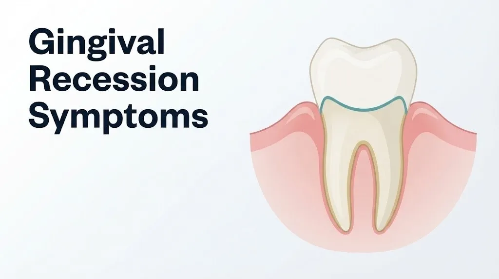
The observable symptoms of gum recession are those a person can detect at home: teeth that look longer than they used to, a thin or absent band of pink gum tissue near the tooth base, and in advanced cases, a distinctly yellowish or brownish surface visible near the gumline where the root becomes exposed.
What makes these visual signs clinically meaningful is understanding what normal gingival anatomy looks like. Healthy gingival tissue ends at the cementoenamel junction (CEJ), the point where the tooth’s white enamel crown meets the yellower cementum-covered root. When the free gingival margin migrates even 1 to 2 millimeters apically (toward the root tip) from this junction, the color change from white enamel to yellowish cementum becomes faintly visible. By the time most people notice the tooth “looking longer,” the free margin has already migrated several millimeters.
The tactile symptoms are often noticed before the visual ones. Many people first notice a rough or sensitive spot at the gumline, a feeling of “exposure” when drinking cold water, or a notch-like groove at the base of a tooth that was not there before. These sensory changes happen because the cementum covering the root surface is thinner (about 50 to 100 micrometers) than the overlying enamel (up to 2.5 millimeters), and once the gum margin migrates below the CEJ, the much thinner cementum wears through quickly, exposing dentin and its fluid-filled tubular channels directly to oral stimuli.
Women tend to notice gingival sensitivity changes earlier than men, partly because estrogen influences the density of gingival nerve endings and partly because women in general report pain symptoms earlier in the disease course than men do, a pattern documented across multiple periodontal epidemiological studies published in the Journal of Periodontology.
What Is Gingival Recession and Why Does It Progress?
Gingival recession is the apical displacement of the free gingival margin beyond the cementoenamel junction, exposing the root surface of the tooth to the oral environment, a process that involves the irreversible loss of the periodontal attachment apparatus including the junctional epithelium, periodontal ligament fibers, and the alveolar bone that supports the tooth.
Understanding why recession progresses requires understanding what is actually being destroyed. Picture the periodontal attachment as a three-layer anchor system. The outermost layer is the gingival epithelium. Below it is the connective tissue attachment, composed of collagen fiber bundles running between the cementum of the root and the alveolar bone. The deepest layer is the alveolar bone itself. When gram-negative anaerobic bacteria including Porphyromonas gingivalis, Treponema denticola, and Fusobacterium nucleatum colonize the subgingival sulcus, their lipopolysaccharide cell wall components activate local macrophages and T-lymphocytes.
These activated immune cells release interleukin-1 beta (IL-1beta) and tumor necrosis factor-alpha (TNF-alpha), which stimulate the production of matrix metalloproteinases (MMPs) including MMP-1, MMP-8, and MMP-13. These enzymes degrade the type I collagen fibers of the periodontal ligament and the connective tissue attachment. Simultaneously, IL-1beta and prostaglandin E2 activate osteoclasts through the RANKL/OPG pathway, driving alveolar bone resorption apically. As bone and connective tissue are lost, the gingival margin has no underlying support structure and migrates apically. The result is the exposed root surface visible as a “longer tooth.”
Think of it like a retaining wall losing its foundation. The gum tissue is the visible wall. The bone and connective tissue are the structural fill behind it. Once the fill is gone, the wall collapses downward, no matter how intact the surface looks from the front.
According to the 2017 AAP/EFP World Workshop on Periodontal and Peri-Implant Diseases and Conditions, periodontitis-related recession is classified within a staging and grading system that accounts for severity (Stage I through Stage IV) and rate of progression (Grade A through Grade C), replacing the older chronic and aggressive periodontitis terminology.
Early Gum Recession Symptoms: What to Watch for First
Early gum recession symptoms are subtle and frequently dismissed. The three most reliably early clinical signs are a very slight increase in cold sensitivity at specific teeth, barely visible lengthening of one or two teeth compared with adjacent teeth, and the faintest beginning of triangular dark spaces at the base of the interdental contacts between teeth.
Most people do not notice these earliest signs because they develop gradually over months to years. The change in tooth appearance is only obvious when comparing photographs taken years apart, or when a dentist measures the distance from the CEJ to the free gingival margin with a calibrated periodontal probe. In a healthy mouth, the free gingival margin sits 1 to 3 mm coronal (above) to the CEJ, or at the CEJ itself. Even 1 mm of recession is clinically detectable and recordable, though often invisible to the untrained eye.
Early-stage recession symptoms to monitor at home:
- A new sensation of cold air or cold water hitting a specific tooth more intensely than it used to
- The appearance of a very faint yellowish tinge at the base of one or two teeth near the gumline
- A slightly rough or grittier surface texture when running your tongue along the gumline at a specific tooth
- Small triangular dark areas (“black triangles”) beginning to form between your upper front teeth, where the gum used to fill that space completely
- Gum tissue that appears slightly thinner or more translucent at a specific tooth compared to neighboring teeth
- Mild soreness or tenderness at the gumline when pressing on the gum tissue, even without brushing
In adolescents and young adults, early recession symptoms are particularly easy to miss because the tissue often appears healthy except for a subtle apical shift of the gingival margin. A 2019 review published in the Journal of Clinical Periodontology reported that recession affecting at least one tooth is present in approximately 88% of adults over 65 and in approximately 50% of adults aged 18 to 64, indicating that early-stage recession is far more common in younger adults than most people assume.
What Does Gum Recession Feel Like From Day to Day?
Day-to-day, gum recession feels different depending on which stage the tissue loss has reached. In the early stages, it feels like nothing at all for much of the day, punctuated by brief, sharp sensitivity when something cold, sweet, or acidic contacts the tooth near the gumline.
As recession progresses to a moderate stage, that intermittent sensitivity becomes more predictable and more intrusive. Cold water triggers it. Sour foods trigger it. Sometimes just breathing cold winter air through the mouth is enough to produce a sharp, fleeting zing at the affected tooth. This sensation is produced by the hydrodynamic theory of dentin hypersensitivity: fluid inside the dentinal tubules (microscopic channels running through the dentin toward the pulp) is displaced by external thermal, osmotic, or tactile stimuli, and this fluid movement activates mechano-sensitive nerve fibers (specifically A-delta and C-fibers) within the dental pulp, producing a brief, sharp pain response.
Think of the dentinal tubules like tiny hydraulic lines running from the root surface to the tooth’s nerve center. When cold or sweet substances hit the exposed root, they shift the fluid inside those lines. That sudden shift rings the alarm bell inside the pulp.
Moderate to advanced gum recession may also produce a constant mild ache at the gumline, sensitivity to pressure when chewing, and discomfort when using a medium or firm toothbrush. Some people describe the sensation as a persistent rawness or tenderness at one or more teeth, even between meals.
Quick Tip:
- Keep a 7-day symptom log noting which tooth is sensitive, what triggered it (cold, heat, sweet, pressure, air), and how long the sensation lasted. This information is directly useful to a dentist assessing recession severity and dentin hypersensitivity impact.
- A sharp sensation lasting less than 30 seconds after a cold stimulus is typical of dentin hypersensitivity. A dull, lingering ache persisting more than 30 seconds after the stimulus is removed can indicate pulpal inflammation, which warrants separate evaluation by a dentist or endodontist.
- For older adults taking blood pressure medications such as amlodipine or nifedipine (calcium channel blockers), drug-induced gingival changes can alter the normal sensitivity pattern and mask recession-related symptoms.
Key Takeaway: Gingival recession symptoms often begin as fleeting cold sensitivity at specific teeth and progress to persistent sensitivity and visible root exposure. The first noticeable sensation is the most actionable moment: that is when intervention is easiest and most effective.
Gum Recession Sensitivity Symptoms: Why Your Teeth Hurt
Gum recession sensitivity symptoms, specifically the sharp or stinging pain experienced at exposed root surfaces, occur because the cementum layer covering the root is thin enough to wear through with minimal abrasion, exposing the underlying dentin and its approximately 15,000 to 65,000 dentinal tubules per square millimeter directly to oral stimuli.
Healthy teeth are covered by enamel on the crown portion and cementum on the root. Enamel has no nerve supply and no tubular structure, which is why enamel does not feel temperature changes the way an exposed root does. Cementum is thinner and softer than enamel. Once the gingival margin recedes and leaves cementum exposed, normal oral function (brushing, acidic food, thermal exposure) wears through cementum within months, leaving dentin directly exposed.
The pattern of sensitivity can help identify the cause of the recession:
| Sensitivity Pattern | Most Likely Cause of Recession |
|---|---|
| Sharp, brief cold sensitivity at one or two teeth | Localized toothbrushing trauma or orthodontic root movement |
| Sensitivity affecting multiple teeth in a specific arch | Toothbrush abrasion pattern, malposition, or chronic acid erosion |
| Sensitivity with visible gum inflammation around it | Periodontitis-driven recession with active infection |
| Sensitivity at a tooth with a groove or notch at the gumline | Abfraction lesion from excessive occlusal loading or bruxism |
| Sensitivity worst in the morning on waking | Bruxism (nighttime tooth grinding) with associated mechanical recession |
| Sensitivity appearing months after braces were removed | Orthodontic-related dehiscence of the labial bone plate |
People with bruxism (involuntary grinding and clenching, ICD-10: F45.8) experience a particularly aggressive form of recession sensitivity because the excessive occlusal forces concentrate tensile stress at the cervical third of the tooth, flexing the enamel-cementum junction and creating or widening abfraction lesions that accelerate root surface exposure. The American Academy of Periodontology recognizes bruxism-related mechanical occlusal trauma as a contributing co-factor in periodontal attachment loss and recession.
Bleeding Gums and Gum Recession: What the Connection Means
Bleeding gums during brushing or flossing, a symptom known clinically as bleeding on probing (BOP) or spontaneous gingival bleeding, is a reliable early indicator of gingival inflammation, and active gingival inflammation is one of the primary drivers of recession when it occurs repeatedly over months and years.
The connection between bleeding and recession works like this. Gram-negative anaerobic bacteria in subgingival plaque biofilm produce LPS that penetrates the sulcular epithelium, triggering capillary dilation, increased vascular permeability, and leukocyte infiltration in the underlying connective tissue. This inflamed, engorged tissue bleeds easily when contacted by a toothbrush bristle or floss. That same inflammatory state, sustained over time, produces the cytokine and MMP cascade described earlier, which destroys the connective tissue attachment and drives recession.
It is important to separate two distinct bleeding patterns. Bleeding that occurs consistently during brushing and resolves within 30 seconds, is bright red, and is present at multiple sites likely indicates generalized gingivitis that has not yet produced significant attachment loss. This type of bleeding is potentially reversible with improved plaque control. Bleeding that occurs spontaneously, is accompanied by swelling, redness, or a bad taste, is present at deeper areas around specific teeth, and is accompanied by any degree of tooth movement likely indicates active periodontitis with attachment loss and recession that has already begun.
The American Academy of Periodontology states that a bleeding on probing score above 25% across the dentition (meaning more than one in four teeth bleed when gently probed) indicates generalized gingival disease requiring professional evaluation and management.
Critically, people who smoke may not bleed despite active periodontal inflammation because nicotine causes vasoconstriction of the gingival capillaries, masking the bleeding sign. For smokers, the absence of bleeding is not reassurance. It can be a false negative.
Symptoms of Receding Gums Getting Worse: The Progression Pattern
The symptoms of receding gums getting worse follow a recognizable escalation pattern: increasing root surface exposure, worsening sensitivity that becomes harder to manage, interdental papilla loss, and eventually changes in the way teeth feel when biting, which signals alveolar bone loss is occurring alongside the tissue recession.
The Miller Classification (published in the Journal of Periodontology in 1985 by Dr. Preston D. Miller Jr.) provides the foundational framework for understanding how recession progresses in measurable stages:
| Miller Class | Description | Key Symptom Indicator |
|---|---|---|
| Class I | Recession not extending to the mucogingival junction. No bone or soft tissue loss in the interdental area. | Mild sensitivity, slight visual lengthening of one tooth |
| Class II | Recession extending to or beyond the mucogingival junction. No bone or soft tissue loss in the interdental area. | Moderate sensitivity, visible root exposure, no papilla loss |
| Class III | Recession extending to or beyond the mucogingival junction. Bone or soft tissue loss in the interdental area is present. | Visible black triangles, tooth sensitivity, slight mobility possible |
| Class IV | Recession extending to or beyond the mucogingival junction. Severe interdental bone and soft tissue loss present. Tooth malposition may be a factor. | Significant root exposure, mobility, severe sensitivity, tooth loss risk |
Class I and II recession have the best prognosis for complete root coverage with surgical treatment. Class III recession can achieve partial coverage. Class IV recession cannot be fully covered surgically. This progression pattern makes early detection genuinely consequential.
According to a longitudinal study published in the Journal of Clinical Periodontology, untreated recession progressed at a mean rate of approximately 0.1 mm per year in participants without active periodontitis, and substantially faster in those with concurrent periodontal infection or parafunctional habits.
Key Takeaway: Recession that crosses the mucogingival junction and produces interdental papilla loss (black triangles) represents a clinically important threshold. At this point, the prognosis for full surgical root coverage changes, making professional evaluation no longer optional.
Black Triangles Between Teeth and Gum Recession
Black triangles between teeth, the dark triangular spaces that appear at the base of the contacts between upper or lower front teeth when interdental papilla height is reduced, are one of the most recognizable and aesthetically distressing symptoms of moderate to advanced gum recession.
The interdental papilla is the small triangular wedge of gingival tissue that normally fills the contact area between adjacent teeth. Its height is maintained by the underlying alveolar bone and the connective tissue attachment on the adjacent tooth surfaces. When periodontal inflammation destroys the interdental bone and connective tissue, or when gingival recession causes the papilla to shrink apically, the space between the teeth that was once filled with pink tissue becomes an open dark triangle visible when smiling or speaking.
Black triangles are not only an aesthetic concern. They indicate that the interdental attachment apparatus has been compromised. Food trapping in these spaces increases the plaque burden in the interdental area, which accelerates further periodontal inflammation and can worsen the recession that created the triangle in the first place. This feedback loop is one of the reasons moderate-stage recession tends to progress faster than early-stage recession without intervention.
Black triangles can also result from orthodontic tooth movement that separates previously close teeth without adequate time for papilla to regenerate, or from crown placement or veneer preparations that alter the contact point relationship between teeth. In these mechanical or restorative cases, the papilla loss is not driven by periodontal infection but the clinical appearance is similar.
For older adults, black triangle formation is compounded by the natural reduction in papilla height that occurs with age even in periodontally healthy individuals, a phenomenon documented in the Journal of Periodontology. This means black triangles in adults over 65 require careful clinical evaluation to determine whether they reflect age-related physiological change, active periodontal disease, or both.
Gum Recession and Tooth Mobility: A Late-Stage Warning
Tooth mobility in the context of gum recession is a late-stage symptom that indicates substantial loss of alveolar bone support, and it should be evaluated by a licensed periodontist promptly once detected.
A tooth is held in its socket by the periodontal ligament, a network of collagen fiber bundles running between the cementum of the root and the inner wall of the alveolar bone socket. The alveolar bone provides the structural housing. When the inflammatory cascade of chronic periodontitis has destroyed enough alveolar bone and periodontal ligament fibers, the tooth no longer has adequate support against lateral forces. The result is detectable movement when the tooth is pressed horizontally or vertically, measured clinically on a Tooth Mobility Index scale from Grade I (slight, less than 1 mm horizontal movement) through Grade III (severe mobility in multiple directions including vertical displacement).
The critical clinical distinction is between physiological and pathological mobility. All teeth have a tiny degree of physiological mobility (0.1 to 0.2 mm) because the periodontal ligament fibers act as a hydraulic cushion. Mobility beyond this range indicates structural compromise.
Recession-associated tooth mobility symptoms to recognize:
- A tooth that feels slightly “springy” or gives slightly when you press it with your tongue
- A feeling that the tooth moves when biting down firmly
- A visible wobbling of the tooth when touched
- A tooth that has shifted position noticeably relative to adjacent teeth
- Pain or discomfort when biting on that tooth that was not present previously
The American Academy of Periodontology staging system classifies periodontitis involving tooth mobility of Grade II or greater at its most severe stage (Stage IV), which requires complex multidisciplinary treatment planning involving a periodontist, and potentially an oral surgeon or prosthodontist.
For patients taking bisphosphonate medications (alendronate, zoledronic acid) for osteoporosis or cancer treatment, tooth mobility in the context of any dental procedure carries an additional risk: medication-related osteonecrosis of the jaw (MRONJ). This population should disclose their medication history to their dentist before any periodontal evaluation or treatment planning.
Key Takeaway: Tooth mobility associated with gum recession means alveolar bone loss is severe enough to compromise structural tooth support. This is the clinical threshold at which a periodontist referral is not a suggestion but a clinical necessity.
Hormone-Related Gum Recession Symptoms
Hormone-related gum recession symptoms are driven primarily by the effects of progesterone and estrogen on gingival vascular permeability, prostaglandin sensitivity, and epithelial keratinization, and they are most pronounced during pregnancy, puberty, menstrual cycles, and the perimenopausal transition.
During pregnancy, rising progesterone levels amplify the gingival tissue’s inflammatory response to plaque bacteria. Progesterone increases vascular permeability in the gingival capillaries, increases sensitivity to prostaglandin E2, and promotes growth of certain Bacteroides species (including Prevotella intermedia) that thrive in a progesterone-rich environment. The result is a condition called pregnancy gingivitis, affecting up to 70% of pregnant women according to a review published in the Journal of Clinical Periodontology. Without adequate plaque control, sustained pregnancy gingivitis can progress to attachment loss and the beginning of recession.
During puberty, similar hormonal shifts produce exaggerated gingival responses to even minimal plaque accumulation. Adolescents may experience bleeding and gingival swelling disproportionate to the amount of plaque present, a phenomenon called puberty gingivitis, which can contribute to early recession if plaque control is not established.
During the perimenopausal and postmenopausal periods, declining estrogen levels reduce the keratinization and thickness of the gingival epithelium, making it more vulnerable to mechanical trauma and less resistant to bacterial penetration. Postmenopausal women on hormone replacement therapy (HRT) have been shown in observational studies published in the Journal of Periodontology to have lower rates of clinical attachment loss compared with those not on HRT, suggesting an estrogen-protective effect on periodontal tissue integrity.
Women noticing increased gum sensitivity, increased bleeding, or visible gum changes that correlate with specific points in their menstrual cycle, or that appeared or worsened during pregnancy, should raise these observations specifically with their dentist and their obstetrician-gynecologist at the same visit, as both the dental and hormonal dimensions require coordinated management.
Diabetes and Gum Recession Symptoms
Gum recession in people with diabetes mellitus tends to present more severely, progress faster, and respond less predictably to conventional treatment than recession in people without diabetes, due to the specific impairment of immune cell function caused by chronic hyperglycemia.
The mechanism is specific and well-established. Chronically elevated blood glucose promotes advanced glycation end-products (AGEs), which bind to receptors (RAGE) on gingival macrophages and endothelial cells, driving excessive and dysregulated production of IL-1beta, TNF-alpha, and IL-6. Simultaneously, the hyperglycemic environment impairs neutrophil chemotaxis and oxidative burst, meaning the white blood cells responsible for clearing bacterial biofilm from the gingival sulcus do not function effectively. The result is a compromised host defense that allows periodontal bacteria to drive deeper inflammation and faster connective tissue destruction.
According to the National Institute of Dental and Craniofacial Research, people with poorly controlled type 2 diabetes (HbA1c above 7.0%) are approximately three times more likely to have severe periodontitis compared with people without diabetes. Periodontitis, in turn, worsens glycemic control by increasing systemic inflammatory burden, creating a bidirectional relationship.
In people with diabetes, gum recession symptoms may be complicated by:
- Xerostomia (dry mouth) from reduced salivary flow, which accelerates root surface caries on newly exposed cementum
- Delayed wound healing after any periodontal procedure, including scaling and root planing
- More rapid progression from early to moderate recession staging compared with non-diabetic peers
- Greater tooth mobility at equivalent recession depths due to more extensive alveolar bone loss
- Susceptibility to periodontal abscess formation, which can present as acute swelling and pain at a recessed site
Any person with type 2 diabetes who is told they have gum recession should inform both their dentist and their primary care physician or endocrinologist, and ask that periodontal status be part of their routine diabetes management review. A licensed periodontist experienced with diabetic patients should be involved in treatment planning.
Gum Recession in Young Adults and After Braces
Gum recession in young adults and in people who have completed orthodontic treatment is more common than most patients and even many general dentists expect, and the mechanism is distinctly different from the periodontitis-driven recession that dominates in older adults.
In young adults without significant periodontal disease, the most common recession mechanism is toothbrush abrasion combined with anatomical thin-bone phenotype. Some individuals have naturally thin alveolar bone covering the roots of their lower front teeth or upper canines, called a dehiscence (a gap in the labial bone plate) or fenestration (a window-like defect). These thin bone areas provide minimal soft tissue support. When combined with aggressive horizontal brushing technique, recession at these specific teeth can begin in the late teenage years and progress steadily through the 20s and 30s.
After orthodontic treatment, recession risk increases for specific anatomical reasons:
- Orthodontic tooth movement that expands the dental arch or moves teeth buccally (outward) beyond their natural bone envelope can thin or breach the labial bone plate.
- Teeth moved rapidly through bone may outpace the bone’s remodeling capacity, leaving areas of thin or absent labial bone.
- Increased overbite correction that changes tooth angulation can place the root in proximity to the labial cortical plate.
- Post-treatment without a retainer allowing teeth to shift back can create stress forces at the gingival margin.
Research published in the Journal of Clinical Periodontology found that 10 to 25% of orthodontically treated patients develop clinically visible recession at one or more teeth within five years of completing treatment, most commonly at the lower anterior teeth.
Young adults noticing increased cold sensitivity at one or two lower front teeth after orthodontic treatment, or seeing one tooth appear slightly longer than its neighbors in that region, should bring this to the attention of their general dentist, who can measure the degree of recession and refer to a licensed periodontist for evaluation if progression is suspected.
Gum Recession Causing Loose Teeth: What It Signals
When gum recession reaches the stage where teeth feel loose, the underlying issue is no longer just the gingival margin position. The alveolar bone, the jaw bone that forms the socket housing each tooth root, has been resorbed to a degree that the remaining bone surface area cannot adequately distribute normal biting and chewing forces.
The relationship between bone loss and recession is not always linear. Moderate recession at the gingival margin does not necessarily mean the bone immediately beneath the gum has been equally resorbed. In some cases, the tissue recedes faster than the bone is lost (a pattern seen in mechanical trauma-driven recession). In other cases, bone resorption drives tissue recession from below (the pattern typical of periodontitis). When tooth mobility appears alongside recession, it means bone loss has become clinically relevant regardless of which process came first.
Loose teeth from recession-associated bone loss may present as:
- A subtle springiness when pressing a tooth with your fingertip horizontally
- A tooth that has visibly drifted or tilted from its original position over weeks to months
- A change in the way teeth come together when biting (shift in occlusion, new food trapping between specific teeth)
- Pain when biting on a specific tooth that previously felt stable
- A tooth that feels “higher” than usual, which can mean the bone is no longer providing adequate vertical support
Tooth mobility in the context of recession is a particularly serious finding in adults over 50, where it can indicate Stage III or Stage IV periodontitis according to the 2017 AAP/EFP World Workshop classification. At Stages III and IV, tooth loss risk is substantial without active treatment by a licensed periodontist and potentially an oral surgeon.
For patients on corticosteroid therapy for autoimmune conditions, bone density may already be compromised systemically, making gingival recession-associated bone loss progress faster and mobility appear earlier in the disease course.
Key Takeaway: Tooth mobility appearing alongside visible gum recession is a signal that alveolar bone loss is clinically significant and that a licensed periodontist needs to evaluate the case for staging and treatment planning without delay.
When to See a Dentist for Gum Recession
See a dentist for gum recession as soon as you notice any of the following: consistent cold sensitivity at a specific tooth that persists for more than two weeks, visible lengthening of one or more teeth, a notch or groove at the base of a tooth, triangular dark spaces forming between teeth, or any degree of tooth movement that you can detect.
The right time to seek care is earlier than most people think. By the time gingival recession is clearly visible to an untrained eye in the bathroom mirror, it has typically been progressing for months to years. The 1 to 2 millimeters of recession that produces visible aesthetic change represents a tissue loss that has already crossed the threshold where the body will not restore it spontaneously.
When contacting a dental provider, prioritize finding a dentist who will measure your recession clinically with a calibrated millimeter probe and document the recession depth at specific tooth surfaces. Ask specifically whether a referral to a licensed periodontist (a dentist who has completed an additional three years of accredited specialty training in the diagnosis and treatment of periodontal disease) is appropriate for your situation.
Schedule an appointment with your general dentist within two to four weeks for:
- New cold sensitivity at a specific tooth or teeth
- First-time noticing teeth appear longer
- Bleeding that has persisted for more than two weeks despite improved brushing and flossing
- A slight rough spot or groove at the base of a tooth
Request an expedited appointment (within one to three days) for:
- Visible swelling or pus at the gumline
- A tooth that has suddenly become mobile
- Pain at a specific tooth that is worsening day by day
- A tooth that has visibly shifted position
Adults with systemic conditions including type 2 diabetes, rheumatoid arthritis, or Sjogren’s syndrome (which causes profound xerostomia) should not use a standard self-monitoring approach for gum health. These conditions accelerate periodontal destruction. A proactive schedule of three to four periodontal maintenance visits per year is the appropriate clinical standard for this population, as stated in AAP clinical practice guidelines.
How Dentists Diagnose Gingival Recession
Dentists diagnose gingival recession through a structured clinical examination that measures the position of the free gingival margin relative to the cementoenamel junction at multiple sites per tooth, combined with radiographic assessment of the underlying alveolar bone.
The standard diagnostic tool is the calibrated periodontal probe, a slender metal instrument with millimeter markings. The dentist or dental hygienist places the probe into the gingival sulcus (the shallow groove between the tooth and the gingival margin) at six designated locations around each tooth: mesiobuccal, mid-buccal, distobuccal, mesiolingual, mid-lingual, and distolingual. The measurements recorded include the probing pocket depth (how deep the probe goes before resistance) and the recession depth (how far the gingival margin has migrated below the CEJ). Adding these two numbers gives the clinical attachment level (CAL), the most accurate and reproducible measure of total periodontal attachment loss.
The diagnostic process from first visit to confirmed staging typically unfolds as:
- Full-mouth periodontal charting: probing depths, recession depths, and clinical attachment levels recorded at six sites per tooth across the entire dentition
- Bleeding on probing assessment: percentage of sites that bleed within 30 seconds of probing, which indicates sites with active inflammation
- Tooth mobility testing: horizontal and vertical mobility graded at each tooth using a scale of 0 (no detectable mobility) through III (severe mobility)
- Furcation assessment: for multi-rooted teeth (molars), whether bone loss has reached the area where roots divide, graded Class I through Class III
- Periapical dental radiographs: reveal the height of alveolar bone relative to the CEJ, identify bone defects, and show furcation involvement in molars
- Panoramic radiograph: provides an overview of bone levels across the entire dentition and identifies any pathology not visible clinically
- Comprehensive diagnosis and staging: applying the AAP/EFP classification to produce a Stage (I through IV) and Grade (A through C) diagnosis that determines treatment urgency and complexity
Cone beam computed tomography (CBCT) may be ordered in cases of complex bone defect morphology or when surgical planning requires three-dimensional bone architecture data. According to the American Academy of Periodontology’s 2018 clinical practice guidelines, CBCT is appropriate for surgical planning in selected complex cases but is not a routine screening tool for recession diagnosis.
Emergency Symptoms: When to Call 911 or Go to the ER
Certain symptoms associated with advanced gingival recession and periodontal infection require immediate emergency evaluation. Do not wait to see if these resolve on their own.
Call 911 or go to the nearest emergency room immediately if you experience:
- Swelling of the floor of the mouth (the area under the tongue), the neck, or the jaw below a painful tooth area, as this can indicate Ludwig’s angina, a rapidly spreading deep space infection of the head and neck that can obstruct the airway within hours
- Difficulty swallowing or breathing, or the sensation that your airway is narrowing, in the context of severe gum pain or dental infection
- High fever (above 101.5 degrees Fahrenheit / 38.6 degrees Celsius) combined with jaw, neck, or throat swelling and severe dental or gum pain
- Trismus (inability to fully open the mouth) developing rapidly in the context of a known dental infection or severe gum disease
- Severe and worsening pain radiating from the jaw to the neck, combined with systemic symptoms of infection (chills, sweating, rapidly worsening illness)
Go to a dental emergency clinic or hospital emergency room within hours (not days) for:
- A dental abscess (localized swelling, pus, severe throbbing pain at a specific tooth area, often associated with advanced periodontal disease) that is not yet spreading to the neck or floor of the mouth but is causing worsening pain and swelling despite any over-the-counter pain relief
- Sudden tooth loss or tooth fracture at a site with known advanced gum recession and significant bone loss
Spreading dental infection that begins in the periodontal tissues of severely recessed teeth can advance quickly into the deep fascial spaces of the head and neck. These presentations can indicate necrotizing fasciitis, mediastinitis, or airway compromise, and require emergency medical assessment, not an urgent care appointment or a phone call to your dentist.
Frequently Asked Questions About Gingival Recession Symptoms
What are the first signs of gum recession?
The first signs of gum recession are brief, sharp sensitivity to cold or sweet stimuli at one or two specific teeth near the gumline, and a very faint visual lengthening of those teeth compared with adjacent teeth.
Many people also notice a slightly rough texture when running their tongue along the gumline where the gum has begun to pull back from the cementoenamel junction.
If you notice either of these changes persisting for two or more weeks, contact your general dentist for a clinical examination with periodontal probing measurements.
Can gum recession happen without any pain?
Gum recession can and frequently does develop completely without pain, particularly when it is driven by mechanical trauma such as aggressive brushing rather than by active periodontal infection.
The absence of pain does not mean the tissue loss is not occurring or progressing: recession that is painless at an early stage can still reach advanced staging and compromise tooth stability over years without treatment.
The only reliable way to detect painless recession is clinical measurement of recession depth at each tooth surface by a dentist or dental hygienist using a calibrated periodontal probe.
How quickly does gum recession progress if untreated?
The rate of gum recession progression varies by cause and individual risk factors, but observational data published in the Journal of Clinical Periodontology indicates that untreated recession progresses at approximately 0.1 mm per year on average in patients without active periodontitis, and significantly faster in those with concurrent periodontal infection, tobacco use, uncontrolled diabetes, or bruxism.
Even slow progression of 0.1 mm per year accumulates to 1 mm over a decade, which is clinically meaningful when the starting point is already 1 to 2 mm of recession.
Active periodontitis-driven recession can progress at rates well above this average during periods of acute infection, making active monitoring essential.
What does gum recession look like at the early stage?
At the early stage, gum recession looks like one or two teeth that appear very slightly longer than the teeth on either side, with a faint transition visible at the gumline where the white enamel crown meets the slightly yellower or duller root surface.
The gum tissue itself may appear slightly thinner or more translucent at the affected tooth, and tiny triangular dark spaces may be barely visible at the base of the contact point between the affected tooth and its neighbor.
These changes are subtle enough that most people do not notice them without side-by-side comparison of dental photographs taken one to two years apart.
Can receding gums grow back on their own?
Receding gums cannot grow back on their own once recession has occurred, because the cells that form the periodontal attachment apparatus (junctional epithelium, periodontal ligament fibroblasts) do not regenerate spontaneously once they have been destroyed by inflammation or mechanical trauma.
The gingival margin does not regenerate apical tissue by healing: the only natural response to recession is stabilization (no further progression if the cause is controlled), not reversal.
Surgical techniques performed by a licensed periodontist, including connective tissue grafting, free gingival grafting, and the pinhole surgical technique, can restore gum tissue coverage over exposed root surfaces, but these require specialist evaluation and cannot be replicated by home care alone.
When is gum recession serious enough to need a periodontist?
Gum recession is serious enough to require a referral to a licensed periodontist when recession measures 2 mm or more from the cementoenamel junction at any tooth surface, when recession is accompanied by any degree of tooth mobility, when black triangles have formed between teeth indicating interdental papilla loss, or when gum sensitivity is severe enough to interfere with eating or normal oral hygiene.
A licensed periodontist should also be involved if recession is progressing despite corrected brushing technique and professional dental cleanings, if the patient has systemic conditions that accelerate periodontal destruction (type 2 diabetes, rheumatoid arthritis, HIV), or if surgical root coverage is being considered.
General dentists can and should perform initial recession assessment and monitoring, but surgical treatment planning and complex periodontal staging require the expertise of a board-certified periodontist.
Putting It All Together
Gingival recession is a progressive, irreversible tissue loss that announces itself through specific, recognizable symptoms at every stage. Cold sensitivity at a specific tooth, teeth that look slightly longer than before, thin or disappearing gum tissue at the gumline, black triangles between front teeth, and any degree of tooth mobility are all part of a clinical story that gets easier to treat the earlier it is caught.
The most actionable thing you can do right now is take stock of what you are noticing in your own mouth. If any of the symptoms described in this article sound familiar, particularly sensitivity that has appeared in the past few weeks or teeth that have visually changed, make an appointment with your general dentist and ask specifically for a full-mouth periodontal chart with recession measurements. That 20-minute examination generates more useful clinical information than any symptom checker can provide.
The goal of early detection is not to alarm you. It is to give you the clinical information to act before the window for the best outcomes closes.