L4-L5 Pain Symptoms: What They Mean and What to Do
L4-L5 pain symptoms range from a deep ache in the lower back to electric, shooting leg pain, foot numbness, toe weakness, and in serious cases, loss of bladder or bowel control that requires emergency surgery the same day. The specific symptoms you experience depend on exactly which structure is damaged at the L4-L5 spinal level and whether the L4 nerve root, the L5 nerve root, or the entire bundle of nerve roots below this level is being compressed.
The National Institute of Neurological Disorders and Stroke (NINDS) identifies lumbar disc herniation at L4-L5 as one of the most frequently occurring spinal disorders in adults, with the L4-L5 and L5-S1 levels together accounting for the majority of all lumbar disc herniations requiring clinical attention. This spinal segment bears the greatest mechanical load in the entire lumbar spine due to its position at the lumbosacral transition zone.
This article covers every symptom pattern produced by L4-L5 pathology, the specific anatomical reason each symptom occurs, how to distinguish L4 nerve root involvement from L5 nerve root involvement, population-specific differences in how these symptoms present, the one emergency pattern that cannot wait, and clear guidance on when to monitor at home versus when to seek care.
L4-L5 Pain Symptoms: What They Are and Why They Happen
L4-L5 pain symptoms are produced by damage to or compression of the structures at the fourth and fifth lumbar spinal level, including the intervertebral disc, the nerve roots passing through the spinal canal at this level, and the facet joints and surrounding ligaments.
The symptoms divide into two broad categories. The first is local spinal pain: a deep, aching pain in the lower lumbar region produced by stimulation of nociceptors (pain-sensing nerve endings) in the annulus fibrosus, facet joint capsules, and paraspinal muscles. The second is radicular pain and neurological deficit: symptoms that travel into the buttock, thigh, lower leg, or foot because a nerve root exiting the spinal canal is being mechanically compressed or chemically irritated.
What makes L4-L5 symptoms clinically distinct is the specific set of nerves at risk. The L4 nerve root and L5 nerve root supply distinct areas of the lower limb with sensation, movement, and reflex function. When either root is compromised, the resulting symptoms follow predictable anatomical patterns, called dermatomal (sensory), myotomal (motor), and reflex arc distributions. Understanding which pattern you are experiencing tells a clinician which nerve root is involved.
Key L4-L5 symptoms across all pathology types:
- Lower lumbar back pain localized between the beltline and the base of the spine
- Unilateral (one-sided) or bilateral (both sides) buttock pain
- Pain radiating down one leg along the sciatic nerve distribution
- Numbness or tingling in the thigh, lower leg, or foot
- Weakness of specific leg muscles depending on which nerve root is compressed
- Reduced or absent patellar tendon reflex (knee jerk) with L4 root involvement
- Difficulty lifting the foot or toes (foot drop) with L5 root involvement
- In severe central disc herniation: new loss of bladder or bowel control (emergency)
Sedentary individuals who spend more than six hours daily sitting have consistently elevated intradiscal pressure at L4-L5 compared to those who alternate sitting with standing and walking, because the lumbar spine in a seated position loses its natural lordotic curve and increases compressive and shear loading on the posterior disc and annulus fibrosus.
The L4-L5 Spinal Segment: Anatomy and Why It Fails So Often
The L4-L5 intervertebral disc is the most mechanically stressed intervertebral disc in the lumbar spine, positioned at the point where the mobile lumbar column meets the relatively fixed pelvis and sacrum.
The disc itself consists of two distinct structures. The central nucleus pulposus is a gel-like structure composed of water (approximately 80% in young adults), proteoglycans (principally aggrecan), and type II collagen. It functions as a hydrostatic pressure distributor, spreading compressive loads evenly across the vertebral endplates. The outer annulus fibrosus is a series of concentric collagen fiber rings (type I collagen) oriented alternately at 30-degree angles, providing the tensile strength that contains the nucleus under compression. The two structures work together like a sealed water balloon: the nucleus absorbs compression and the annulus prevents it from bulging outward.
At L4-L5 specifically, the mechanical demands are the highest in the lumbar spine for two reasons. First, this level bears the full compressive weight of the upper body, both in static posture and during dynamic loading from bending, lifting, and rotation. Second, the transition from the mobile lumbar vertebral column to the fixed sacrum concentrates motion at L4-L5 and, to a lesser extent, L5-S1. The segment must absorb the full angular change between lumbar movement and sacral fixation in every flexion-extension cycle.
Think of the L4-L5 disc like the hinge point on a door that opens thousands of times a day. Every other part of the door is rigid, so the full wear concentrates at the hinge. Over years, the annular fibers fatigue, the nucleus dehydrates, and the disc loses height, changing how load is distributed to the facet joints and nerve root canals.
According to the North American Spine Society (NASS) clinical practice guidelines, posterolateral disc herniation at L4-L5 accounts for the majority of single-level lumbar disc herniations producing radiculopathy in working-age adults, reflecting this segment’s uniquely high mechanical demand.
Women who are pregnant, individuals with BMI above 30, and heavy-lift workers have accelerated mechanical loading at L4-L5 relative to baseline population norms, placing them at elevated risk for both acute herniation and faster progressive disc degeneration.
L4-L5 Lower Back Pain: Location, Character, and Triggers
L4-L5 lower back pain is typically felt as a deep, aching, or pressure-like sensation localized to the midline or just lateral to the midline of the lower lumbar spine, at the level of or just above the beltline, distinguishable from sacroiliac joint pain (which is felt more laterally at the dimples of the back) and from muscular spasm (which is felt more broadly across the paraspinal region).
The character of the pain provides diagnostic information. Pain that is worse with lumbar flexion (bending forward, sitting for prolonged periods) and better with standing or walking in a short arc suggests primarily discogenic pain, consistent with annular irritation or disc herniation, because lumbar flexion increases intradiscal pressure and posteriorly displaces disc material toward the nerve roots. Pain that is worse with lumbar extension (standing, walking, lying flat) and better with lumbar flexion (sitting, leaning forward, cycling) suggests primarily facet joint or stenotic pain, because extension closes the spinal canal and compresses the facet joints.
Acute-onset severe lower lumbar pain following a bending, lifting, or twisting activity, with a sensation the person often describes as “something gave way,” suggests annular tear or acute disc herniation. This typically produces intense local spasm of the erector spinae muscle group (iliocostalis lumborum, longissimus thoracis) and multifidus muscle as the paraspinal muscles reflexively contract to splint the injured segment, which itself can be more painful than the original disc injury.
Mayo Clinic notes that lower back pain from lumbar disc herniation typically reaches its most severe intensity within 24 to 72 hours of the precipitating event, as the inflammatory cascade from phospholipase A2 and prostaglandin E2 peaks in the epidural space around the injured disc and adjacent nerve root.
Men with occupational heavy loading histories (construction, warehouse, agricultural work) present more frequently with acute L4-L5 disc herniation superimposed on pre-existing degenerative changes, compared to women in equivalent age groups, where the distribution shifts toward degenerative disc disease and facet-mediated pain as the more common presentation.
L4-L5 Leg Pain and Sciatica: The Full Nerve Pathway
Leg pain from L4-L5 pathology travels through the sciatic nerve, the longest and widest peripheral nerve in the human body, formed by the convergence of nerve roots from L4 through S3 in the lumbosacral plexus posterior to the piriformis muscle.
When the L4 or L5 nerve root is compressed or chemically irritated by a herniated disc, the pain signal does not stay at the spine. It travels the full length of the nerve root and its downstream nerve fibers. This is called radicular pain, distinct from referred pain: radicular pain follows the specific anatomical path of the nerve root, while referred pain is a broader, less well-defined aching that does not follow a precise nerve distribution.
The radicular pain from L4-L5 herniation is often described as sharp, burning, electric, or shooting. It typically worsens with positions that increase disc pressure on the nerve root: coughing, sneezing, bearing down (Valsalva maneuver), and sustained sitting. It tends to improve with positions that reduce nerve root tension: lying with hips and knees slightly flexed, walking slowly (in cases where stenosis is not the primary cause), or lying on the side with a pillow between the knees.
The distinction between L4 and L5 radicular pain distribution is clinically precise:
| Feature | L4 Radicular Pattern | L5 Radicular Pattern |
|---|---|---|
| Pain location | Anterior thigh, medial lower leg, medial foot | Lateral lower leg, dorsum of foot, first to third toes |
| Sensory loss | Anterior thigh and medial lower leg | Lateral lower leg and dorsum of foot |
| Reflex affected | Patellar tendon reflex (diminished or absent) | No consistent reflex loss in most cases |
| Motor weakness | Quadriceps (knee extension), tibialis anterior (foot dorsiflexion) | Extensor hallucis longus (great toe extension), peroneus longus (foot eversion), gluteus medius (hip abduction) |
Pregnant women in the third trimester frequently develop L4-L5 radicular pain because the gravid uterus tilts the pelvis anteriorly, increasing lumbar lordosis and compressive loading on the posterior disc. Most cases resolve postpartum as pelvic alignment normalizes.
Key Takeaway: L4-L5 leg pain follows a specific nerve root pattern: L4 compression causes anterior thigh and medial lower leg symptoms with a diminished knee jerk reflex, while L5 compression causes lateral lower leg and top-of-foot symptoms with great toe weakness. Knowing which pattern you have tells your doctor which nerve root is involved.
L4 Nerve Root Compression: Specific Symptoms and Deficits
L4 nerve root compression produces a specific triad of symptoms: pain and sensory change in the anterior thigh and medial lower leg, weakness of the quadriceps femoris muscle (which extends the knee), and a diminished or absent patellar tendon reflex (knee jerk reflex).
The L4 nerve root exits the lumbar spinal canal through the L4-L5 intervertebral foramen, traveling anterolaterally to join the lumbar plexus and eventually contribute to the femoral nerve (via L2, L3, L4) and the sciatic nerve. When a posterolateral disc herniation at L4-L5 compresses the exiting L4 root, it disrupts the conduction of both sensory and motor signals along this specific neural pathway.
Sensory symptoms from L4 compression include numbness, tingling (paresthesia), or burning pain along the anterior (front) aspect of the thigh, continuing down the medial (inner) side of the lower leg to the medial foot and big toe. These symptoms may be intermittent at first, appearing with prolonged sitting or specific positions, before becoming more constant as compression worsens.
The motor weakness produced by L4 root compression affects knee extension, because the quadriceps femoris is the primary knee extensor and receives its principal motor supply from the L4 root via the femoral nerve. Clinically, this presents as difficulty rising from a chair without using the arms, difficulty going up stairs one leg at a time, or in severe cases, the knee buckling unexpectedly during walking because the quadriceps cannot maintain joint stability under load.
The American Academy of Orthopaedic Surgeons states that measurable motor weakness (less than four out of five muscle power on manual muscle testing) at presentation is one of the primary factors that accelerates the clinical decision toward surgical consultation, as persistent motor weakness beyond four weeks significantly reduces the probability of complete neurological recovery.
Older adults with pre-existing knee osteoarthritis may misattribute L4-mediated quadriceps weakness to their knee joint condition, delaying recognition of the neurological deficit. Any knee weakness appearing alongside lower back or thigh pain warrants spinal evaluation rather than knee-focused treatment alone.
L5 Nerve Root Compression: Specific Symptoms and Deficits
L5 nerve root compression produces the most functionally limiting neurological deficit associated with L4-L5 pathology: weakness of great toe extension, foot eversion, and hip abduction, combined with sensory changes over the lateral lower leg and the dorsum (top) of the foot, and notably, no consistent change in deep tendon reflexes.
The L5 nerve root traverses the lateral recess of the spinal canal at the L4-L5 level before exiting at the L5-S1 foramen. This traversing position makes it the nerve root most commonly compressed by a posterolateral or far-lateral L4-L5 disc herniation. The L5 root contributes to the sciatic nerve and is the primary motor supply to the extensor hallucis longus (EHL, which lifts the great toe), peroneus longus and brevis (which evert the foot), and gluteus medius (which abducts the hip and stabilizes the pelvis during walking).
The absence of a reliable reflex change with L5 compression is a clinically important point. The Achilles tendon reflex (ankle jerk) tests the S1 root, not L5. The patellar tendon reflex tests L4. There is no easily testable deep tendon reflex supplied primarily by L5, which means the neurological examination for L5 compression relies more heavily on manual muscle testing and sensory mapping than reflex testing. This is why L5 nerve root compression can be underdiagnosed if the clinician does not specifically test EHL strength and lateral lower leg sensation.
L5 compression also produces a distinctive gait disturbance called steppage gait, in which the person lifts the knee higher than normal with each step to clear the foot from the ground, compensating for the foot’s inability to dorsiflex adequately. This gait pattern is visible and can be observed by the person’s family or caregivers before the person themselves recognizes it.
Athletes in sports requiring explosive cutting, jumping, and lateral movement (basketball, football, soccer) who develop L5 compression may report it primarily as “hip weakness” or “hip instability” during lateral movements, because the gluteus medius dysfunction from L5 involvement reduces pelvic stability during single-leg loading.
Numbness and Tingling From L4-L5 Problems
Numbness and tingling from L4-L5 pathology occur because nerve root compression reduces or disrupts the normal conduction of sensory signals along the compressed nerve root’s axons, producing an abnormal sensory experience in the skin territory supplied by that root.
The specific sensation depends on the degree of nerve root compression. Partial compression, where the nerve root is irritated but not fully blocked, typically produces paresthesia: an active, spontaneous sensory experience of tingling, pins and needles, burning, or electrical buzzing. Complete or severe compression, where axonal conduction is substantially blocked, produces hypoesthesia or frank anesthesia: a reduction or complete absence of sensation in the affected skin territory. Both patterns can occur in the same patient at different stages of the same condition.
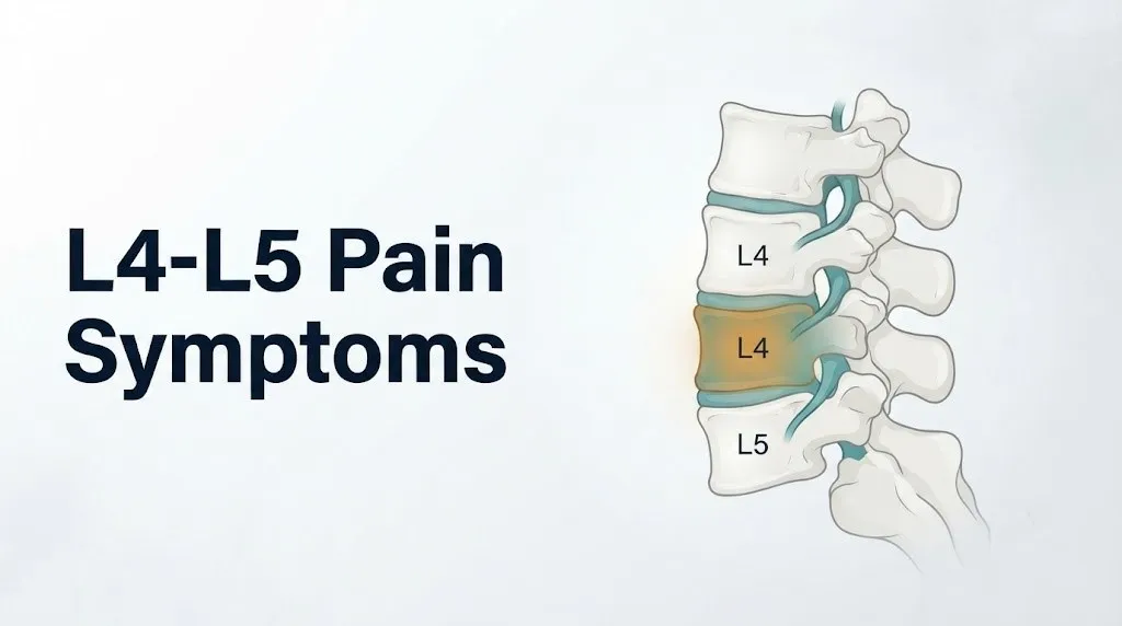
Paresthesia from L4 compression is felt in the anterior thigh, crossing the kneecap area, continuing along the inner surface of the lower leg to the inner ankle and medial foot. Paresthesia from L5 compression is felt over the outer surface of the lower leg, crossing the outer ankle, spreading across the top of the foot to the first through third toes.
The timing and constancy of numbness carries clinical information. Numbness that appears only with specific positions (prolonged sitting, forward bending) and resolves quickly with position change suggests intermittent compression of a nerve root that is not yet persistently compressed. Constant numbness that is present regardless of position suggests more severe and sustained nerve root compression, or in some cases, early axonal damage (axonopathy) that requires more urgent evaluation.
Quick Tip:
- Numbness in both feet simultaneously, or a stocking distribution of numbness in both lower limbs, is not typical of single-level L4-L5 pathology and should prompt evaluation for bilateral compression, peripheral neuropathy (e.g., from diabetes mellitus), or spinal cord pathology.
- Spreading or ascending numbness (starting in the foot and moving upward over hours or days) is not typical of disc herniation and warrants urgent neurological evaluation to exclude spinal cord compression or Guillain-Barre syndrome.
- Postmenopausal women with both lumbar disc disease and peripheral neuropathy from diabetes may have overlapping sensory symptoms that make dermatomal localization more difficult, requiring EMG/NCV testing for accurate root-level identification.
Key Takeaway: Numbness that is constant regardless of position and not improving over two to three weeks, or numbness that is spreading rather than stable, is beyond the self-monitoring threshold and warrants evaluation by a primary care physician or board-certified physiatrist within one to two weeks, not several months.
Muscle Weakness and Foot Drop From L4-L5 Compression
Muscle weakness from L4-L5 compression is the symptom that most determines the urgency of clinical evaluation, because weakness that persists beyond four weeks carries a higher risk of incomplete neurological recovery than pain or sensory symptoms.
Foot drop is the most clinically recognizable and functionally disabling weakness pattern produced by L4-L5 pathology. It occurs when L5 nerve root compression sufficiently impairs motor conduction to the tibialis anterior and extensor hallucis longus muscles to prevent active dorsiflexion of the foot and extension of the great toe. Without foot dorsiflexion, the foot hangs downward during the swing phase of walking, causing the person to drag the toe or adapt with the steppage gait pattern described in the L5 section.
Foot drop from L4-L5 disc herniation is a surgical urgency. The North American Spine Society clinical practice guidelines indicate that foot drop representing a new-onset motor deficit is one of the primary indications for expedited surgical consultation, typically within days to two weeks of onset rather than the standard six to eight week conservative management trial.
Assessing your own leg strength at home can help you communicate clearly with a provider:
To self-assess for L4-L5 motor weakness before your appointment:
- Stand barefoot on a flat floor and try to stand on your heels with your toes lifted off the ground. Inability to do this with one foot suggests possible tibialis anterior weakness (L4-L5, primarily L5).
- Try to extend (lift) your great toe upward toward your shin against gentle resistance from your own hand. Weakness here compared to the other foot suggests extensor hallucis longus weakness (L5 primary).
- Try single-leg squats: lower yourself slowly on each leg and notice whether one knee buckles or the hip drops. Knee buckling suggests quadriceps weakness (L4); hip dropping suggests gluteus medius weakness (L5).
- Walk across the room noticing whether one foot clears the floor normally or tends to slap down or drag.
- Write down which specific movements feel weak and bring this description to your appointment with a primary care physician, board-certified physiatrist, or board-certified orthopedic spine surgeon.
Individuals with diabetes mellitus should note that peripheral neuropathy from uncontrolled blood glucose can independently produce foot weakness and sensory changes, making it clinically important to distinguish lumbar radicular weakness from peripheral nerve weakness. EMG and nerve conduction velocity testing can reliably differentiate these two conditions.
Acute vs. Chronic L4-L5 Symptoms: How the Presentation Changes
Acute and chronic L4-L5 symptoms differ in their underlying pathological mechanism, their typical trajectory, and their response to treatment, and conflating the two leads to both mismanagement and missed opportunities for recovery.
Acute L4-L5 symptoms develop over hours to days, typically following a precipitating mechanical event (lifting, twisting, bending) or without a clear trigger in some cases. The dominant mechanism is acute herniation of the nucleus pulposus, where nucleus material extrudes through a tear in the annulus fibrosus and makes contact with a nerve root. The pain is intense, often rated seven to ten on the Visual Analog Scale, and is accompanied by severe paraspinal muscle spasm. Radicular symptoms (leg pain, numbness, weakness) may appear simultaneously or develop over the following 24 to 72 hours as the inflammatory response around the extruded disc material peaks.
The natural history of acute disc herniation is genuinely favorable in the majority of patients without severe neurological deficit. The Annals of Internal Medicine published findings from the Maine Lumbar Spine Study showing that the majority of patients with acute lumbar disc herniation and radiculopathy experienced substantial improvement in symptoms over the first three months without surgical intervention. The extruded disc material undergoes enzymatic degradation by macrophages over weeks to months, reducing its size and the degree of nerve root compression it produces.
Chronic L4-L5 symptoms develop insidiously over months to years, reflecting progressive disc degeneration (loss of disc height, desiccation, annular fissuring), facet joint arthrosis, and ligamentum flavum hypertrophy. The pain pattern is different: lower intensity but persistent, often bilateral, with less dramatic radicular symptoms but more consistent activity limitation. The episodic nature is characteristic: periods of relative comfort alternating with flare-ups triggered by specific activities, postures, or weather changes.
| Feature | Acute L4-L5 Presentation | Chronic L4-L5 Presentation |
|---|---|---|
| Onset | Hours to days | Months to years |
| Pain intensity | Severe (7 to 10/10 VAS) | Moderate and variable (3 to 6/10 VAS) |
| Radicular symptoms | Often prominent from onset | Intermittent, less severe |
| Provocating factors | Single mechanical event common | Accumulation of activity, posture |
| Natural history | Favorable in 80 to 90% without surgery | More variable, less complete resolution |
| Typical pathology | Nucleus pulposus herniation | Degenerative disc disease, facet arthropathy, stenosis |
L4-L5 Spinal Stenosis and Neurogenic Claudication Symptoms
L4-L5 spinal stenosis produces a symptom pattern called neurogenic claudication that is fundamentally different from disc herniation symptoms, and recognizing the difference determines which diagnostic pathway and treatment approach applies.
Neurogenic claudication is produced by ischemic compression of the cauda equina nerve roots within the narrowed central spinal canal or lateral recesses at L4-L5. As the spinal canal narrows from ligamentum flavum hypertrophy and facet joint overgrowth (osteophyte formation from degenerative arthrosis), the available space for the nerve roots decreases, particularly during lumbar extension and walking, when the ligamentum flavum folds inward further and the facet joints approximate. The nerve roots become transiently ischemic during these postures, producing the characteristic symptom pattern.
The neurogenic claudication pattern has a highly characteristic presentation:
- Bilateral leg pain, heaviness, aching, or weakness that develops after walking a specific distance (the “walking distance threshold,” which may be as short as half a city block in severe stenosis)
- Symptoms that are absent or minimal at rest and in sitting but develop reliably with standing and walking
- Relief from lumbar flexion: sitting down, leaning on a shopping cart, walking uphill (which produces lumbar flexion), or assuming the “shopping cart sign” posture (leaning forward while walking)
- Absence of relief from simply stopping and standing (standing in lumbar extension does not open the canal)
- May involve both lower limbs, both buttocks, or both thighs rather than following a precise single-nerve-root dermatome
Think of neurogenic claudication like a garden hose being kinked in two places. Walking squeezes the kink tighter. Sitting down releases it. The nerve roots downstream of the kink get enough blood flow when you sit, then get starved again when you stand and walk. This is mechanically, fundamentally different from a disc herniation pressing on a single root.
Adults over 60 are the primary population affected by lumbar spinal stenosis at L4-L5, because the ligamentum flavum hypertrophy and facet arthrosis that produce stenosis are age-related degenerative processes. Stenosis and disc herniation can coexist at L4-L5 in older adults, producing a mixed clinical picture that requires MRI to characterize.
Key Takeaway: If your leg symptoms develop only after walking a specific distance and consistently improve by sitting or leaning forward, this is the neurogenic claudication pattern of lumbar spinal stenosis, not disc herniation radiculopathy. These are different conditions requiring different evaluations.
L4-L5 Symptoms in Women: Pregnancy, Hormones, and Age-Related Changes
L4-L5 pain symptoms in women differ from those in men across three distinct life phases: the reproductive years (including pregnancy), the perimenopausal transition, and the postmenopausal period, each driven by specific hormonal and mechanical changes affecting the L4-L5 disc.
During pregnancy, the gravid uterus progressively shifts the center of gravity anteriorly, compensated by an increase in lumbar lordosis (the natural inward curve of the lower back). This exaggerated lordosis increases compressive and shear loading on the posterior elements of the L4-L5 segment, including the facet joints, the posterior annulus fibrosus, and the intervertebral foramen through which the L4 and L5 nerve roots exit. The American College of Obstetricians and Gynecologists notes that lower back pain affects approximately 50% of pregnancies, with lumbar radicular symptoms affecting a subset of those. The relaxin hormone released during pregnancy also increases ligamentous laxity throughout the pelvis and lumbar spine, altering the stability of the L4-L5 segment and its mechanical behavior under load.
During perimenopause and the postmenopausal period, declining estrogen levels accelerate intervertebral disc degeneration at L4-L5 through a specific mechanism: estrogen receptors are present in the nucleus pulposus cells, and estrogen stimulates the synthesis of proteoglycans (principally aggrecan) that maintain the water content and hydrostatic pressure-distributing capacity of the nucleus pulposus. As estrogen falls, proteoglycan synthesis decreases, the nucleus pulposus loses water content, disc height decreases, and the annulus fibrosus bears more load, accelerating its degeneration. Research published in the Spine Journal has documented that postmenopausal women show faster progressive lumbar disc height loss compared to premenopausal women of equivalent age and BMI.
Postmenopausal women taking bisphosphonates for osteoporosis should be aware that vertebral fragility fractures at L4-L5, while distinct from disc herniation, can produce acute-onset severe lower back pain that mimics disc pathology. MRI distinguishes these conditions.
L4-L5 Symptoms in Older Adults and Athletes
L4-L5 symptoms in older adults and in athletes differ from the general population presentation because the underlying pathological mechanism and the mechanical stresses acting on the L4-L5 segment are specific to each group.
In older adults (generally those over 60), the dominant L4-L5 pathology shifts from acute disc herniation toward degenerative spondylolisthesis and lumbar spinal stenosis. Degenerative spondylolisthesis at L4-L5 involves the anterior slipping of the L4 vertebral body on L5, produced by facet joint arthrosis that loses the normal posterior restraining geometry and allows forward translation. This produces bilateral stretching of the L5 nerve roots in the neural foramina at L5-S1, combined with central canal stenosis at L4-L5. The resulting symptoms are often bilateral leg heaviness, aching, or weakness with walking, more diffuse than the sharply defined single-root radiculopathy typical of acute disc herniation in younger adults.
Older adults presenting with gradual-onset bilateral leg symptoms, urinary urgency (distinct from the frank retention of cauda equina syndrome), and lower back pain that has worsened over years rather than appearing suddenly should specifically request an MRI of the lumbar spine with attention to the degree of canal stenosis and the presence of spondylolisthesis at L4-L5.
In athletes, the L4-L5 presentation depends on the sport’s mechanical demands. Gymnasts, cricket fast bowlers, and athletes in hyperextension-dominant sports have a higher rate of isthmic spondylolisthesis at L5-S1 but also increased stress fractures (spondylolysis) at L4, which can produce L4-L5 level instability and bilateral L5 root symptoms. Weightlifters and powerlifters who perform heavy deadlifts and squats face acute disc herniation risk during maximal loading efforts. Cyclists, who maintain sustained lumbar flexion for extended periods, develop posterior annular stress at L4-L5 from prolonged elevated intradiscal pressure in flexion.
Athletes under 30 with acute L4-L5 symptoms during or after competitive activity should receive evaluation by a primary care sports medicine physician or board-certified orthopedic spine surgeon within one to two weeks, because the combination of high mechanical demand and young disc tissue means the herniation pattern and recovery trajectory may differ from the middle-aged presentation.
Cauda Equina Syndrome: The L4-L5 Emergency You Cannot Miss
Cauda equina syndrome (CES) is a spinal cord emergency caused by severe compression of the cauda equina nerve roots at or below the L4-L5 level, producing a specific constellation of symptoms that require same-day emergency surgical decompression to prevent permanent neurological damage.
The cauda equina is the collection of nerve roots descending from the conus medullaris (the terminal end of the spinal cord, located at approximately L1-L2 in adults) through the lumbar and sacral spinal canal. These roots include the nerve fibers supplying the bladder, bowel, sexual organs, and the perineal and lower limb sensory and motor function. A large central disc herniation at L4-L5, or a massive extrusion of disc material into the central canal, can compress multiple cauda equina roots simultaneously, producing the CES syndrome.
The clinical hallmarks of CES are:
- New-onset urinary retention (the inability to urinate despite a full bladder, often described as the sensation of needing to urinate urgently but being unable to start the stream) or urinary incontinence (loss of bladder control)
- New-onset fecal incontinence or loss of sensation of bowel fullness
- Saddle anesthesia: numbness in the perineum, inner thighs, buttocks, and scrotum or labia, the anatomical territory that would contact a horse saddle
- Bilateral leg weakness appearing acutely in conjunction with the above symptoms
- Reduction or absence of the bulbocavernosus reflex on clinical examination
The surgical urgency window for cauda equina syndrome is narrow. Research published in the Spine Journal has consistently demonstrated that surgical decompression performed within 24 to 48 hours of complete CES onset produces better neurological outcomes (bladder and bowel recovery) than surgery delayed beyond 48 hours. This is not a condition to watch and wait on.
Go to the nearest emergency room immediately if you experience any new combination of urinary retention or incontinence, fecal incontinence, or numbness in the perineal area alongside lower back or leg pain. Do not call your primary care physician and wait for a callback. Do not go to urgent care. Go to the emergency room.
How L4-L5 Problems Are Diagnosed
L4-L5 problems are diagnosed through a combination of clinical history, physical examination with specific neurological tests, and imaging, with MRI being the most informative single diagnostic study for soft tissue pathology at this level.
The clinical examination for L4-L5 pathology includes several named maneuvers with specific diagnostic utility:
The straight leg raise (SLR) test is performed with the patient lying flat: the examiner slowly lifts the straight leg upward, dorsiflexing the foot simultaneously. Reproduction of radicular leg pain (not just back pain or hamstring tightness) at between 30 and 60 degrees of elevation is a positive result, with strong sensitivity for L4-L5 and L5-S1 disc herniation compressing the L4 or L5 root. The crossed straight leg raise (contralateral leg lift reproducing ipsilateral radicular pain) has lower sensitivity but very high specificity for large central or paracentral disc herniation.
The femoral nerve stretch test is performed with the patient prone: the examiner flexes the knee toward the buttock while extending the hip. Reproduction of anterior thigh pain is a positive result, indicating L4 root involvement via the femoral nerve distribution.
| Diagnostic Tool | What It Identifies | When It Is Ordered |
|---|---|---|
| MRI lumbar spine | Disc herniation, stenosis, nerve root compression, spondylolisthesis | First-line imaging for symptoms beyond four to six weeks or with neurological deficit |
| CT scan lumbar spine | Bony anatomy, osteophytes, foraminal narrowing | When MRI is contraindicated or for surgical planning |
| CT myelogram | Nerve root compression with better bony detail | Before spinal surgery or when MRI quality is inadequate |
| EMG and NCV study | Confirms nerve root damage, distinguishes root from peripheral nerve | When diagnosis is uncertain or neurological deficit needs characterization |
| X-ray with flexion-extension | Dynamic instability, spondylolisthesis translation | When spondylolisthesis is suspected or before surgery |
| Discography | Identifies symptomatic disc at specific level | Reserved for surgical planning in selected cases |
According to the American Academy of Orthopaedic Surgeons, routine imaging (MRI, CT, or X-ray) is not indicated for acute lower back pain without neurological symptoms in the first four to six weeks, because the natural history of acute disc herniation is favorable and early imaging often reveals findings (disc bulges, degeneration) that may not be clinically relevant.
Individuals with a history of prior lumbar surgery, spinal instrumentation, or known spinal deformity should disclose this history to their ordering provider, as metallic instrumentation requires modified MRI protocols or CT myelogram for adequate imaging.
Key Takeaway: An MRI of the lumbar spine is the most informative diagnostic test for L4-L5 pathology, but it is not indicated in the first four to six weeks of acute symptoms without neurological deficit. A positive straight leg raise test with radicular pain between 30 and 60 degrees of leg elevation is one of the most clinically reliable physical examination findings for L4-L5 disc herniation.
When to Stop Waiting and See a Doctor for L4-L5 Pain
The threshold for seeking professional evaluation of L4-L5 pain depends on the specific combination of symptoms, the trajectory (improving, stable, or worsening), and the presence of any neurological deficits.
Self-monitoring at home is appropriate for up to four to six weeks for otherwise healthy adults with: lower back pain and mild radicular symptoms (tingling or mild aching in the thigh or lower leg) that are not worsening, no measurable weakness, no bladder or bowel symptoms, and a functional life that is inconvenienced but not disabled.
Prompt evaluation within one to two weeks by a primary care physician is appropriate when: symptoms are not improving at all after two weeks of conservative management, radicular pain is severe and preventing sleep or basic daily function, tingling or numbness is spreading to new areas rather than staying stable, or there is any weakness noticed during walking or daily tasks.
Urgent evaluation within days by a board-certified orthopedic spine surgeon or board-certified neurosurgeon is appropriate when: there is measurable foot or toe weakness (foot drop or great toe extension weakness), the patellar tendon reflex is absent on one side, or symptoms have been severe and unremitting for four weeks without any improvement.
Red flags requiring emergency evaluation the same day:
- New loss of bladder or bowel control in any combination with lower back or leg symptoms
- Saddle anesthesia (numbness in the perineum, inner thighs, or buttocks)
- Acute bilateral leg weakness appearing suddenly
- Any symptom of cauda equina syndrome as described in the previous section
To prepare for your first appointment with a primary care physician or spine specialist for L4-L5 symptoms:
- Write down when the pain started and what you were doing at onset.
- Map your symptoms: draw on a body diagram which areas have pain, tingling, numbness, or weakness and in which leg.
- Note which positions or activities worsen symptoms and which relieve them.
- List all prior spine imaging results and bring copies if available.
- Note any bladder or bowel changes, even mild ones you are uncertain about.
Treatment Approaches for L4-L5 Pain Symptoms
Treatment for L4-L5 pain is stratified by the severity of symptoms, the specific underlying pathology, the presence or absence of neurological deficit, and whether the condition is acute or chronic in nature. No single treatment applies to all presentations.
Conservative management is the first-line approach for acute disc herniation without significant neurological deficit, because the favorable natural history of most lumbar disc herniations means many patients improve substantially over six to twelve weeks without surgical intervention. Conservative care includes structured physical therapy focusing on lumbar stabilization exercises (specifically targeting the multifidus, transversus abdominis, and erector spinae muscles), activity modification to minimize positions that increase intradiscal pressure, and pain management with anti-inflammatory medications (for context only, not as a specific recommendation).
Epidural steroid injection (corticosteroid injected into the epidural space under fluoroscopic guidance) is an option for acute radiculopathy that has not responded to four to six weeks of conservative management. The evidence base, reviewed in a Cochrane systematic review, indicates that epidural steroid injection provides meaningful short-term (one to three month) reduction in radicular pain, with less consistent benefit beyond three months. It does not alter the natural history of disc resorption but can reduce pain enough to allow functional rehabilitation.
Surgical options include microdiscectomy (minimally invasive removal of the herniated disc fragment) for disc herniation with persistent neurological deficit or intractable pain beyond six to eight weeks of conservative care, and decompressive laminectomy (with or without spinal fusion) for lumbar spinal stenosis producing neurogenic claudication that is limiting daily function despite conservative management.
| Treatment Approach | Evidence Basis | Primary Provider | When Applicable |
|---|---|---|---|
| Physical therapy and stabilization exercise | Well-established by randomized controlled trials | Physical therapist with spine specialization | Acute and chronic presentations without surgical deficit |
| Epidural steroid injection | Short-term benefit: well-established; long-term: limited | Board-certified interventional pain physician | Radiculopathy not responding at 4 to 6 weeks |
| Microdiscectomy | Well-established for appropriate surgical candidates | Board-certified orthopedic spine surgeon or neurosurgeon | Neurological deficit, foot drop, failed conservative care |
| Decompressive laminectomy | Well-established for stenosis with claudication | Board-certified orthopedic spine surgeon or neurosurgeon | Spinal stenosis limiting walking distance |
| Lumbar fusion | Variable evidence; case-specific | Board-certified spine surgeon | Spondylolisthesis with instability, recurrent stenosis |
The SPORT trial, published in the New England Journal of Medicine, compared surgical versus non-surgical treatment for lumbar disc herniation and found that both groups improved over two years, but patients treated surgically improved faster in the first three to six months. This evidence supports offering surgery to patients with significant, life-disrupting symptoms rather than mandating a prolonged conservative trial in every case.
Individuals with spinal cord stimulators, cardiac pacemakers, or cochlear implants should disclose these devices to their spine provider before any imaging or procedural planning, as MRI compatibility requirements vary by device generation.
Emergency Symptoms: When to Call 911 or Go to the ER
Certain symptoms associated with L4-L5 pathology require immediate emergency evaluation. Do not wait to see if these resolve on their own.
Call 911 or go to the nearest emergency room immediately if you experience:
- New-onset inability to urinate (urinary retention) combined with any lower back or leg pain, even if the back pain has been present for weeks
- Urinary incontinence (loss of bladder control) that is new and appearing alongside back or leg symptoms
- New loss of bowel control (fecal incontinence or inability to feel the urge to defecate)
- Numbness in the perineum (the area between the legs), inner thighs, buttocks, or genitals (saddle anesthesia)
- Both legs becoming weak or heavy suddenly, in the context of lower back pain
- Any combination of two or more of the above symptoms appearing together or within hours of each other
These presentations can indicate acute cauda equina syndrome, produced by severe central disc herniation compressing multiple cauda equina nerve roots at L4-L5. This requires emergency MRI and surgical decompression within hours, not an urgent care appointment or a next-day primary care call.
Frequently Asked Questions About L4-L5 Pain Symptoms
What does L4-L5 pain feel like?
L4-L5 pain typically feels like a deep, aching pressure in the lower lumbar spine at or just above the beltline, often accompanied by sharp, burning, or electric pain that travels into the buttock and down one or both legs.
The local back pain is produced by irritation of nociceptors in the annulus fibrosus and facet joints, while the leg pain (radicular pain) is produced by compression or chemical irritation of the L4 or L5 nerve root.
The pain typically worsens with prolonged sitting, bending forward, coughing, or sneezing, and may improve with walking short distances or lying with hips and knees slightly flexed.
How do I know if my back pain is from L4-L5?
The most reliable clinical indicator that lower back pain originates from the L4-L5 level is the pattern of associated leg symptoms: pain, tingling, or numbness traveling into the thigh, lower leg, or foot in the specific distribution of the L4 or L5 nerve root.
A straight leg raise test that reproduces radicular leg pain (not just back pain) at between 30 and 60 degrees of leg elevation is a positive finding consistent with L4-L5 or L5-S1 disc herniation, but confirmation requires MRI of the lumbar spine.
A primary care physician, board-certified physiatrist, or board-certified orthopedic spine surgeon can perform the relevant clinical examination maneuvers and interpret imaging to confirm the level of pathology.
What are the symptoms of L4-L5 nerve damage?
L4 nerve root damage produces numbness and weakness along the anterior thigh and medial lower leg, with weakness of the quadriceps (knee extension) and a diminished or absent patellar tendon reflex.
L5 nerve root damage produces numbness over the lateral lower leg and dorsum of the foot, with weakness of the extensor hallucis longus (great toe extension) and peroneus longus (foot eversion), without a consistent reflex change.
Persistent nerve damage symptoms (weakness, constant numbness, reflex loss) that are not improving after four to six weeks warrant evaluation by a board-certified orthopedic spine surgeon or board-certified neurosurgeon, as EMG and nerve conduction velocity testing can quantify the degree of nerve injury.
Can L4-L5 problems cause groin or hip pain?
L4-L5 problems can produce groin or inner thigh pain through L4 nerve root involvement, because the L4 root contributes to the femoral nerve, which carries sensory fibers to the anterior thigh and groin region.
This referred pain pattern can be mistaken for hip joint pathology (osteoarthritis, labral tear) or groin strain, but its key distinguishing feature is that it is accompanied by lower back pain, reproduces with lumbar provocation tests, and follows the medial thigh distribution rather than the deep groin discomfort of true hip joint pathology.
A femoral nerve stretch test performed during clinical examination (prone knee flexion with hip extension reproducing anterior thigh pain) is a specific indicator of L4 nerve root involvement rather than hip joint pathology.
When is L4-L5 pain a medical emergency?
L4-L5 pain becomes a medical emergency when it is accompanied by new loss of bladder or bowel control, saddle anesthesia (numbness in the perineum, inner thighs, and buttocks), or sudden bilateral leg weakness.
These symptoms indicate cauda equina syndrome, produced by acute severe compression of the cauda equina nerve roots, which requires emergency surgical decompression within 24 to 48 hours to maximize the chance of bladder and bowel function recovery.
Go to the nearest emergency room immediately for any of these symptoms. Do not wait for a primary care appointment or go to urgent care.
Does L4-L5 disc herniation heal on its own?
The majority of L4-L5 disc herniations in patients without severe neurological deficit improve substantially with conservative management over six to twelve weeks, because the extruded nucleus pulposus material undergoes enzymatic degradation by macrophages and decreases in size over time.
The Maine Lumbar Spine Study, published in the Annals of Internal Medicine, documented that the majority of patients with acute lumbar disc herniation and radiculopathy reported substantial improvement in function and pain over one to two years without surgery, though surgical patients improved faster in the first six months.
Herniations associated with significant motor weakness (foot drop, quadriceps weakness) or cauda equina syndrome do not resolve safely without surgical intervention and require prompt specialist evaluation.
The most important distinction in all of L4-L5 medicine is between symptoms you can reasonably monitor and symptoms you cannot wait on. A deep lower back ache with occasional thigh tingling that has been present for two weeks and is not worsening is in the first category. New bladder or bowel changes alongside any spinal symptom is in the second category, without exception.
If you are experiencing leg weakness, a foot that is dropping, or numbness spreading into new territory, move from monitoring to evaluation. Contact a primary care physician or board-certified physiatrist this week, not this month, and bring a written list of exactly which movements are weak and which skin areas feel different.
The anatomy is specific, the patterns are predictable, and the difference between L4 and L5 root involvement is clinically meaningful. You now have the information to describe your symptoms precisely and ask the right questions at your appointment.