What Causes Feet to Peel: 15 Reasons and When to Act
What causes feet to peel ranges from common, self-resolving conditions like dry skin and sunburn to infections like athlete’s foot, chronic skin diseases like psoriasis and eczema, and serious systemic illnesses including toxic shock syndrome and Kawasaki disease, each disrupting the foot’s outer skin layer through a distinct biological mechanism. The specific pattern of peeling, where it appears on the foot, whether it itches or hurts, and whether other symptoms accompany it are the clinical clues that point toward the correct cause.
The American Academy of Dermatology (AAD) estimates that skin conditions affecting the feet are among the most frequently encountered dermatological complaints, with tinea pedis (athlete’s foot) alone affecting roughly 15 to 25% of the global population at any given time. The plantar surface of the foot bears the thickest epidermis in the human body, measuring up to 1.4 millimeters, yet that thick outer layer remains vulnerable to a wide range of disrupting forces.
This article covers every recognized cause of peeling feet with the actual cellular and physiological mechanism behind each one, specific pattern differences that help distinguish one cause from another, the populations most affected by each, and a clear framework for when peeling feet require urgent medical attention rather than home care.
What Causes Feet to Peel: The Full Clinical Picture
The causes of feet peeling fall into four biological categories: fungal enzymatic attack on skin protein, barrier failure from inflammation or immune dysregulation, accelerated keratinocyte turnover from immune-driven conditions, and moisture-induced disruption of the skin’s intercellular bonding layer.
Understanding which category your peeling falls into is the fastest route to identifying the likely cause. Each category produces a distinctly different peeling pattern. Fungal peeling tends to start between the toes and is almost always itchy. Barrier-failure peeling in eczema produces weeping, then crusting, then scaling. Immune-driven psoriatic peeling produces thick, silvery, adherent scale on the heels or soles. Moisture maceration peeling produces softened, white, shredding skin in constantly wet areas.
The primary causes of peeling feet by clinical frequency category:
- Most frequent: tinea pedis (athlete’s foot), dry skin, atopic dermatitis (eczema), contact dermatitis, hyperhidrosis
- Moderately frequent: palmoplantar psoriasis, sunburn, juvenile plantar dermatosis, medication side effects
- Less frequent: trench foot/immersion foot, allergic shoe contact dermatitis
- Least frequent (but clinically serious): Kawasaki disease, toxic shock syndrome, scarlet fever, diabetic foot skin changes
People with immunocompromising conditions, including those living with HIV, organ transplant recipients, and individuals on long-term systemic corticosteroids, face a different frequency distribution. In these groups, tinea pedis tends to be more extensive, more resistant to standard treatment, and more likely to involve nail structures (onychomycosis), requiring prescription systemic antifungal therapy rather than over-the-counter topical agents.
How Skin Normally Sheds on the Feet
Skin normally sheds on the feet through a tightly regulated process called desquamation, in which fully differentiated, anucleate skin cells called corneocytes in the outermost epidermal layer (the stratum corneum) are released one by one through enzymatic cleavage of the protein junctions holding them together.
The plantar stratum corneum is the body’s thickest, with up to 30 to 40 layers of corneocytes stacked in a brick-and-mortar arrangement. The “mortar” is a ceramide-rich intercellular lipid lamella secreted by lamellar bodies from the stratum granulosum below. These ceramide layers prevent water loss and maintain the structural integrity that keeps the skin from peeling prematurely. The “bricks” are the corneocytes themselves, connected by proteinaceous structures called corneodesmosomes.
Two serine proteases, kallikrein-5 (KLK5) and kallikrein-7 (KLK7), gradually digest corneodesmosomes as corneocytes rise toward the skin surface. Their activity is controlled by a natural inhibitor called LEKTI (encoded by the SPINK5 gene) and by the skin’s slightly acidic pH (normally 4.5 to 5.5). When this system runs correctly, shedding is invisible. You don’t notice it.
Filaggrin (encoded by the FLG gene) is a structural protein synthesized in the stratum granulosum and processed into natural moisturizing factor (NMF) components including urocanic acid and pyrrolidone carboxylic acid. These NMF molecules retain water within corneocytes, keeping them supple enough to shed normally. When filaggrin is absent or deficient, corneocytes become brittle and the stratum corneum barrier fails.
According to research published in the Journal of Investigative Dermatology, loss-of-function mutations in the FLG gene are present in approximately 10% of people of European ancestry and are the single strongest known genetic risk factor for atopic dermatitis, directly explaining why barrier disruption and subsequent skin shedding are central to that condition.
Athlete’s Foot as a Cause of Peeling Feet
Athlete’s foot (tinea pedis) is caused by dermatophyte fungi, most commonly Trichophyton rubrum and Trichophyton mentagrophytes, that colonize the dead corneocyte layer of the stratum corneum and enzymatically digest keratin proteins using secreted keratinases, producing inflammation, barrier disruption, and visible peeling as the degraded skin sheds.
The dermatophytes are uniquely adapted to the foot environment. They cannot survive in living tissue with an intact immune response, so they confine themselves to the protein-rich but immunologically quiet stratum corneum. Their secreted keratinase enzymes break down the structural keratin proteins within corneocytes, weakening the cornified cell wall and disrupting corneodesmosomes from within. The immune response to fungal antigens in the dermis below then amplifies local inflammation, producing the redness, itching, and accelerated skin shedding that characterizes the condition.
Tinea pedis presents in three classic patterns. The interdigital type causes peeling, itching, and maceration between the toes, particularly the fourth and fifth toe web spaces. The moccasin type produces diffuse, fine, silvery-white scaling of the entire plantar surface and sides of the foot. The vesicular type produces blisters on the midplantar surface that rupture, leaving peeling patches behind.
According to the AAD, tinea pedis is significantly more prevalent in athletes and military personnel who wear occlusive footwear for extended periods, because the warm, humid environment created inside closed shoes provides ideal conditions for dermatophyte growth. Men are affected at higher rates than women in most epidemiological studies, attributed to higher rates of communal showering and closed-shoe occupational footwear use.
Quick Tip:
- Peeling between toes that itches reliably points toward tinea pedis more than any other cause.
- Peeling that spreads from between the toes to the sole of the foot suggests moccasin-type tinea pedis.
- People with diabetes should not self-treat suspected athlete’s foot with over-the-counter antifungals without first confirming the diagnosis with a primary care physician, as their impaired immune response increases the risk of secondary bacterial infection.
Key Takeaway: Athlete’s foot produces peeling because Trichophyton fungi enzymatically digest the keratin in your outer skin cells, and the itching that accompanies it is the clearest clinical signal separating this cause from non-infectious peeling like dry skin or psoriasis.
Dry Skin and Peeling Feet
Dry skin causes feet to peel when transepidermal water loss from the stratum corneum exceeds the skin’s capacity to retain moisture, leading to corneocyte dehydration, reduced plasticity of the skin surface, and fragmentation of the intercellular ceramide lamellae that normally hold the stratum corneum together as a continuous sheet.
The plantar skin, despite being the thickest in the body, contains no sebaceous glands (the oil-producing glands present almost everywhere else on the skin surface). This means the plantar foot relies entirely on the ceramide-rich intercellular lipid matrix and filaggrin-derived NMF for moisture retention, without the supplementary lipid coat that sebum provides on other body areas. When these systems are stressed, the plantar skin dries out faster than most other sites.
Environmental factors that accelerate plantar dryness include low ambient humidity (particularly in winter with central heating), prolonged hot water exposure during bathing or showering (which removes the intercellular lipid layer), and footwear materials that reduce breathability and create friction against the plantar skin. Occupational standing for prolonged hours on hard floors increases the shear stress on plantar skin, accelerating corneocyte shedding beyond the normal rate.
Dry skin peeling on the feet is characteristically not itchy, not inflamed, and not associated with blisters or vesicles. The peeling tends to be fine, flaky, and diffuse rather than thick or concentrated in specific zones. These features distinguish it clinically from tinea pedis (usually itchy), eczema (usually itchy and inflamed), and psoriasis (thicker, adherent scale).
Older adults are disproportionately affected by plantar dry skin peeling because ceramide synthesis declines with age, sebaceous gland activity (on non-plantar skin areas) decreases, and the skin’s ability to generate and retain NMF components diminishes. Postmenopausal women experience an additional accelerated decline in skin hydration capacity linked to estrogen withdrawal, because estrogen upregulates hyaluronic acid production and enhances filaggrin expression in the skin.
Eczema Causing Feet to Peel
Atopic dermatitis (eczema) causes feet to peel through a two-stage process: first, a filaggrin-deficient or cytokine-disrupted skin barrier allows irritants and allergens to penetrate the epidermis, triggering a Th2-polarized immune response that produces inflammatory edema and vesicle formation; second, when that acute inflammation resolves, the damaged skin surface sheds as peeling or scaling.
The immune mechanism in atopic dermatitis is well-characterized. In susceptible individuals (particularly those with FLG loss-of-function mutations), the stratum corneum barrier is structurally deficient. Reduced filaggrin means reduced NMF, increased transepidermal water loss, and a more permeable barrier to environmental antigens. When antigens penetrate, dendritic cells in the epidermis present them to naive T-lymphocytes, which differentiate preferentially into Th2 cells in atopic individuals. These Th2 cells release interleukin-4 (IL-4) and interleukin-13 (IL-13), which further suppress FLG gene expression (worsening the barrier defect) and drive mast cell activation and IgE production.
The result on the foot surface is a cycle of acute flares (itching, redness, vesicle formation, weeping) followed by subacute and chronic phases (scaling, thickening, and peeling of damaged skin). Plantar eczema specifically can be difficult to distinguish clinically from psoriasis and fungal infection without a dermatological evaluation.
According to the National Institute of Arthritis and Musculoskeletal and Skin Diseases (NIAMS), atopic dermatitis affects approximately 7% of adults in the United States. Plantar involvement (foot eczema) is a recognized presentation that produces significant pain and functional disability because the plantar skin bears full body weight.
People with known atopic dermatitis who develop new plantar peeling should be aware that the inflamed, barrier-compromised plantar skin is significantly more susceptible to secondary tinea pedis infection than healthy skin. A clinical evaluation by a board-certified dermatologist is appropriate before assuming plantar peeling in an eczema patient is purely eczematous in origin.
Psoriasis on the Feet Causing Peeling
Palmoplantar psoriasis causes peeling through immune-driven acceleration of keratinocyte proliferation, producing skin cells that are generated at 5 to 10 times the normal rate and shed before completing full maturation, resulting in thick, silvery, adherent scale on the plantar surface rather than the fine flaking of dry skin or the weeping-then-scaling pattern of eczema.
Normal keratinocytes take approximately 28 days to differentiate from a basal cell to a fully cornified corneocyte and shed from the surface. In psoriatic skin, this cycle is compressed to as few as 4 to 7 days. The driving force is a Th17-cell and macrophage-mediated inflammatory cascade: activated Th17 cells in the dermis release interleukin-17 (IL-17), which together with TNF-alpha from macrophages stimulates keratinocytes to proliferate rapidly and produce additional inflammatory cytokines in a self-amplifying loop. Interleukin-23 (IL-23) from dendritic cells sustains and drives Th17 cell expansion.
The peeling this produces on the foot is distinctively different from other causes. Palmoplantar psoriasis produces thick, yellowish or silvery plaques with a firmly adherent scale that bleeds at pinpoint locations (the Auspitz sign) when the scale is removed. On the plantar surface, it can appear as dry, fissured, hard-edged plaques on the heel and arch with surrounding fine scale. Palmar and plantar pustular psoriasis (a variant) produces tiny yellow pustules that dry and peel in addition to the scaling.
The AAD notes that palmoplantar psoriasis is a subtype that disproportionately affects the hands and feet while sparing the body, making diagnosis challenging for clinicians unfamiliar with its presentation, particularly when it occurs without classic plaque psoriasis elsewhere on the body. Women may develop palmoplantar pustular psoriasis at higher rates than men, particularly during or after pregnancy when immune system shifts occur.
Key Takeaway: Psoriasis on the feet produces thick, adherent, silvery-edged scale that bleeds at pinpoints when lifted, which is the single most reliable feature distinguishing it from the fine flaking of dry skin, the itchy web-space peeling of athlete’s foot, and the weeping-then-scaling pattern of eczema.
Hyperhidrosis and Peeling Feet
Hyperhidrosis causes feet to peel by producing excessive plantar sweating from the eccrine sweat glands of the foot (which number approximately 620 per square centimeter on the plantar surface, the highest density in the body), creating a chronically moist environment that hydrolyzes the ceramide-rich intercellular lipid lamellae of the stratum corneum and weakens corneodesmosomes through a process called maceration.
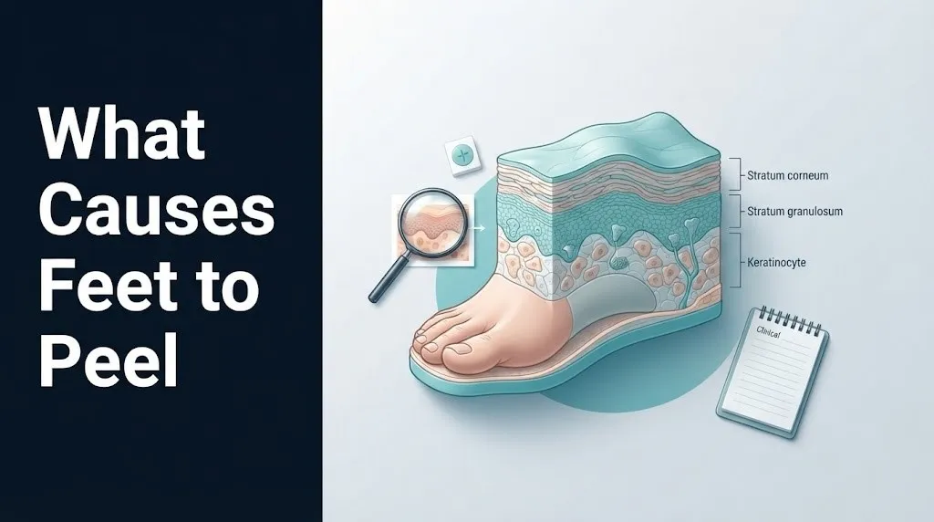
Think of the intercellular lipid lamellae in the stratum corneum like a laminated waterproofing system. Prolonged water exposure degrades that lamination from the surface inward. The lipid layers swell and delaminate, the corneodesmosomes soften and fail, and the result is a white, waterlogged, peeling skin surface that sheds in irregular sheets rather than fine flakes. This is maceration, and it is exactly the same process that causes the skin on your fingers to wrinkle and peel after a long bath, amplified and sustained by the foot’s unusually high sweat output.
Hyperhidrosis-related peeling on the feet tends to produce soft, white, shredding skin between the toes and on the plantar surface. The affected skin may appear water-soaked and peel in larger, thicker sheets than the fine flaking of dry skin. The moisture environment created by hyperhidrosis also dramatically increases the risk of secondary tinea pedis infection, because dermatophytes thrive in the warm, humid microenvironment produced by persistent foot sweat.
According to the International Hyperhidrosis Society, primary hyperhidrosis (excessive sweating without an identifiable medical cause) affects approximately 4.8% of the US population. Secondary hyperhidrosis (from an underlying condition) can be caused by thyroid disease, menopause, infection, and certain medications including antidepressants and antipyretics. Plantar hyperhidrosis in adolescents often follows a different course than in adults: autonomic nervous system maturation during puberty can produce transient hyperhidrosis that improves spontaneously in some teenagers by their early 20s.
Sunburn and Allergic Reactions Causing Feet to Peel
Sunburn causes feet to peel by triggering UV-B radiation-induced keratinocyte apoptosis (programmed cell death) in the epidermis; when these killed cells are simultaneously shed en masse several days after the burn, the result is the characteristic large-sheet peeling that follows a sunburn, including on the tops of the feet.
UV-B radiation (wavelengths 280 to 315 nanometers) penetrates the epidermis and induces DNA photodamage in keratinocytes. Cells with sufficient DNA damage activate apoptotic pathways mediated by p53 tumor suppressor protein signaling, leading to organized cell death. These apoptotic keratinocytes (sometimes called “sunburn cells” histologically) are processed out of the epidermis over the following 3 to 7 days as a synchronized wave of shedding, producing peeling that often occurs in sheets rather than fine flakes.
Sunburn-related foot peeling is almost exclusively confined to the dorsal (top) surface and toes, because these are the areas exposed to direct sunlight during activities like beach walking or outdoor sports without sunscreen. The peeling begins 3 to 5 days after the sun exposure, following the initial red, painful, warm phase of the burn itself.
Allergic contact dermatitis causes peeling through a different mechanism entirely: a type IV delayed hypersensitivity reaction mediated by sensitized T-lymphocytes, triggered by contact with a hapten (typically a shoe material component such as rubber accelerators, chromate in leather tanning, or nickel in shoe hardware). The immune response produces dermal and epidermal inflammation with vesicle formation in the acute phase, followed by peeling and scaling as the inflammation resolves.
According to the Contact Dermatitis journal, shoe-related allergic contact dermatitis most commonly presents on the dorsum of the foot and the plantar forefoot, corresponding to the areas of greatest contact with sensitizing shoe materials. Women who wear open-toed sandals with rubber or synthetic straps may develop a patterned contact dermatitis corresponding exactly to the strap distribution.
Medication Side Effects Causing Feet to Peel
Certain medications cause feet to peel by directly disrupting keratinocyte function, interfering with epidermal growth signaling pathways, altering the skin’s immune response, or producing drug-induced hypersensitivity reactions that manifest on the plantar skin surface.
Targeted cancer therapies, particularly EGFR inhibitors (epidermal growth factor receptor inhibitors such as erlotinib and cetuximab) and multikinase inhibitors (such as sorafenib and sunitinib), produce a condition called hand-foot syndrome (palmar-plantar erythrodysesthesia) as a recognized adverse effect. The palmar and plantar skin, which bears the highest mechanical load and has the highest density of eccrine sweat glands, is particularly susceptible to the cytotoxic and vascular effects of these agents. This produces painful erythema, blistering, and peeling that can range from mild to dose-limiting.
Retinoids (vitamin A derivatives used in acne and psoriasis treatment, including isotretinoin and acitretin) cause plantar peeling as a direct pharmacological effect of their mechanism of action: retinoids bind retinoic acid receptors (RARs) in keratinocytes, altering differentiation and cornification programs in ways that reduce scale formation in psoriasis but simultaneously produce fine peeling on the palms and soles as a side effect of normalized keratinocyte turnover acceleration.
Statins (cholesterol-lowering medications) have been associated with plantar skin changes in some individuals, though the mechanism is less clearly established than for retinoids or EGFR inhibitors.
Things to tell your doctor if you suspect medication-related foot peeling:
- The full name and dose of every prescription and over-the-counter medication you are taking
- When you started any new medication relative to when the peeling began
- Whether the peeling is painful (suggests hand-foot syndrome from targeted therapy) or painless (suggests retinoid effect)
- Whether the peeling also affects your palms (bilateral palmoplantar involvement increases the likelihood of a systemic drug effect)
- Whether you have developed any blistering, raw areas, or difficulty walking
Older adults taking multiple medications (polypharmacy) represent the population at highest risk for medication-related plantar skin changes, because drug interactions can amplify individual medication effects on the skin, and reduced renal clearance prolongs drug exposure in older individuals.
Key Takeaway: If your feet began peeling within weeks of starting a new medication, particularly a cancer therapy, retinoid, or a drug you know affects the skin, tell your prescribing physician before trying any topical treatment, because some medication-induced foot peeling requires a dose adjustment rather than a dermatological intervention.
Systemic Illness Causing Feet to Peel
Systemic illnesses including Kawasaki disease, toxic shock syndrome (TSS), and scarlet fever all cause peeling of the feet as part of their clinical presentation, but the mechanism in each case involves a systemic immune or inflammatory process rather than a local skin condition, and the foot peeling always occurs alongside other systemic symptoms that indicate a medical emergency.
Kawasaki disease (mucocutaneous lymph node syndrome) is a systemic vasculitis affecting medium-sized blood vessels, most commonly in children under 5. Its cause remains incompletely understood, but current evidence points to an abnormal immune response to a preceding infectious trigger. The cutaneous manifestations, including peeling of the fingers and toes (desquamation beginning in the periungual region), occur during the subacute phase, typically 1 to 3 weeks after the onset of fever and other diagnostic features.
Toxic shock syndrome (TSS) is caused by superantigen toxins, most commonly TSST-1 (from Staphylococcus aureus) or streptococcal pyrogenic exotoxins (from Group A Streptococcus). Superantigens bypass the normal antigen-presentation pathway and directly activate up to 20% of all T-lymphocytes simultaneously, producing a massive cytokine storm. The resulting systemic inflammatory response produces multiorgan effects including a characteristic desquamation of the palms and soles 1 to 2 weeks after the acute illness.
Scarlet fever (caused by Group A Streptococcus pyogenes producing pyrogenic exotoxins) follows a similar post-infectious desquamation pattern. The feet and hands peel in sheets 1 to 3 weeks after the pharyngeal or skin infection, following the characteristic sandpaper rash and strawberry tongue of the acute phase.
The Centers for Disease Control and Prevention (CDC) reports that Kawasaki disease affects approximately 19 per 100,000 children under 5 years in the United States annually. It is the leading cause of acquired heart disease in children in the US.
Peeling Skin Between the Toes
Peeling skin between the toes is most commonly caused by interdigital tinea pedis (athlete’s foot in the web spaces), but can also result from erythrasma (a bacterial infection), interdigital maceration from hyperhidrosis, or contact dermatitis from shoe or sock materials, with the location and associated features distinguishing these causes from one another.
The fourth and fifth toe web spaces are the most common sites for interdigital tinea pedis because this space is the narrowest, receives the least air circulation, and retains the most moisture of all toe web spaces. The fungal enzymatic activity of Trichophyton species in this moist environment produces the characteristic softened, whitish, peeling skin with a surrounding rim of erythema and itching. The macerated skin may develop fissures that are painful and at risk of secondary bacterial infection.
Erythrasma is a superficial bacterial infection caused by Corynebacterium minutissimum, which also produces interdigital skin changes including peeling, scaling, and brown discoloration. Unlike tinea pedis, erythrasma fluoresces coral-red under Wood’s lamp (UV-A light) due to coproporphyrin III production by the organism. This Wood’s lamp examination is a simple, non-invasive clinical test that a dermatologist or primary care physician can perform at the bedside to distinguish erythrasma from fungal infection, because the two conditions require different treatments.
Interdigital maceration without infection produces white, softened, shredding skin between the toes that lacks the erythema and itch of fungal infection and the brown discoloration of erythrasma. It is purely a moisture-excess problem and tends to resolve when the foot is dried more thoroughly after bathing and footwear breathability is improved.
People with diabetes mellitus who develop peeling between the toes must have the area evaluated by a primary care physician or podiatrist regardless of the suspected cause, because the combination of peripheral neuropathy (reduced pain sensation) and impaired peripheral circulation in diabetic foot creates conditions where a simple maceration or superficial infection can progress to secondary bacterial cellulitis or deeper infection without producing the pain that would normally prompt the person to seek care.
Peeling Heels and the Bottom of the Feet
Peeling of the heel and plantar surface of the foot is most often caused by dry skin with chronic mechanical pressure (the combination of moisture loss and friction producing heel fissures and plantar scaling), palmoplantar psoriasis, or moccasin-type tinea pedis, each producing a distinct visual pattern that a clinician can use to differentiate them.
The heel is the highest pressure point on the plantar foot during standing and walking. The calcaneus (heel bone) transmits a substantial proportion of ground reaction force through a relatively small surface area during the heel-strike phase of gait. The plantar heel skin responds by developing a thickened keratin layer (callus) through normal pressure adaptation. When this thickened skin dries out, it cracks and peels along the pressure lines, producing the characteristic heel fissure pattern that affects a large proportion of adults who spend extended periods standing on hard surfaces.
Plantar heel fissures that extend into the dermis (deep fissures with bleeding or pain at the crack margins) represent a breakdown of the skin’s protective function. This is more than cosmetic peeling: it is a physical breach of the primary integumentary barrier. Bacterial organisms can enter through fissures, and in people with compromised circulation or immune function (older adults, those with diabetes or peripheral vascular disease), deep plantar fissures can lead to cellulitis or deeper soft tissue infection.
| Heel/Plantar Peeling Pattern | Most Likely Cause | Key Distinguishing Feature |
|---|---|---|
| Fine white flaking, diffuse, no itching | Dry skin | No inflammation, no demarcated borders |
| Thick silvery adherent scale with sharp borders | Palmoplantar psoriasis | Auspitz sign, may have nail pitting |
| Fine diffuse scaling extending from heel to arch and sides | Moccasin tinea pedis | Positive KOH preparation or culture |
| Hard, cracked edges with painful fissures | Pressure callus with dry skin | Fissures follow weight-bearing pressure lines |
| Yellowish, waxy thickening with callus | Hereditary palmoplantar keratoderma | Presents from childhood, family history |
Athletes who train in shoes with inadequate cushioning at the heel absorb higher repetitive impact forces through the calcaneus and overlying plantar fat pad, accelerating the mechanical shear that drives plantar peeling and callus formation. A sports medicine physician or podiatrist can assess whether footwear modification or custom orthotics are appropriate for athletes with persistent plantar peeling related to mechanical loading patterns.
Peeling Feet in Children
Peeling feet in children are caused by several conditions that are either specific to or disproportionately prevalent in the pediatric age group, including Kawasaki disease (primarily under age 5), juvenile plantar dermatosis (ages 3 to 14), scarlet fever, and atopic dermatitis, and the accompanying systemic or pattern-specific features are the key to distinguishing which condition is responsible.
Juvenile plantar dermatosis is a condition almost exclusively affecting children between 3 and 14 years of age, characterized by a shiny, smooth, cracked, peeling surface on the weight-bearing plantar forefoot and toes, without the web space involvement seen in tinea pedis. Its mechanism involves repeated cycles of wetting and drying of the plantar forefoot combined with occlusion by synthetic shoe materials, producing a friction-irritant disruption of the plantar stratum corneum. The condition tends to improve spontaneously at or after puberty, possibly due to changes in sweat gland activity and altered gait patterns that accompany adolescent growth.
The peeling of Kawasaki disease appears in the second to third week of illness, primarily around the nail folds of fingers and toes, and occurs in a child who has previously had a high fever lasting at least 5 days, conjunctivitis, red cracked lips, lymphadenopathy, and a polymorphous rash on the trunk. It is never an isolated foot peeling without these other features.
According to the American Academy of Pediatrics (AAP), prompt diagnosis and treatment of Kawasaki disease with intravenous immunoglobulin (IVIG) is necessary within the first 10 days of illness to reduce the risk of coronary artery aneurysm, the most serious complication of the condition. A child with peeling around the toenails following a recent high fever illness should be evaluated by a pediatrician the same day.
Children with atopic dermatitis are also at higher risk for foot peeling than adults because the Th2-skewed immune response is more pronounced during childhood and early adolescence. Research published in the British Journal of Dermatology has documented that FLG gene mutations present from birth make the plantar skin more susceptible to barrier failure in children with atopic dermatitis than in those with normal FLG expression.
Key Takeaway: A child with peeling feet who also had a fever in the past 2 to 3 weeks, red eyes, cracked lips, or a skin rash on their trunk needs to be seen by a pediatrician the same day, because Kawasaki disease is a treatable pediatric emergency that produces characteristic foot peeling as a late feature.
Peeling Feet and Diabetes
Diabetes mellitus causes or significantly worsens peeling of the feet through three concurrent mechanisms: peripheral neuropathy reducing the sensation that normally prompts protective foot care, peripheral vascular disease reducing the blood supply that maintains skin integrity, and advanced glycation end-products (AGEs) accumulating in skin collagen and keratin structures and altering their mechanical properties.
The plantar skin in people with poorly controlled diabetes undergoes structural changes that make it fundamentally different from non-diabetic plantar skin. AGEs accumulate in the extracellular matrix and within keratinocytes themselves, cross-linking collagen fibers and reducing skin elasticity. The resulting skin is stiffer, less pliable, and more prone to cracking and fissuring under normal mechanical loading. Reduced sweat gland function from autonomic neuropathy additionally impairs plantar skin hydration, worsening the baseline dryness that contributes to peeling.
Peripheral neuropathy in diabetes removes the discomfort that would normally draw attention to plantar skin problems early. A person with full sensation notices a heel fissure immediately because it is uncomfortable. A person with significant diabetic peripheral neuropathy may not feel the fissure at all, allowing it to deepen into the dermis or become secondarily infected without producing the pain signal that would prompt medical attention.
According to the Mayo Clinic, foot complications are among the most serious and costly consequences of diabetes, with diabetic foot ulcers affecting approximately 15% of people with diabetes during their lifetime and accounting for more than 60% of non-traumatic lower limb amputations in the United States. This epidemiological reality means that peeling, cracking, or any skin change on the foot of a person with diabetes requires professional evaluation rather than home management, regardless of how minor it appears.
Things people with diabetes should report to their primary care physician or podiatrist regarding foot peeling:
- Any new areas of peeling, cracking, or callus formation on the feet
- Changes in skin color or temperature between the two feet
- Any area that is not healing or appears to be breaking down after two weeks
- Swelling, warmth, or discharge from any area of the foot, with or without pain
- New odor from the foot skin or between the toes
When to See a Doctor for Peeling Feet
The right time to see a doctor for peeling feet depends on the pattern, accompanying symptoms, and the person’s underlying health status, with specific combinations of features requiring same-day evaluation and others appropriately managed with primary care follow-up at a standard appointment.
Most peeling feet caused by dry skin, mild hyperhidrosis, or early tinea pedis can be monitored at home for 2 to 4 weeks with appropriate self-care measures before seeking professional evaluation. The window narrows considerably when specific warning signs are present.
See a primary care physician within one week if:
- Peeling feet have not improved after 2 to 4 weeks of over-the-counter antifungal treatment
- The peeling is spreading beyond the initial area or involving the nails
- Peeling is accompanied by pain, redness, swelling, or warmth
- You are not certain what is causing the peeling and it has persisted for more than 3 to 4 weeks
- You have an immune-compromising condition or take immunosuppressive medications
See a board-certified dermatologist within 2 to 4 weeks if:
- Peeling is thick, adherent, and silvery-edged and matches the psoriasis pattern described above
- Peeling follows a distribution that corresponds exactly to shoe or sock material contact areas (suggesting allergic contact dermatitis requiring patch testing)
- Plantar peeling has recurred multiple times without a clear triggering cause
See a podiatrist if:
- Heel fissures are deep, painful, or bleeding
- There are calluses or pressure-related plantar changes that are not responding to emollient therapy
| Clinical Situation | Provider to See | Urgency |
|---|---|---|
| Child with peeling toes after fever illness | Pediatrician | Same day |
| Suspected toxic shock syndrome | Emergency room | Immediately, call 911 |
| Peeling with active fever, rash, and systemic symptoms | Emergency room | Immediately, call 911 |
| Diabetic foot with any skin breakdown | Primary care or podiatrist | Within 48 hours |
| Tinea pedis not responding after 4 weeks | Primary care physician | Within 1 week |
| Suspected psoriasis or contact dermatitis | Dermatologist | Within 2 to 4 weeks |
| Dry skin and heel fissures without systemic features | Primary care or podiatrist | Routine appointment |
Pregnant women who develop new plantar peeling should mention the change at their next prenatal appointment. Hormonal changes during pregnancy alter skin hydration, immune function, and inflammatory responses in ways that can trigger both new-onset atopic dermatitis flares and new presentations of palmoplantar psoriasis, neither of which should be managed with standard over-the-counter products without obstetric or dermatologic guidance on what is safe during pregnancy.
Emergency Symptoms: When to Call 911 or Go to the ER
Certain symptoms associated with conditions that cause feet to peel require immediate emergency evaluation. Do not wait to see if these resolve on their own.
Call 911 or go to the nearest emergency room immediately if peeling feet are accompanied by any of the following:
- High fever (above 103°F / 39.4°C) in a child under 5 with red eyes, cracked red lips, swollen lymph nodes, or a rash on the body: this combination can indicate Kawasaki disease, and treatment within the first 10 days is necessary to prevent coronary artery complications.
- Sudden high fever, low blood pressure, vomiting, diarrhea, diffuse sunburn-like rash, and confusion or dizziness: this pattern can indicate toxic shock syndrome, a life-threatening systemic infection requiring emergency hospitalization and intravenous antibiotic therapy.
- Sore throat, fever, and a sandpaper-texture rash on the trunk with subsequent peeling of the feet in a child: this can indicate scarlet fever with post-streptococcal desquamation, requiring prompt antibiotic treatment to prevent rheumatic fever and rheumatic heart disease.
- A rapidly spreading area of redness, warmth, swelling, and pain from a plantar fissure or wound, particularly in a person with diabetes or peripheral vascular disease: this can indicate bacterial cellulitis or a deeper soft tissue infection requiring emergency antibiotic treatment and possible surgical evaluation.
These presentations can indicate life-threatening systemic infections or pediatric vasculitic emergencies and require emergency medical assessment at a hospital, not an urgent care appointment or a phone call.
Frequently Asked Questions About Peeling Feet
What causes the bottom of feet to peel with no itching?
Peeling on the bottom of the feet without itching most commonly results from dry skin, palmoplantar psoriasis, moccasin-type tinea pedis (a non-itchy subtype), or a reaction to medication.
Dry skin peeling is fine, diffuse, and flaky, while psoriatic peeling produces thicker, adherent, silvery-edged scale with sharply defined borders on the heel and arch.
If bottom-of-foot peeling persists for more than 3 to 4 weeks without an identifiable cause or with scale that is thicker than expected for dry skin, evaluation by a board-certified dermatologist is appropriate to distinguish between these conditions.
What is the most common cause of peeling feet?
Tinea pedis (athlete’s foot) and dry skin are the two most frequently encountered causes of peeling feet in the general adult population, with the American Academy of Dermatology noting that tinea pedis affects approximately 15 to 25% of people globally at any given time.
The pattern and presence of itching are the most useful initial features for distinguishing between them: athlete’s foot almost always itches and begins between the toes, while dry skin peeling is typically non-itchy and diffuse.
Both conditions are initially manageable with appropriate over-the-counter products, though persistent or spreading cases of either require evaluation by a primary care physician or dermatologist.
Can diabetes cause feet to peel?
Yes, diabetes mellitus causes peeling on the feet through three concurrent mechanisms: peripheral neuropathy reducing protective sensation, peripheral vascular disease reducing skin blood supply, and advanced glycation end-products altering the structural properties of plantar skin.
People with diabetes are also at higher risk for dry skin because autonomic neuropathy impairs eccrine sweat gland function, reducing plantar skin hydration.
Any skin change on the foot of a person with diabetes, including peeling, fissuring, or callus formation, should be evaluated by a primary care physician or podiatrist within 48 hours rather than monitored at home.
When should I worry about peeling feet?
Peeling feet require immediate emergency evaluation when they are accompanied by fever, systemic symptoms like rash or conjunctivitis, low blood pressure, or confusion, because these combinations can indicate Kawasaki disease, toxic shock syndrome, or scarlet fever.
Peeling feet in a person with diabetes, peripheral vascular disease, or an immunocompromising condition require professional evaluation within 48 hours even without systemic symptoms, because skin integrity loss in these populations carries a higher risk of secondary infection.
Peeling without systemic features and without an underlying high-risk condition is appropriate for home management initially, with primary care follow-up if it does not improve within 2 to 4 weeks.
Why do my feet peel in summer?
Feet peel more in summer due to a combination of increased UV-B sun exposure on the dorsal foot and toes causing sunburn-related desquamation, higher temperatures creating a warmer shoe interior that promotes tinea pedis growth, and more frequent transitions between wet (pool, beach) and dry environments that strip and disrupt the intercellular ceramide lipid layer of the stratum corneum.
Sandal and flip-flop use, while increasing ventilation, also removes the shoe-based friction control that limits mechanical shear on the plantar skin, and open footwear exposes the plantar forefoot to rough surfaces that accelerate superficial corneocyte shedding.
For people who develop significant plantar peeling reliably every summer without systemic symptoms or itching, the pattern often reflects a combination of sun exposure, pool chemicals, and mechanical friction rather than a fungal or inflammatory skin condition.
What causes peeling skin between the toes?
Peeling skin between the toes is most commonly caused by interdigital tinea pedis (athlete’s foot), which produces itching, white macerated-appearing skin, and a surrounding rim of redness in the fourth and fifth toe web spaces.
Less common causes include erythrasma (a Corynebacterium minutissimum bacterial infection that produces brown discoloration and fluoresces coral-red under Wood’s lamp UV light), interdigital maceration from excessive sweating without infection, and contact dermatitis from synthetic sock or shoe materials.
A primary care physician or dermatologist can perform a simple in-office KOH preparation test (potassium hydroxide microscopic examination) to identify dermatophyte fungal elements and confirm tinea pedis versus other causes.
Making Sense of a Skin Signal
Peeling feet are the skin’s most direct way of communicating that something has disrupted the normal shedding process in the stratum corneum. The disruption may be as straightforward as dry winter air stripping ceramides from heel skin, or as specific as a dermatophyte fungus digesting keratin proteins in the fourth toe web space. The pattern, the itching, the location, and what happened in the weeks before the peeling started all matter.
For most adults, the practical next step is straightforward: itch between the toes suggests a trial of an over-the-counter antifungal. Dry, flaky, non-itchy heels suggest an emollient-based approach and reduction of hot water exposure. Any peeling with systemic symptoms, any peeling in a child following a recent fever illness, and any peeling in a person with diabetes all require professional evaluation rather than self-management.
Your plantar skin is the most mechanically loaded surface in the body. When it starts peeling, the cause is almost always identifiable with the right clinical information, and most causes respond well to treatment once correctly identified.