What Causes Arthritis Flare Ups? Triggers Explained 2026
Arthritis flare ups are caused by a combination of immune system disruptions, lifestyle factors, environmental exposures, and physiological changes that reactivate joint inflammation in ways that range from mildly uncomfortable to acutely debilitating. What causes arthritis flare ups is not random. Each trigger works through a specific biological pathway involving named inflammatory mediators, joint tissue responses, and hormonal or neurological signals that amplify the underlying disease process.
According to the Centers for Disease Control and Prevention (CDC), more than 54 million adults in the United States live with a diagnosed form of arthritis, and a substantial majority experience recurrent flares that disrupt daily functioning, sleep quality, and mental health. The National Institute of Arthritis and Musculoskeletal and Skin Diseases (NIAMS) notes that understanding personal flare triggers is one of the most clinically actionable tools a person with arthritis has for reducing disease burden.
This article explains the biology behind every major flare trigger, how the mechanism differs depending on your arthritis subtype, which triggers are highest-risk for your specific demographic profile, and exactly when a flare crosses from manageable at home to requiring same-day evaluation.
What Causes Arthritis Flare Ups
Arthritis flare ups are caused by any factor that reactivates or amplifies the inflammatory process already present in affected joints, pushing symptoms from a controlled baseline to an acute symptomatic episode. The triggering event does not create new disease. It destabilizes whatever fragile immune or mechanical equilibrium was keeping symptoms suppressed.
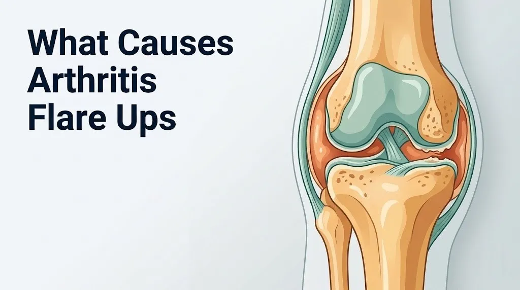
The joint structures involved differ by arthritis type. In rheumatoid arthritis (RA), the primary site is the synovial membrane, a thin tissue lining the joint cavity. During a flare, this membrane becomes acutely inflamed, thickens, and releases waves of pro-inflammatory cytokines into the joint space. In osteoarthritis (OA), flares originate more from mechanical stress on degraded articular cartilage and subchondral bone, triggering a local inflammatory response without the systemic immune activation seen in RA. In gout, flares are triggered by the crystallization of monosodium urate within the joint space, which activates the innate immune system’s NLRP3 inflammasome with striking speed and intensity.
The common thread across all subtypes is the same final output: elevated levels of tumor necrosis factor alpha (TNF-alpha), interleukin-6 (IL-6), and interleukin-1 beta (IL-1beta) flooding the joint space and producing the heat, swelling, pain, and stiffness that define a flare.
Individual responses to triggers vary considerably. A 2021 review published in Annals of the Rheumatic Diseases found that both disease duration and biological sex influence flare susceptibility, with women reporting higher flare frequency than men across RA and PsA populations.
Key triggers include:
- Psychological stress (via HPA axis cortisol dysregulation)
- Infection (via pathogen-driven immune activation)
- Dietary factors (via arachidonic acid-prostaglandin pathways)
- Sleep disruption (via IL-6 elevation and melatonin suppression)
- Physical overexertion (via mechanical joint loading and cartilage debris activation)
- Medication changes (via loss of pharmacological inflammation suppression)
- Hormonal fluctuation (via estrogen’s regulatory effect on synovial inflammation)
What Is an Arthritis Flare
An arthritis flare is a temporary but significant worsening of joint symptoms beyond a person’s usual stable baseline, driven by acute reactivation of the inflammatory processes underlying their specific type of arthritis. The word “flare” in clinical rheumatology refers to a measurable increase in disease activity, not merely subjective discomfort.
Clinically, a flare is distinct from everyday arthritis pain in several ways. Baseline arthritis pain is typically predictable: associated with specific activities, relieved by rest or medication, and stable in character. A flare produces pain that is unpredictable in onset, worsens at rest, appears in joints that were previously quiet, or dramatically exceeds the patient’s usual pain threshold. The American College of Rheumatology (ACR) uses composite measures like the Disease Activity Score DAS28 and the Routine Assessment of Patient Index Data 3 (RAPID3) to define RA flare status clinically. A change of 1.2 points or more on the DAS28 is considered clinically meaningful disease activity increase.
Think of the inflamed joint during a flare like a garden hose with its nozzle partially crimped shut during remission. A flare is someone stomping on the middle of the hose. Water pressure spikes. The inflammation that was slow-dripping at baseline is now flooding the joint capsule all at once.
Duration matters diagnostically. Flares lasting less than 24 hours are often reactive: a brief mechanical provocation, a transient dietary trigger, or a minor physiological disruption. Flares lasting more than 72 hours, particularly those with worsening trajectory, typically indicate a need for clinical reassessment of the current treatment regimen.
People over age 65 often experience longer flares with slower resolution, partly because the adrenal cortex produces lower baseline cortisol levels with advancing age, meaning the body’s internal anti-inflammatory mechanism is already less effective before the trigger event occurs.
Arthritis Types That Cause Flares
Not all forms of arthritis produce the same type of flare, and the distinction matters because the triggers, warning signs, and management approaches differ meaningfully by subtype.
| Arthritis Type | Primary Affected Structure | Flare Mechanism | Most Common Flare Trigger |
|---|---|---|---|
| Rheumatoid Arthritis (RA) | Synovial membrane | Autoimmune T-cell and B-cell reactivation, TNF-alpha surge | Infection, stress, missed medication |
| Osteoarthritis (OA) | Articular cartilage, subchondral bone | Cartilage debris activates NLRP3 inflammasome, local IL-1beta release | Overexertion, joint injury, weight gain |
| Gout | Metatarsophalangeal joint (typically) | Monosodium urate crystal deposition, neutrophil-driven IL-1beta release | High purine foods, alcohol, dehydration |
| Psoriatic Arthritis (PsA) | Synovium and enthesis | TNF-alpha and IL-17 overexpression, skin-joint immune crosstalk | Skin flare, infection, stress |
| Ankylosing Spondylitis (AS) | Sacroiliac joint, spinal entheses | IL-17-driven enthesitis and vertebral inflammation | Physical inactivity, infection, missed biologic therapy |
RA affects approximately 1.3 million adults in the United States, according to NIAMS, and carries a remitting-relapsing disease pattern where periods of low disease activity are punctuated by flares of varying severity. OA, by contrast, affects more than 32 million adults in the US (CDC data) and has a more progressive trajectory with flares superimposed on a background of ongoing structural joint damage.
Gout is the most common form of inflammatory arthritis in men, according to the Arthritis Foundation, and its flares are typically acute, severe, and reach maximum intensity within 12 to 24 hours of onset.
People with more than one arthritis type, such as OA concurrent with RA, often have more frequent and harder-to-characterize flares because triggers from both pathways can be active simultaneously. A rheumatologist is the appropriate specialist to distinguish which type is driving a given flare episode in this population.
Most Common Arthritis Flare Triggers
The most common triggers of arthritis flare ups are psychological stress, physical infection, dietary factors, overexertion, sleep disruption, and medication changes. Each acts through a specific biological mechanism rather than vaguely “causing inflammation.”
Understanding the mechanism matters because it changes what you can actually do about it. Knowing that stress triggers flares through cortisol dysregulation, for example, points toward specific interventions targeting the HPA axis, not generic relaxation advice.
The seven most clinically recognized flare triggers, ranked by frequency in RA population studies:
- Psychological stress (reported by approximately 80% of RA patients in flare surveys published in Arthritis and Rheumatology)
- Physical infection, particularly upper respiratory viral or bacterial infections
- Physical overexertion or joint trauma
- Dietary indiscretion specific to disease type (high purines in gout, inflammatory dietary patterns in RA)
- Sleep deprivation or disrupted sleep architecture
- Missed or changed medication doses
- Hormonal changes (menstrual cycle in women, perimenopause)
A 2022 analysis published in Arthritis and Rheumatology found that patients who tracked their personal trigger patterns and adjusted behavior accordingly had meaningfully lower flare frequency over a 12-month follow-up period compared with patients who did not. Trigger identification is a clinical tool, not just a lifestyle exercise.
People with obesity face an elevated baseline flare risk because adipose tissue acts as an endocrine organ, secreting adipokines including leptin and adiponectin that interact with the same TNF-alpha and IL-6 pathways driving synovial inflammation. Each additional unit of body mass index above 30 correlates with higher circulating IL-6 levels independent of disease activity, according to data from the British Society for Rheumatology Biologics Register.
Key Takeaway: Arthritis flares are not random. Each trigger activates a specific inflammatory pathway in the joint, and identifying your personal triggers with the help of a rheumatologist is one of the most effective ways to reduce flare frequency.
Stress and Arthritis Flares
Psychological stress causes arthritis flare ups by disrupting the body’s own cortisol-mediated anti-inflammatory control system through sustained activation of the hypothalamic-pituitary-adrenal (HPA) axis. This is one of the most well-documented trigger mechanisms in rheumatology.
Here is how the pathway works. Acute stress causes the hypothalamus to release corticotropin-releasing hormone (CRH), which signals the pituitary gland to release adrenocorticotropic hormone (ACTH), which then drives the adrenal cortex to produce cortisol. Under normal circumstances, cortisol binds to glucocorticoid receptors on immune cells and suppresses the production of TNF-alpha, IL-6, and IL-1beta. Short-term, this system works effectively. But during chronic or severe psychological stress, cortisol receptor sensitivity in immune cells decreases through a process called glucocorticoid resistance. The anti-inflammatory signal becomes less effective even when cortisol levels are not low. The result: pro-inflammatory cytokines that were previously suppressed begin to rise, and the synovial membrane becomes progressively more reactive.
A 2019 study published in Annals of the Rheumatic Diseases found that RA patients who experienced high psychological stress in a given week had a statistically higher likelihood of reporting a flare in the following one to two weeks, consistent with the biological lag time of the HPA axis dysregulation pathway.
Beyond cortisol, stress also activates the sympathetic nervous system (SNS), causing the adrenal medulla to release norepinephrine. Norepinephrine acts on beta-2 adrenergic receptors on T-lymphocytes. Depending on which T-cell population is engaged, this can either suppress or amplify immune activity. In RA, the net effect appears to favor pro-inflammatory T-helper 17 cell activity and increased autoantibody production, according to mechanistic research reviewed in Arthritis and Rheumatology.
Women with RA appear to be disproportionately affected by stress-triggered flares compared with men, which some researchers attribute to sex differences in HPA axis reactivity. Women show a more prolonged cortisol response to psychosocial stressors, extending the window of glucocorticoid receptor desensitization and the associated inflammatory rebound.
Stress management approaches with rheumatological relevance (for discussion with your care team):
- Mindfulness-based stress reduction (MBSR): a 2020 trial in Arthritis Care and Research found associations between MBSR practice and lower CRP levels in RA patients
- Cognitive behavioral therapy (CBT): addresses the catastrophizing thought patterns that amplify stress physiology
- Sleep hygiene (overlaps with the sleep section below, as stress and sleep disruption are bidirectionally linked)
- Physical activity at appropriate intensity: moderate aerobic activity reduces baseline IL-6 levels; intensity matters (covered in the overexertion section)
Infection and Arthritis Flare
Infections are among the most reliable and physiologically clear-cut causes of arthritis flare ups, particularly in autoimmune arthritis types like RA and PsA. Any pathogen, bacterial, viral, or fungal, that activates the innate immune system can secondarily reactivate the autoimmune processes driving joint inflammation.
The mechanism begins with pathogen recognition. Immune cells throughout the body carry toll-like receptors (TLRs), which detect specific molecular patterns on bacteria and viruses. When a TLR binds its target, the immune cell activates the NF-kB signaling pathway, which drives production of TNF-alpha, IL-6, and IL-1beta. In a person without inflammatory arthritis, this response is localized and resolves. In a person with RA or PsA, the same systemic cytokine surge reaches the already-sensitized synovial membrane and can trigger acute joint inflammation within 24 to 72 hours of infection onset.
Upper respiratory infections are the most common infection-related flare trigger in RA, according to the Arthritis Foundation. Urinary tract infections, gastrointestinal infections, and skin infections also carry flare risk. Even the immune response to a vaccine can trigger a transient flare in some people with RA, though this is typically mild and brief. The long-term benefit of vaccination (preventing the more severe flare that a full infection would cause) vastly outweighs this short-term risk, per ACR guidance on vaccination in immunosuppressed patients.
People taking disease-modifying antirheumatic drugs (DMARDs) or biologic therapies such as TNF inhibitors have a double concern during infections: the infection triggers a flare while simultaneously being harder to clear because the immune system is pharmacologically suppressed. If you are on a biologic therapy and develop a fever with signs of systemic infection, contact your rheumatologist the same day. Do not hold your biologic dose without specific guidance from your prescribing physician.
Older adults with RA who develop infections are particularly vulnerable to severe flares because both infection-induced cytokine elevation and the blunted cortisol response seen in older adults converge simultaneously to reduce joint disease control.
Diet and Arthritis Flare Ups
Diet causes arthritis flare ups primarily by shifting the body’s inflammatory mediator balance through the arachidonic acid cascade, the gut microbiome-intestinal permeability pathway, and in gout specifically, through uric acid load management. These are three distinct mechanisms, and they apply differently depending on arthritis type.
The arachidonic acid mechanism works like this: arachidonic acid is an omega-6 fatty acid found predominantly in animal fats, processed foods, and refined vegetable oils. Once absorbed, it is converted by cyclooxygenase-2 (COX-2) into prostaglandin E2, a potent pro-inflammatory lipid mediator that drives joint pain, swelling, and synovial membrane sensitization. A diet high in arachidonic acid substrates keeps prostaglandin E2 production elevated chronically, lowering the threshold at which a flare can be triggered.
Conversely, omega-3 fatty acids (found in oily fish, walnuts, and flaxseed) compete with arachidonic acid for the same enzymatic pathways, producing less inflammatory resolvins and protectins instead. A 2021 systematic review published in Arthritis Care and Research found that omega-3 supplementation was associated with modest but consistent reductions in morning stiffness and tender joint count in RA patients, though this did not replace DMARD therapy.
The gut microbiome pathway is an emerging but increasingly well-supported mechanism. Intestinal dysbiosis, an imbalance in gut bacterial populations, increases intestinal permeability. This allows lipopolysaccharide (LPS), a bacterial endotoxin from gram-negative gut bacteria, to enter systemic circulation. LPS binds TLR-4 receptors on circulating monocytes, activating NF-kB and driving systemic TNF-alpha and IL-6 elevation. Research from a 2022 study in Arthritis and Rheumatology found distinct gut microbiome composition differences between RA patients in remission versus flare, suggesting that gut bacterial composition is a real-time modifier of disease activity.
For gout specifically, high-purine foods (organ meats, shellfish, red meat, alcohol particularly beer and spirits) elevate serum uric acid levels. When serum uric acid exceeds approximately 6.8 mg/dL, monosodium urate crystal deposition becomes thermodynamically favorable in cooler peripheral joints. Fasting, rapid weight loss, and dehydration can also precipitate gout flares by raising uric acid concentration acutely.
Sleep Deprivation and Arthritis Flares
Sleep deprivation directly elevates pro-inflammatory cytokine levels in the blood, and this cytokine elevation is one of the most mechanistically direct causes of arthritis flare ups. Sleep is not passive recovery for the immune system. It is the period when immune homeostasis is actively maintained.
The mechanism centers on two parallel pathways. First, adequate sleep is required for the pineal gland to produce melatonin at appropriate circadian levels. Melatonin has pleiotropic immune-regulatory effects, including the suppression of IL-6 and TNF-alpha production in synovial macrophages. When sleep is disrupted or shortened, melatonin production drops and this suppressive effect is lost. Second, during deep slow-wave sleep (stages 3 and 4 of non-REM sleep), the pituitary gland releases growth hormone, which has anti-inflammatory properties in joint tissue. Sleep fragmentation preferentially disrupts slow-wave sleep architecture, removing this protective signal at exactly the time joint inflammation needs to be checked.
A 2020 study published in JAMA Internal Medicine found that people with RA who reported poor sleep quality were significantly more likely to report higher disease activity the following day, even after controlling for pain, mood, and physical activity. The relationship was bidirectional: pain also disrupted sleep, creating a clinically important reinforcing cycle.
Clinically, even one night of significant sleep deprivation is associated with measurable IL-6 elevation within 24 hours, according to controlled sleep restriction studies published in Sleep Medicine Reviews. For arthritis patients already operating near their inflammatory threshold, this cytokine spike can be sufficient to tip a controlled disease state into a symptomatic flare.
People with RA who also have obstructive sleep apnea (OSA) face compounded risk. OSA causes intermittent nocturnal hypoxia, which independently activates NF-kB and drives TNF-alpha production from airway and systemic immune cells. OSA prevalence is higher in RA patients than in the general population, and untreated OSA is associated with worse RA disease control. If you have RA and your sleep quality is consistently poor despite adequate pain control, ask your primary care physician about a formal sleep study.
Key Takeaway: Sleep deprivation raises IL-6 and TNF-alpha through two independent pathways within 24 hours, and in someone with RA or PsA, this inflammatory rise can be sufficient to trigger a full flare even without any other identifiable cause.
Weather Changes and Arthritis Pain
Weather changes are associated with arthritis flares through barometric pressure fluctuation, temperature change, and humidity shifts that affect joint tissue mechanics and peripheral nerve sensitization, though the evidence is more observational than mechanistic. This is a real phenomenon, not a placebo effect, but the biology is more nuanced than “cold weather causes arthritis.”
The most well-supported mechanism involves barometric pressure. The joint capsule and surrounding soft tissues contain fluid-filled spaces that respond to external pressure changes much like a sealed balloon would. When barometric pressure drops (before a rainstorm or during a cold front), the surrounding atmospheric pressure on joint tissues decreases. Tissues that are already inflamed or edematous expand slightly. This expansion increases pressure within the joint capsule and stimulates mechanosensitive nerve endings, called nociceptors, on the joint capsule and synovial membrane. People with more joint damage have more exposed nociceptors and report more weather sensitivity as a result.
Temperature change adds a second layer. Cold temperatures cause local vasoconstriction in joint-supplying blood vessels, reducing synovial fluid production and increasing viscosity. Thicker, less-mobile synovial fluid means less joint lubrication and more friction-generated cartilage stress with movement, particularly relevant for OA joints.
A 2019 Dutch study involving 2,658 participants, published in npj Digital Medicine, used continuous weather and pain reporting data via a smartphone app and found that increased pain was significantly associated with higher humidity, higher wind speed, and lower atmospheric pressure. The relationship was modest but consistent across the study population.
Not everyone is equally affected by weather. People with more advanced joint damage report stronger weather sensitivity, which fits the nociceptor exposure hypothesis. People with OA of the knee and hip, joints with large capsular surface areas, tend to report weather effects more frequently than those with small-joint predominant disease.
Extreme cold is a particular concern for people with Raynaud’s phenomenon, which is common in systemic autoimmune arthritis conditions. Raynaud’s involves episodic vasospasm of digital arteries in response to cold, producing finger blanching, pain, and color change. For these patients, weather-triggered vascular events can precede or coincide with joint flare activity.
Overexertion and Arthritis Flare
Physical overexertion causes arthritis flares by mechanically stressing damaged joint structures beyond their current load tolerance, generating cartilage debris, synovial irritation, and subsequent local inflammatory cascade activation. The key word is “over” exertion. Appropriate physical activity is anti-inflammatory and joint-protective. The dose makes the poison.
In OA, the overexertion mechanism is primarily mechanical. Articular cartilage that has already lost proteoglycan density and collagen architecture is less capable of distributing compressive load evenly across the joint surface. Activities that exceed the cartilage’s load tolerance, such as prolonged high-impact exercise, heavy lifting with poor mechanics, or activities involving repetitive joint loading without adequate rest, generate fragments of cartilage matrix called extracellular matrix degradation products. These fragments are recognized by synovial macrophages as danger signals, activating the NLRP3 inflammasome and triggering IL-1beta release, which drives acute synovitis even in a joint that was relatively quiet before the activity.
In RA, overexertion acts differently. RA joints have already-inflamed synovium and often pannus formation. Mechanical stress on these tissues causes microtrauma to blood vessels within the pannus, releasing local damage-associated molecular patterns (DAMPs) that reactivate macrophages and amplify TNF-alpha output. The joint doesn’t need the same mechanical threshold to flare as an OA joint does.
Guidelines for activity modification during and between arthritis flares:
- During a flare, reduce joint-loading activity but do not stop moving entirely. Complete rest accelerates muscle weakness and joint stiffness.
- Switch to low-impact alternatives: swimming, water aerobics, and cycling place approximately 50 to 80% less compressive load on hip and knee joints than walking on pavement.
- After a flare resolves, return to previous activity levels gradually over one to two weeks rather than resuming at pre-flare intensity immediately.
- Apply the “two-hour pain rule” used in ACR physical therapy guidance: if joint pain persists for more than two hours after a given activity, that activity exceeded safe load for your current joint status.
- Tell your physical therapist or rheumatologist about your activity patterns at each visit. Exercise prescription in arthritis is individualized and should be adjusted based on current disease activity, not a fixed routine.
Older adults with OA should be aware that proprioceptive deficits common after age 60 increase fall risk and uncontrolled joint loading during everyday activities. A physical therapist can assess proprioceptive function and prescribe balance training that reduces accidental overloading.
Medication Changes and Arthritis Flare
Changes to arthritis medications, whether missed doses, dose reductions, drug switches, or abrupt discontinuation, are a direct and common cause of arthritis flares because they remove the pharmacological control keeping the underlying inflammatory process suppressed. This trigger is distinct from all others because it is fully predictable and largely preventable.
In RA and PsA, disease-modifying antirheumatic drugs (DMARDs), including conventional DMARDs like methotrexate and hydroxychloroquine, and biologic DMARDs like TNF inhibitors (adalimumab, etanercept, infliximab), work by blocking specific steps in the inflammatory cascade at the molecular level. Missing even one or two doses of a biologic can allow circulating drug levels to drop below the therapeutic threshold. When drug levels fall, the cytokine targets these medications were blocking (TNF-alpha for TNF inhibitors, IL-6 for tocilizumab, IL-1beta for anakinra) resume signaling in the synovium, often producing a flare within days to two weeks.
Think of biologic therapy as a thumb held firmly over a garden hose nozzle. The water pressure (inflammation) is still there. The thumb (medication) is what keeps it controlled. Remove the thumb suddenly, and the pressure is released all at once.
For gout, stopping or reducing urate-lowering therapy (ULT) such as allopurinol or febuxostat can allow serum uric acid to rise above crystal deposition thresholds within weeks, setting up conditions for the next acute gout attack. Paradoxically, initiating or adjusting ULT doses can also trigger a flare in the short term because of crystal mobilization, which is why colchicine or low-dose NSAIDs are often prescribed concurrently during ULT dose changes.
For corticosteroid-dependent patients with any arthritis type, rapid tapering of oral prednisone or prednisolone can precipitate a severe rebound flare. The adrenal cortex requires time to resume normal cortisol production after sustained corticosteroid use, and during this gap period, the body lacks both exogenous and endogenous anti-inflammatory glucocorticoid support.
If your medication is being changed, your dose is being adjusted, or cost or supply issues are interrupting your current regimen, tell your rheumatologist before stopping any dose. There are bridging strategies available for most arthritis medications.
Key Takeaway: Missed biologic or DMARD doses can trigger an arthritis flare within days because drug levels fall below the threshold needed to suppress TNF-alpha or IL-6, and the inflammatory cascade resumes with little delay.
Sex Hormones and Arthritis Flares
Sex hormones are a biologically significant but frequently under-explained modifier of arthritis flare frequency and severity. Women with RA experience higher flare rates than men overall, and flare clustering around hormonal transitions (menstruation, pregnancy, the postpartum period, and perimenopause) points directly to estrogen’s regulatory role in synovial inflammation.
Estradiol, the predominant form of estrogen, binds to estrogen receptor alpha (ERa) on synovial fibroblast-like synoviocytes (FLS), which are the primary producers of TNF-alpha, IL-6, and matrix metalloproteinases in the inflamed RA joint. At physiologically normal estrogen levels, ERa activation generally suppresses the inflammatory output of these cells. When estrogen levels drop sharply, as they do during the luteal-to-follicular transition of the menstrual cycle or during perimenopause, this suppressive signal is withdrawn. The FLS produce more cytokines, the synovial membrane becomes more reactive, and flare threshold lowers.
A 2023 analysis published in Annals of the Rheumatic Diseases found that women with RA reported higher disease activity scores during the days immediately preceding menstruation, consistent with the timing of the late luteal phase estrogen drop. The magnitude of this cyclical flare risk was greater in women who also reported high psychological stress, suggesting that the HPA axis and hormonal fluctuation pathways interact additively.
Pregnancy produces a distinctive pattern. Many women with RA experience significant disease improvement during pregnancy, particularly in the second and third trimesters, when estrogen and progesterone levels are at their highest. However, the postpartum period, when sex hormones fall sharply, is associated with a high rate of RA flares and even new-onset RA, suggesting that the hormonal withdrawal unmasks the disease in susceptible individuals.
Testosterone is not irrelevant in this context. Testosterone has documented anti-inflammatory properties, acting partly by suppressing Th17 cell differentiation and IL-17 production. Men with RA have lower testosterone levels on average compared to age-matched controls, and lower testosterone correlates with higher disease activity scores in several observational studies reviewed by NIAMS.
Postmenopausal women with both OA and RA face an additional mechanism. Estrogen withdrawal after menopause accelerates subchondral bone remodeling and reduces synovial fluid viscosity, directly worsening the mechanical environment in OA joints. This contributes to the sharp increase in OA prevalence and symptom severity observed in women between ages 50 and 65.
Rheumatoid Arthritis vs Osteoarthritis Flare Mechanisms
Rheumatoid arthritis and osteoarthritis produce flares through fundamentally different biological mechanisms, and confusing the two means using the wrong management strategy and missing the correct warning signs. Both cause joint pain and swelling during a flare, but what is happening inside the joint is not the same.
In RA, a flare is an autoimmune event. The immune system’s CD4+ T-helper cells encounter an antigen (often precipitated by one of the triggers already discussed) and activate B-cells to produce anti-CCP antibodies and rheumatoid factor. These autoantibodies form immune complexes that deposit in the synovial membrane, activating complement and macrophages. Synovial macrophages release TNF-alpha, which drives fibroblast-like synoviocyte proliferation, pannus formation (an invasive inflammatory tissue that erodes cartilage and bone from the inside), and a systemic inflammatory response that can include fever, fatigue, and elevated CRP and ESR.
In OA, a flare is primarily a mechanically-initiated local event. Degraded cartilage releases fragments of type II collagen and aggrecan into the joint space. These fragments are recognized by toll-like receptors on synovial macrophages as damage-associated molecular patterns (DAMPs), triggering NLRP3 inflammasome activation and local IL-1beta release. The inflammatory response is largely confined to the joint and does not typically produce systemic symptoms like fever or elevated inflammatory markers in blood tests.
| Feature | RA Flare | OA Flare |
|---|---|---|
| Primary mechanism | Autoimmune (T/B cell, autoantibodies) | Mechanical (DAMP, NLRP3, local IL-1beta) |
| Systemic symptoms | Fatigue, fever, malaise: common | Typically absent |
| Inflammatory markers (CRP, ESR) | Usually elevated during flare | Often normal or mildly elevated |
| Joint distribution | Symmetric, small joints (MCP, PIP, wrists) | Asymmetric, weight-bearing joints (knee, hip) |
| Duration of flare | Days to weeks | Hours to days |
| Key associated trigger | Infection, stress, missed DMARD | Overexertion, injury, weight gain |
| Treatment approach | DMARDs, biologics, short-course steroids | Activity modification, topical NSAIDs, intraarticular injection |
This distinction matters particularly for older adults, who often have both OA and RA concurrently. When they flare, identifying which disease is driving the episode determines the treatment approach. A rheumatologist can use synovial fluid analysis, inflammatory marker patterns, and joint distribution to determine the dominant active process.
Gout and Psoriatic Arthritis Flare Causes
Gout and psoriatic arthritis produce flares through mechanisms that are distinct from both RA and OA, and both conditions are frequently misdiagnosed or undertreated when clinicians rely on the standard RA or OA framework.
Gout flares are caused by the acute deposition of monosodium urate (MSU) crystals in the joint space, most commonly the first metatarsophalangeal joint (the big toe joint), though ankles, knees, wrists, and elbows are also commonly affected. The deposition occurs when serum uric acid levels exceed the saturation threshold of approximately 6.8 mg/dL. MSU crystals are recognized by synovial macrophages as foreign particles, triggering NLRP3 inflammasome activation and a massive, rapid IL-1beta-driven neutrophil recruitment. Neutrophils attempt to phagocytose the crystals, release reactive oxygen species (ROS) and additional cytokines in the process, and produce the characteristic excruciating pain, swelling, warmth, and erythema of an acute gout attack.
Gout flare triggers by mechanism:
- Purine-rich foods (organ meats, anchovies, sardines, shellfish): directly raise uric acid load via purine catabolism to uric acid
- Alcohol (especially beer and spirits): reduces renal uric acid excretion by increasing lactic acid, which competes with uric acid for tubular secretion
- Dehydration: concentrates serum uric acid and reduces renal uric acid excretion
- Fasting and rapid weight loss: trigger ketosis, and ketoacids compete with uric acid for renal excretion
- Certain medications: loop diuretics (furosemide), thiazide diuretics, low-dose aspirin, cyclosporine all raise serum uric acid by reducing renal excretion
Psoriatic arthritis flares follow a different logic. PsA involves both synovitis and enthesitis (inflammation at the sites where tendons and ligaments attach to bone). The driving mediators are TNF-alpha and IL-17A, produced by dysregulated Th17 cells that are activated by antigen presentation in both skin and joint tissues. Skin disease activity and joint disease activity are linked but not perfectly correlated: some PsA patients have minimal skin involvement with severe arthritis, while others have extensive psoriasis with mild joint disease.
PsA flares are frequently triggered by the same triggers that worsen psoriasis: skin infections (particularly streptococcal), physical trauma to skin or joints (the Koebner phenomenon), psychological stress, and abrupt withdrawal of biologic therapy. According to the ACR/National Psoriasis Foundation joint guidelines, TNF inhibitors and IL-17 inhibitors are first-line biologic therapies for active PsA, and missing doses is among the strongest predictors of flare.
Men are affected by gout at roughly four times the rate of premenopausal women, because estrogen promotes renal uric acid excretion. After menopause, this protective effect disappears, and gout rates in women approach those in men.
Symptoms of an Arthritis Flare
The symptoms of an arthritis flare are increased joint pain beyond the patient’s usual baseline, joint swelling and warmth, prolonged morning stiffness lasting more than 45 minutes, fatigue, and in RA, systemic symptoms such as low-grade fever and malaise.
Recognizing a flare accurately is clinically important because it determines the management pathway. Not every increase in joint pain is a flare. Overexertion-related pain that resolves after rest within a few hours is not a flare. A flare persists, worsens through a rest period, and often affects joints beyond those typically symptomatic.
Key symptoms by flare severity:
| Severity Level | Joint Symptoms | Systemic Symptoms | Action |
|---|---|---|---|
| Mild | Increased pain in 1 to 2 joints, minimal swelling, resolves within 24 to 48 hours | None | Rest, ice, topical NSAID, monitor |
| Moderate | Increased pain in 3 or more joints, visible swelling, morning stiffness greater than 60 minutes, not improving at 48 to 72 hours | Fatigue, mild malaise | Contact rheumatologist within 2 to 3 days |
| Severe | Intense pain in multiple joints, significant swelling limiting function, not responding to usual medications | Fever, chills, marked fatigue | Contact rheumatologist same day or go to urgent care |
| Emergency | Single joint acutely hot, red, swollen, with fever above 38.5°C | High fever, rigors, systemic illness | Go to emergency room immediately: septic arthritis exclusion required |
Morning stiffness duration is a particularly useful clinical gauge. The ACR identifies morning stiffness lasting more than 45 minutes in multiple joints as a marker of active RA disease. OA-related morning stiffness typically resolves within 30 minutes. A sudden worsening of morning stiffness duration is one of the earliest objective signals that a flare is developing.
Older adults may have blunted pain perception due to comorbid peripheral neuropathy or analgesic medication use, meaning their subjective pain reports may underestimate actual flare severity. Caregivers and clinicians evaluating older adults should pay closer attention to functional changes (difficulty dressing, reduced grip, limping) as flare indicators in this group.
Emergency Symptoms: When to Call 911 or Go to the ER
Certain symptoms associated with arthritis flares require immediate emergency evaluation. Do not wait to see if these resolve on their own.
Call 911 or go to the nearest emergency room immediately if you experience:
- A single joint that becomes acutely hot, red, swollen, and intensely painful with fever above 38.5°C (101.3°F): this presentation can indicate septic arthritis, a bacterial joint infection that causes permanent joint destruction within 24 to 48 hours and can progress to life-threatening sepsis if untreated
- Severe chest pain, shortness of breath, or palpitations during an arthritis flare: people with RA face a two-fold higher cardiovascular risk than the general population (per NIAMS data), and cardiac events can occur during periods of high systemic inflammation
- High fever (above 39°C/102°F) with joint pain and a widespread skin rash: this combination may indicate systemic juvenile idiopathic arthritis or adult-onset Still’s disease, both of which require urgent rheumatological assessment
- Sudden inability to bear weight on a previously functional joint with no recent trauma: this can indicate fracture of bone weakened by chronic inflammation or corticosteroid use
- Severe jaw pain with headache and scalp tenderness in adults over 50 with known RA or other systemic inflammatory conditions: this combination warrants emergency evaluation to rule out giant cell arteritis (temporal arteritis), which can cause sudden permanent vision loss
These presentations can indicate medical emergencies distinct from a standard arthritis flare, and they require emergency room assessment, not an urgent care appointment or a next-day phone call.
When to See a Doctor for Arthritis Flare
You should contact your rheumatologist or primary care physician when an arthritis flare does not improve within 48 to 72 hours of self-management, when its severity is substantially beyond your usual baseline, when it occurs more frequently than previous patterns, or when you develop any new symptoms not typical of your established arthritis history.
The right provider and the right timing depend on your specific situation. Here is a structured framework:
When to seek care and who to contact:
- Within 24 hours, call your rheumatologist: Fever with joint pain, any suspicion of infection, flare while on a biologic that has never produced this level of response before, any joint that is dramatically more swollen and hot than others.
- Within 48 to 72 hours, schedule with your rheumatologist or primary care physician: Flare that is not responding to your current medication regimen after 48 hours, flare affecting joints not typically involved for you, significant functional limitation affecting work or self-care.
- At your next scheduled appointment: Mild flares that respond to your usual management but are becoming more frequent, a sense that your baseline disease control has worsened over the past month.
- When preparing for your appointment, document: When the flare started, what you were doing in the 24 to 48 hours before onset (diet, stress level, sleep, physical activity, any illness), which joints are affected, your current medications and whether any doses were missed, your pain score on a 0-to-10 scale at rest and with movement, and what has helped or not helped.
- Request these specific assessments: Updated inflammatory markers (CRP and ESR), a review of current DMARD or biologic drug levels if applicable, discussion of whether your maintenance therapy needs adjustment.
- For gout specifically: Request serum uric acid measurement. A value above 6 mg/dL between attacks indicates inadequate urate-lowering therapy and predicts future flare risk regardless of current symptom status.
For people who do not yet have a rheumatologist, a primary care physician is the appropriate first contact for a new or escalating arthritis flare. Ask for a referral to a board-certified rheumatologist if your flares are recurring, if your current treatment is not controlling your disease, or if the diagnosis has not been formally confirmed.
Key Takeaway: A single hot, red, swollen joint with fever is not an arthritis flare until septic arthritis has been ruled out by joint aspiration and synovial fluid culture. This distinction requires emergency room evaluation, not a home remedy.
Frequently Asked Questions About Arthritis Flare Ups
What is the most common cause of an arthritis flare up?
Psychological stress is the most commonly reported trigger of arthritis flare ups, particularly in rheumatoid arthritis, according to patient survey data published in Arthritis and Rheumatology.
Stress triggers flares by disrupting cortisol’s anti-inflammatory control of the immune system through HPA axis dysregulation, allowing TNF-alpha and IL-6 to rise in the synovial membrane.
Infection is the second most commonly identified trigger, particularly upper respiratory infections, which activate TLR signaling pathways that secondarily reactivate synovial inflammation in autoimmune arthritis types.
How long does an arthritis flare up usually last?
An arthritis flare typically lasts between a few days and several weeks, depending on the arthritis type, severity of the trigger, and adequacy of the current treatment regimen.
Gout flares usually peak within 12 to 24 hours and resolve within 7 to 10 days with appropriate treatment, while RA flares can persist for weeks if the triggering cause is not addressed.
A flare lasting more than two weeks without improvement warrants evaluation by a rheumatologist to determine whether the maintenance treatment plan needs to be adjusted.
Can stress really cause arthritis to flare up?
Yes, psychological stress causes arthritis flares through a specific biological mechanism involving the hypothalamic-pituitary-adrenal axis and cortisol resistance in immune cells, not through a vague stress-pain relationship.
When chronic stress desensitizes glucocorticoid receptors on immune cells, the cortisol signal that normally suppresses TNF-alpha and IL-6 production becomes less effective, allowing joint inflammation to escalate.
A 2019 study published in Annals of the Rheumatic Diseases confirmed a consistent lag of one to two weeks between periods of high psychological stress and subsequent flare onset in RA patients.
What foods trigger arthritis flare ups?
Foods that trigger arthritis flare ups include high-purine foods (organ meats, shellfish, red meat) in gout, and pro-inflammatory dietary patterns high in arachidonic acid (processed meats, refined oils) in rheumatoid arthritis and osteoarthritis.
Alcohol, particularly beer and spirits, is a strong gout flare trigger because it reduces renal uric acid excretion by competing with urate for tubular secretion.
A 2021 systematic review in Arthritis Care and Research found that Mediterranean-style dietary patterns, high in omega-3 fatty acids and low in refined carbohydrates, were associated with modest reductions in RA disease activity markers.
How do I know if my arthritis flare is serious?
An arthritis flare is serious and requires same-day medical evaluation if it includes fever above 38.5°C (101.3°F) with a hot, swollen joint, significant systemic symptoms like chills or marked fatigue, or pain and swelling that is rapidly worsening rather than plateauing.
A flare that does not respond to your usual medications after 48 to 72 hours, affects joints not typically involved for you, or is substantially more severe than your prior baseline pattern also warrants prompt evaluation.
Any single acutely hot and swollen joint with fever requires emergency room evaluation to rule out septic arthritis, which is a medical emergency distinct from an inflammatory arthritis flare.
Can weather really make arthritis worse?
Yes, weather changes are associated with arthritis pain through barometric pressure fluctuation, which causes fluid-filled joint tissues to expand slightly when external pressure drops, stimulating nociceptors on the joint capsule.
A 2019 Dutch study published in npj Digital Medicine, involving more than 2,600 participants, found consistent associations between increased pain and lower atmospheric pressure, higher humidity, and higher wind speed.
The effect is stronger in people with more advanced joint damage because greater cartilage loss exposes more nociceptive nerve endings to pressure changes.
Putting It Together
Every arthritis flare has a cause, and that cause operates through a specific biological pathway. Stress rewires your cortisol response. Infection hijacks the same cytokine machinery driving your disease. Missed medication removes the pharmacological dam holding back the inflammatory flood. Diet, sleep, hormones, weather, and physical activity each play a distinct mechanistic role. Knowing which pathway applies to your flare is not just intellectually satisfying. It is the foundation of a practical prevention strategy.
Start tracking your flares with dates, triggers, duration, and severity. Share that data at your next rheumatologist appointment along with your current medication adherence pattern, your sleep quality, and any recent illness or life stress. This information does not just help your doctor. It helps you build a picture of your personal flare biology that no generic article can give you.
If your flares are becoming more frequent, more severe, or less responsive to your current treatment, that is a signal your disease activity has changed and your treatment plan may need updating. A board-certified rheumatologist is the appropriate specialist to guide that reassessment. You deserve a clear explanation for why your joints are flaring, and you now have the biological framework to ask the right questions.