What Causes Rib Flare? Causes, Risks, and When to Act
What causes rib flare is, in most cases, a breakdown in the muscular and respiratory forces that normally hold the lower ribs inward against the thoracic wall, with diaphragm dysfunction and abdominal muscle weakness being the two most clinically important functional drivers. In a smaller proportion of people, rib flare reflects structural changes to the rib cage itself, including scoliosis, connective tissue laxity, and congenital rib cage morphology.
Rib flare is not a trivial cosmetic finding. According to research in the Journal of Applied Physiology examining thoracic wall mechanics, the position and tone of the lower rib cage directly affects respiratory efficiency, intra-abdominal pressure regulation, and spinal load distribution through the thoracolumbar fascia. When the lower ribs are persistently elevated and flared, the mechanical consequences extend well beyond appearance.
This article covers every named cause of rib flare, from the most common functional mechanisms to pregnancy-related changes, pediatric presentations, connective tissue disorders, and scoliosis. You’ll also find out which presentations require professional assessment and exactly which provider type fits each clinical circumstance.
What Causes Rib Flare: An Overview of the Main Mechanisms
Rib flare is caused by any condition that reduces the inward mechanical restraint normally applied to the lower ribs by the diaphragm and abdominal wall muscles, or by structural changes to the rib cage, costal cartilage, or thoracic spine that alter the geometry of the lower thorax. Every specific cause described in this article acts through one or both of these two pathways.
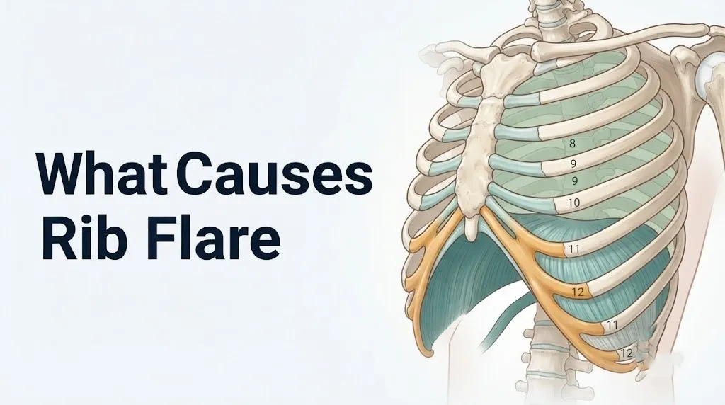
The lower ribs (ribs 7 through 12) are the most mobile segment of the rib cage. Unlike the upper true ribs (1 through 7), which attach directly to the sternum via costal cartilage, the lower false ribs (8 through 10) attach to each other’s cartilage before reaching the sternum, and the floating ribs (11 and 12) have no anterior attachment at all. This architecture gives the lower rib cage both its respiratory mobility and its susceptibility to positional change when stabilizing forces are reduced.
Two distinct categories account for the full range of causes:
- Functional rib flare: the rib cage anatomy is normal, but the muscular and respiratory forces that hold it in position are insufficient. This includes diaphragm dysfunction, transversus abdominis weakness, and thoracic kyphosis.
- Structural rib flare: the ribs, costal cartilage, thoracic vertebrae, or connective tissue are anatomically altered. This includes scoliosis, Ehlers-Danlos syndrome, pectus deformities, and congenital wide infrasternal angles.
| Category | Primary Mechanism | Reversibility | Evaluation Needed |
|---|---|---|---|
| Functional (diaphragm/core) | Reduced inward mechanical restraint on lower ribs | Often improvable with rehabilitation | Physical therapist assessment |
| Functional (postural/kyphosis) | Altered rib cage geometry from spinal curvature | Partially improvable | Physical therapist or sports medicine |
| Structural (scoliosis) | Vertebral and rib rotation altering thoracic shape | Not reversible without treatment | Orthopedic surgeon evaluation |
| Structural (connective tissue) | Costal cartilage laxity reducing rib stability | Not reversible | Clinical assessment required |
| Structural (congenital) | Inherently wide infrasternal angle | Not reversible; may be normal variant | Clinical monitoring |
| Pregnancy-related | Uterine growth and relaxin-mediated laxity | Typically resolves postpartum | Obstetric provider monitoring |
Individual variation note: People with chronic obstructive pulmonary disease (COPD) or asthma experience chronic hyperinflation of the lungs, which flattens the diaphragm and reduces the zone of apposition, producing rib flare through the same physiological mechanism as diaphragm dysfunction in an otherwise healthy person. Their rib flare reflects disease severity, not simply postural or muscular habits.
What Is Rib Flare and How Is It Identified Clinically
Rib flare is the visible or palpable protrusion of the lower ribs beyond the plane of the abdomen and lower thorax, typically assessed by measuring the infrasternal angle, with an angle consistently exceeding 90 degrees considered outside the normal range in most clinical and physical therapy assessment frameworks. The term describes a postural and positional finding, not a single named diagnosis.
The infrasternal angle is the angle formed at the xiphoid process between the two costal margins (the lower edges of the rib cage formed by the cartilaginous borders of ribs 7 through 10). In most adults, this angle sits between 60 and 80 degrees at rest when standing upright. An infrasternal angle above 90 degrees indicates that the lower rib cage is flared outward relative to the midline.
Rib flare is identified clinically by observation and palpation. Viewed from the front, the lower rib margins extend visibly beyond the waistline contour on one or both sides. Viewed from the side, the lower ribs protrude anteriorly beyond the abdomen. A trained clinician can confirm the finding by placing their hands along the lower costal margins and estimating the infrasternal angle in standing and in various postures.
It is worth noting that the infrasternal angle is not an absolute fixed normal: people with naturally wide rib cages and shorter torsos tend toward wider angles than people with narrow, long torsos. The clinical concern arises when the angle is wider than expected for the individual’s body type, when it changes with position or activity, or when it is associated with symptoms such as low back pain, shoulder discomfort, or breathing pattern changes.
Individual variation note: Male and female rib cage morphology differs measurably. Research published in the American Journal of Physical Anthropology has found that female rib cages are proportionally narrower relative to overall thoracic height than male rib cages, which affects baseline infrasternal angle norms. Clinicians interpreting infrasternal angle measurements should apply sex-specific reference ranges rather than a single universal threshold.
Rib Cage Anatomy and the Structures Involved in Rib Flare
The anatomical structures directly involved in rib flare are the lower ribs (7 through 12), their costal cartilages, the costochondral junctions connecting them, the infrasternal angle they form at the xiphoid process, and the thoracic vertebrae whose alignment determines the three-dimensional position of the rib cage. Understanding these structures explains why specific conditions produce flaring in predictable ways.
The true ribs (ribs 1 through 7) articulate directly with the sternum via their own costal cartilage. The false ribs (ribs 8 through 10) have costal cartilages that join the cartilage of rib 7 rather than the sternum directly, forming the costal margin. The floating ribs (11 and 12) have no anterior attachment whatsoever, terminating freely in the posterior abdominal musculature. This segmental architecture means that flaring affects primarily ribs 7 through 12, where mechanical restraint is architecturally weaker than in the upper thorax.
Think of the lower rib cage like the opening of a basket whose sides are held inward by internal bands. The transversus abdominis and the diaphragm’s costal fibers are those bands. Remove or weaken them, and the sides of the basket spread outward under the influence of their own weight and the outward pressure of the contents within.
The costochondral junctions are the cartilage-to-bone interfaces where each rib transitions from its bony shaft to its costal cartilage. These junctions are susceptible to inflammation (costochondritis, Tietze syndrome) and, in connective tissue disorders, to excess compliance that allows ribs to move more freely than normal, contributing to positional instability.
Individual variation note: Children have proportionally larger amounts of cartilaginous tissue in their rib cages relative to adults. This makes their rib cages more compliant and more susceptible to deformation from postural and mechanical influences during growth, which is one reason pediatric rib flare presentations can be more pronounced and more responsive to early intervention than adult presentations.
Key Takeaway: Rib flare divides cleanly into functional causes (diaphragm and abdominal muscle insufficiency) and structural causes (scoliosis, connective tissue laxity, congenital anatomy). Knowing which category applies to your situation directly determines what can be done about it.
Diaphragm Dysfunction as a Primary Cause of Rib Flare
Diaphragm dysfunction causes rib flare by reducing or eliminating the inward mechanical pull that the diaphragm’s costal fibers normally apply to the lower ribs, allowing them to flare outward when the opposing muscular forces from the abdominal wall are insufficient to maintain their position. This is the most physiologically precise explanation for the connection between diaphragm function and lower rib position, and it is consistently missing from most health content on this topic.
The diaphragm has two anatomically and functionally distinct fiber groups. The crural fibers originate from the lumbar vertebrae (L1 through L3) and primarily close the esophageal hiatus during swallowing. The costal fibers originate from the inner surfaces of ribs 7 through 12 and their costal cartilages. These costal fibers run vertically upward to insert on the central tendon. In the resting state, the portion of the costal fibers that runs vertically against the inner lower rib cage wall forms what respiratory physiologists call the zone of apposition.
The zone of apposition is the anatomical segment where the diaphragm lies in direct contact with the lower inner rib cage, creating a column of pressure between the diaphragm and the thoracic wall. Research by De Troyer and colleagues, published in the Journal of Applied Physiology, established that the costal fibers within the zone of apposition generate an inward and caudal (downward) pull on the lower ribs, actively stabilizing the lower rib cage against outward flaring. When the zone of apposition is reduced (because the diaphragm has been pushed upward by increased intra-abdominal pressure, flattened by hyperinflation, or is functioning in a shortened position due to poor resting posture), this inward pull diminishes.
Individual variation note: People with COPD experience chronic lung hyperinflation that flattens the diaphragm and dramatically reduces the zone of apposition. This is a primary anatomical reason that barrel-chested appearance with flared lower ribs is a recognized physical sign of advanced COPD, as noted in respiratory medicine literature. These patients’ rib flare reflects pulmonary disease severity rather than a correctable postural or muscular deficit.
Transversus Abdominis and Abdominal Muscle Weakness Causing Rib Flare
Weakness or insufficient activation of the transversus abdominis is one of the most clinically common functional causes of rib flare, because this muscle is the primary generator of intra-abdominal pressure and its circumferential fibers directly pull the lower costal margin inward during activation. Without adequate transversus abdominis tension, the lower ribs lack their primary anterior muscular anchor.
The transversus abdominis originates from the thoracolumbar fascia posteriorly, the iliac crest, and the inguinal ligament, and inserts anteriorly into the linea alba, xiphoid process, and pubic crest. Its fibers run horizontally across the abdomen, making it the only abdominal muscle that functions purely as a girdle rather than a flexor or rotator of the trunk. When it contracts, it reduces the circumference of the abdominal cylinder, generating intra-abdominal pressure that acts inward on the lateral and lower rib cage.
Research by Hodges and Richardson, published in Spine in 1996, established that the transversus abdominis activates before limb movements in healthy individuals, indicating its role as a feedforward stabilizer of the trunk. When this feedforward activation is delayed or absent (as observed in people with chronic low back pain, post-partum women, and sedentary individuals), the lower rib cage loses its anticipatory inward restraint during movement, allowing it to flare outward under load.
The external obliques, internal obliques, and rectus abdominis also contribute to rib cage position. However, the external obliques specifically depress the lower ribs and increase intra-abdominal pressure when they contract against a stable pelvis. When the pelvis is anteriorly tilted (as is extremely common in people with lumbar hyperlordosis), the external obliques are placed in a lengthened position that reduces their mechanical advantage for rib depression.
Individual variation note: Postpartum women frequently experience transversus abdominis dysfunction and diastasis rectus abdominis (separation of the two halves of the rectus abdominis at the linea alba) simultaneously. Both conditions reduce the mechanical capacity of the abdominal wall to restrain the lower rib cage, making persistent rib flare one of the most frequently observed postpartum postural changes. Evaluation by a pelvic floor physical therapist is appropriate for women experiencing rib flare in combination with core weakness after delivery.
Poor Posture and Thoracic Kyphosis as Causes of Rib Flare
Thoracic kyphosis, the exaggerated forward rounding of the upper and middle back, causes rib flare by rotating the thoracic vertebrae in a way that tilts the attached ribs anteriorly and laterally, altering the three-dimensional geometry of the lower rib cage and increasing the infrasternal angle beyond its resting position. Posture-related rib flare is the most common presentation seen in sedentary adults and desk workers.
The thoracic vertebrae (T1 through T12) each articulate with two ribs at their costovertebral and costotransverse joints. The angle at which each rib exits the thoracic vertebra is determined by the vertebra’s rotational position in three dimensions. In normal thoracic alignment, the lower ribs angle downward and outward at approximately 45 degrees. When thoracic kyphosis increases, the vertebrae flex forward and the ribs attached to them rotate, changing from their normal downward angle to a more horizontal or anteriorly directed position. This change in rib orientation produces the visible forward and outward protrusion of the lower rib cage.
Thoracic kyphosis also indirectly affects the diaphragm. As the thoracic spine rounds, the chest wall collapses anteriorly, reducing the resting length of the diaphragm’s costal fibers and diminishing the zone of apposition. The two mechanisms (altered rib geometry from kyphosis and reduced diaphragm zone of apposition from chest collapse) work simultaneously to produce rib flare in the same individual.
According to the American Academy of Orthopaedic Surgeons, thoracic kyphosis that exceeds 40 to 45 degrees on lateral radiograph is classified as hyperkyphosis, a range associated with increased susceptibility to rib position changes, thoracic pain, and respiratory restriction. Many people with functional rib flare fall below this radiographic threshold but still have enough postural kyphosis to affect rib position functionally.
Individual variation note: Adolescents are susceptible to postural thoracic kyphosis from extended periods of screen use and heavy backpack carrying during critical growth years. A study published in the European Spine Journal found higher rates of thoracic hyperkyphosis in adolescents with sedentary screen-based activity patterns compared to physically active controls. Addressing postural kyphosis early during adolescence may reduce the likelihood of developing persistent functional rib flare in adulthood.
Key Takeaway: Thoracic kyphosis causes rib flare through two simultaneous mechanisms: it changes the three-dimensional angle of rib attachment to the vertebrae, and it reduces the diaphragm’s zone of apposition by collapsing the anterior chest, making postural correction one of the most clinically impactful interventions for functional rib flare.
Scoliosis as a Cause of Asymmetric Rib Flare
Scoliosis causes asymmetric rib flare through vertebral rotation that accompanies the lateral spinal curvature, rotating the ribs on the convex side of the curve posteriorly (producing the classic rib hump) and rotating the ribs on the concave side anteriorly, where they become visible as a unilateral rib flare. This is the most clinically important cause of unilateral rib protrusion, and it requires specialist evaluation.
In idiopathic scoliosis, the spine curves laterally in the coronal plane but simultaneously rotates in the axial (horizontal) plane. As a thoracic vertebra rotates toward the convex side of the curve, the ribs attached to its transverse processes rotate with it. On the convex side, the ribs rotate posteriorly, producing the rib hump visible on the patient’s back during forward bending (the Adams forward bend test). On the concave side of the curve, the ribs rotate anteriorly, producing a forward prominence of the rib cage on that side that can resemble unilateral rib flare.
The Cobb angle (the standard radiographic measurement of scoliosis severity) correlates with the degree of vertebral rotation and therefore with the severity of rib deformity. Research published in Spine has found that thoracic Cobb angles above 25 to 30 degrees are associated with clinically measurable rib cage asymmetry in most patients. Below this threshold, rib asymmetry may be subtle enough to be mistaken for a postural or muscular cause.
Any person who notices that one side of their lower rib cage protrudes more than the other, particularly if this asymmetry is present at rest and does not change with postural correction, should be evaluated for scoliosis. A primary care physician can assess for scoliosis with the Adams forward bend test and refer for spinal radiographs if indicated.
Individual variation note: Adolescent idiopathic scoliosis affects girls approximately four to eight times more often than boys, and curves in girls are more likely to progress during the adolescent growth spurt. According to the American Academy of Orthopaedic Surgeons, girls with scoliosis and a Cobb angle above 25 degrees at the time of diagnosis and with remaining skeletal growth have the highest risk of curve progression, making early identification of rib asymmetry during growth a clinically meaningful finding.
Connective Tissue Disorders and Hypermobility Causing Rib Flare
Connective tissue disorders, including Ehlers-Danlos syndrome and hypermobility spectrum disorder, cause rib flare by increasing the mechanical compliance of the costal cartilages, costal ligaments, and costochondral junctions, allowing the lower ribs to move further outward from their normal position than the typical rib cage restraint system would permit. This category is frequently overlooked as a cause of rib flare, particularly in younger adults and adolescents presenting without obvious structural deformity.
Ehlers-Danlos syndrome (EDS) encompasses a group of heritable connective tissue disorders characterized by abnormal collagen structure or production. The hypermobile subtype (hEDS) is the most prevalent form. In hEDS, the collagen throughout the body is less mechanically stiff than normal, including in the costal cartilages and the costochondral junctions that anchor ribs to their cartilage. The reduced stiffness allows the ribs to yield more readily under the outward forces generated by breathing, posture, and movement, producing a persistently flared rib position at rest.
Hypermobility spectrum disorder (HSD) refers to symptomatic generalized joint hypermobility without meeting full EDS diagnostic criteria. People with HSD often report rib pain, clicking or popping sensations from the costochondral junctions, and visible rib flare that worsens with activity. These rib symptoms are a recognized feature of hypermobility presentations, as noted in the clinical literature on connective tissue hypermobility published in the American Journal of Medical Genetics.
People with Marfan syndrome, another heritable connective tissue disorder affecting fibrillin-1 protein, frequently present with wide infrasternal angles and rib cage deformity (including pectus carinatum or pectus excavatum) as recognized features of the syndrome.
Individual variation note: Connective tissue hypermobility is more clinically apparent in females, with research consistently finding higher rates of symptomatic hypermobility in women compared to men, likely reflecting both hormonal influences on collagen stiffness (particularly estrogen’s effect on collagen laxity across the menstrual cycle) and potentially ascertainment bias. Women with rib flare who also report joint hypermobility, frequent joint pain, or skin hyperextensibility should specifically mention these features to their primary care physician when discussing rib flare evaluation.
Congenital and Structural Rib Cage Causes of Rib Flare
Congenital rib cage conditions, including naturally wide infrasternal angles representing the outer range of normal anatomical variation, pectus carinatum, and certain skeletal dysplasias, cause rib flare through inherent structural features of the rib cage that are present from birth rather than acquired through posture or muscle weakness. Identifying a congenital structural cause is important because it changes both the management approach and the prognosis.
Pectus carinatum (pigeon chest) is a chest wall deformity where the sternum and attached costal cartilages protrude anteriorly. It occurs due to abnormal growth of the costal cartilages, causing the anterior chest wall to bow outward rather than sitting flush with the thorax. While primarily a sternal deformity, the outward protrusion of the anterior chest can be accompanied by lateral lower rib flaring. According to the American Academy of Orthopaedic Surgeons, pectus carinatum occurs in approximately 1 in 1,500 people and is more common in males than females, typically becoming more apparent during the adolescent growth spurt.
Some individuals simply have a naturally wide infrasternal angle as a normal variant of their skeletal anatomy. In people with wide rib cage morphology, short torsos, or broader thoracic frames, an infrasternal angle above 90 degrees may be their baseline normal without any associated muscular, respiratory, or connective tissue dysfunction. Distinguishing this normal variant from pathological flare requires clinical assessment of rib cage symmetry, respiratory pattern, and abdominal wall muscle function, not just angle measurement alone.
Skeletal dysplasias and metabolic bone conditions affecting rib cage development (discussed further in the pediatric section) also produce structural rib flare in children that may persist into adulthood if the underlying condition was not treated during growth.
Individual variation note: Pectus carinatum shows its highest rate of progression during adolescent growth spurts, which are earlier and more rapid in females (typically ages 10 to 14) than in males (typically ages 12 to 16). A family history of pectus deformities in a first-degree relative increases the risk by approximately 25 percent, according to observational data in thoracic surgery literature.
Key Takeaway: Unilateral or asymmetric rib flare that is present at rest, does not change with postural correction, and is not explained by a known muscular or respiratory cause should prompt evaluation for scoliosis, connective tissue disorder, or structural rib cage deformity by a primary care physician.
What Causes Rib Flare During Pregnancy
Pregnancy causes rib flare through two simultaneous anatomical mechanisms: the growing uterus displaces the lower rib cage superiorly and laterally as the fundus rises into the upper abdomen, and the hormone relaxin increases the compliance of costal cartilage by promoting collagen remodeling at the costochondral junctions. These two mechanisms together produce one of the most recognized and yet most underexplained presentations of rib flare.
The uterine fundus (the domed upper portion of the uterus) rises progressively through the second and third trimesters. By 36 weeks of gestation, the fundus typically reaches its highest point just below the xiphoid process. As it rises, it directly displaces the lower abdominal contents upward and outward, pushing the lower rib cage outward and widening the infrasternal angle. The American College of Obstetricians and Gynecologists documents progressive changes in thoracic dimensions throughout the third trimester, including increased chest circumference and altered respiratory mechanics as the diaphragm is displaced upward by the enlarging uterus.
Relaxin is a peptide hormone produced first by the corpus luteum and later by the placenta. It acts on relaxin family peptide receptors (RXFP1 and RXFP2) in connective tissue throughout the body, promoting the activity of matrix metalloproteinases that remodel collagen into a more compliant form. This effect, necessary for pubic symphysis widening during delivery, also acts on the costal cartilages. When costal cartilage compliance increases, the lower ribs are less mechanically restrained by their cartilaginous connections and yield more readily to the outward displacement force of the growing uterus.
For most women, pregnancy-related rib flare resolves within several months postpartum as relaxin levels decline and uterine size normalizes. Persistent rib flare after the postpartum period is more likely to reflect a pre-existing functional or structural cause that was exacerbated by pregnancy rather than the pregnancy itself.
Individual variation note: Women carrying multiple gestations (twins, triplets) experience more rapid and pronounced uterine fundal growth, producing earlier and more marked rib displacement. Women with smaller pre-pregnancy thoracic dimensions may experience more discomfort from rib cage expansion because they have less available space for the uterus to displace the rib cage without producing significant pressure and pain at the costal margin.
Rib Flare Causes in Infants and Children
Rib flare in infants and young children most commonly indicates nutritional rickets from vitamin D or calcium deficiency, a condition that causes softening of the rib bones and costal cartilages, allowing them to yield outward under the mechanical forces of breathing and gravity rather than maintaining their normal inward curvature. This is the most clinically urgent cause of pediatric rib flare and the one most likely to require prompt evaluation.
Nutritional rickets results from insufficient vitamin D, calcium, or phosphate needed to mineralize growing bone matrix. In the rib cage, under-mineralized bone is softer and more compliant than normal, and the repeated expansion and contraction forces of breathing can progressively deform the rib cage over weeks to months. Classic radiographic signs of rickets include widening and cupping of the costochondral junctions (the rachitic rosary), broadening of the lower rib cage, and visible rib flare in severe cases.
The American Academy of Pediatrics recommends vitamin D supplementation of 400 IU per day for all breastfed infants beginning in the first days of life, specifically because human breast milk typically contains insufficient vitamin D to prevent deficiency in exclusively breastfed infants without supplementation. Formula-fed infants consuming adequate volumes of vitamin D-fortified formula typically receive sufficient vitamin D through formula alone.
Beyond rickets, other pediatric causes of rib flare include congenital rib cage deformities, Marfan syndrome presenting during childhood, and neuromuscular conditions that weaken the intercostal muscles and diaphragm during a period of active rib cage growth. Any infant under 12 months with visibly flared lower ribs, particularly if accompanied by slow growth, delayed motor milestones, or bony deformity at the wrists or knees (also signs of rickets), warrants evaluation by a pediatrician with measurement of serum 25-hydroxyvitamin D, calcium, phosphate, and alkaline phosphatase.
Individual variation note: Infants born prematurely face higher rickets risk than full-term infants because they miss the final trimester of active placental calcium transfer (which accounts for approximately 80 percent of fetal calcium accretion) and because premature bone is inherently less mineralized at birth. Premature infants in neonatal intensive care require specific calcium, phosphate, and vitamin D supplementation protocols managed by a neonatologist.
Overhead Athletes and Activity-Related Causes of Rib Flare
Overhead athletes, including gymnasts, swimmers, competitive weightlifters, and CrossFit athletes, develop rib flare through training-induced patterns that systematically overload the muscles that extend and rotate the thoracic spine while under-developing the diaphragmatic control and transversus abdominis activation needed to restrain the lower ribs during loaded movement. This is a common presentation in sports medicine and personal training settings.
Gymnastics provides the clearest example. Overhead loading (handstands, rings, bar work) requires repeated thoracic extension under load. Athletes who habitually achieve this thoracic extension by flaring the lower ribs outward rather than through true thoracic spine mobility are trading a motor control shortcut for positional strain on the lower rib cage. Over time, repeated loading in this position can entrench the flared position as the resting baseline.
Competitive weightlifters and powerlifters sometimes deliberately adopt a wide infrasternal flare position as part of their lifting technique, believing it optimizes their bar path or rack position. Research on whether intentional athletic rib flare produces lasting structural changes or significant associated injuries is limited primarily to case series and clinical observation rather than controlled studies. However, sports medicine physicians consistently observe that weightlifters with persistent rib flare and associated thoracic pain benefit from breathing retraining and core control rehabilitation.
Swimmers present a different mechanism. The repeated bilateral arm elevation of freestyle and butterfly strokes requires sustained upward rotation of the scapula and upper rib cage, which, without concurrent lower rib cage stabilization through diaphragm and abdominal wall co-contraction, can gradually habituate the lower ribs into a flared resting position.
Individual variation note: Adolescent female gymnasts face a compounding risk: the combination of training-related rib flare patterns and higher rates of generalized joint hypermobility (which is more prevalent in females and in gymnasts selected for flexibility) means that their rib flare may reflect both functional training patterns and underlying connective tissue laxity simultaneously. Evaluation should address both components rather than attributing the finding to training mechanics alone.
Key Takeaway: Overhead athletes develop rib flare through training patterns that prioritize thoracic extension under load without adequate diaphragm and transversus abdominis co-contraction, and addressing breathing pattern and core control is as clinically important as addressing technique in this population.
Risk Factors That Make Rib Flare More Likely
The primary risk factors for developing rib flare include sedentary occupational posture, a history of pregnancy, participation in overhead or extension-loaded sports, connective tissue hypermobility, adolescent scoliosis, and any condition associated with chronic lung hyperinflation. No single risk factor guarantees rib flare, but combinations of these factors substantially increase its probability.
Risk factors by category:
- Postural and occupational risks: prolonged desk work with forward head posture and thoracic kyphosis; professions requiring sustained overhead arm positioning without trunk stability training
- Sport and training risks: gymnastics, swimming, competitive weightlifting, CrossFit Olympic lifting, rock climbing (which requires sustained thoracic extension under load)
- Reproductive risks: pregnancy (current or recent), particularly multiple gestations and women with smaller thoracic frames; postpartum period before abdominal wall recovery
- Connective tissue risks: personal or family history of Ehlers-Danlos syndrome, hypermobility spectrum disorder, Marfan syndrome, or generalized joint hypermobility
- Spinal risks: existing scoliosis, particularly thoracic curves above 20 degrees Cobb angle; congenital or postural thoracic hyperkyphosis
- Pulmonary risks: COPD, asthma with frequent or poorly controlled exacerbations, chronic mouth breathing patterns that alter diaphragm mechanics over time
- Pediatric risks: premature birth, exclusive breastfeeding without vitamin D supplementation, insufficient sun exposure in infancy, neuromuscular conditions affecting intercostal and diaphragm muscle function
Individual variation note: People with a body mass index in the obese range have higher resting intra-abdominal pressure, which affects diaphragm position and zone of apposition. Elevated abdominal contents push the diaphragm upward into a shortened resting position, reducing its capacity to stabilize the lower rib cage. Weight management in this context is a component of the overall physiological picture rather than a standalone cause, and should be addressed in collaboration with primary care.
When Rib Flare Warrants Medical Evaluation
Rib flare warrants medical evaluation when it is unilateral or asymmetric, when it is accompanied by pain, breathing difficulty, or progressive worsening, when it appears in a child under age 12, or when it develops suddenly after trauma. Symmetric functional rib flare in a healthy adult with clear postural and muscular correlates can be addressed with structured physical therapy, but these specific presentations require professional assessment before any treatment is attempted.
| Presentation | Urgency | Provider to Contact |
|---|---|---|
| Bilateral symmetric flare, no pain, adult, obvious postural cause | Routine | Physical therapist specializing in thoracic and respiratory rehabilitation |
| Bilateral flare with low back pain or shoulder discomfort | Prompt | Primary care physician; sports medicine physician |
| Unilateral or asymmetric flare at rest | Prompt | Primary care physician for scoliosis assessment; spinal radiograph if indicated |
| Rib flare in a child under age 12 | Prompt | Pediatrician; measurement of vitamin D, calcium, phosphate, alkaline phosphatase |
| Rib flare with joint hypermobility symptoms | Prompt | Primary care physician; rheumatology referral if EDS or HSD suspected |
| Rib flare after recent high-energy trauma | Urgent | Emergency department for pneumothorax and rib fracture assessment |
| Sudden new rib asymmetry with chest pain or shortness of breath | Emergency | Emergency department immediately |
Certain presentations associated with rib flare require immediate emergency evaluation. Sudden development of chest asymmetry or rib protrusion following trauma, particularly if accompanied by difficulty breathing, chest pain, or diminished breath sounds on one side, may indicate a pneumothorax (collapsed lung) requiring emergency management.
Call 911 or go to the nearest emergency room immediately if sudden new chest asymmetry or rib protrusion develops after chest trauma and is accompanied by:
- Shortness of breath that worsens rapidly
- Sharp, severe chest pain on one side
- A feeling that air is trapped in the chest
- Visible deviation of the trachea to one side
These presentations can indicate tension pneumothorax, a life-threatening condition where air accumulates in the pleural space under pressure, requiring emergency decompression.
Individual variation note: Older adults who develop sudden new rib cage asymmetry without clear trauma should be evaluated for pathological rib fractures from metastatic bone disease or osteoporosis, as these can occur with minimal force and present as visible chest wall changes rather than the acute severe pain typically expected from traumatic fractures.
How Rib Flare Causes Are Identified and Assessed Clinically
Rib flare causes are identified through a combination of clinical history, postural assessment, breathing pattern observation, muscle activation testing, and targeted imaging, with the specific tests ordered depending on whether the presentation suggests a functional, structural, or systemic cause. No single test identifies all causes, and the assessment should be guided by the clinical picture.
The clinical assessment process for a patient presenting with rib flare typically unfolds as follows:
- Clinical history: onset (gradual vs. sudden), associated pain or symptoms, pregnancy or postpartum status, history of connective tissue disorder, scoliosis, or pulmonary disease, sporting activity and training patterns, occupational posture, family history of skeletal or connective tissue conditions
- Postural observation: assessment of infrasternal angle at rest in standing; comparison of left and right costal margin prominence; observation of rib cage position in sagittal view (lateral protrusion vs. normal contour)
- Breathing pattern assessment: observation of resting breathing pattern (costal vs. diaphragmatic breathing, presence of accessory muscle use, whether the lower rib cage moves outward or inward during inhalation)
- Muscle activation testing: assessment of transversus abdominis activation (palpation medial to the anterior superior iliac spine during voluntary contraction), external oblique tension assessment, and response to cueing of abdominal wall engagement on rib position
- Adams forward bend test: assessment for vertebral rotation and rib hump consistent with scoliosis
- Imaging when indicated: spinal radiograph (posterior-anterior and lateral) for scoliosis assessment; chest X-ray for structural rib cage evaluation; MRI or CT for complex structural deformity or suspected pathological cause
| Assessment Tool | What It Evaluates | Who Performs It |
|---|---|---|
| Infrasternal angle measurement | Degree of lower rib flare at rest | Physical therapist, physician |
| Breathing pattern observation | Diaphragm vs. accessory muscle use | Physical therapist, physician |
| Transversus abdominis activation test | Abdominal wall functional recruitment | Physical therapist |
| Adams forward bend test | Scoliosis-related vertebral rotation | Primary care physician, orthopedist |
| Spinal radiograph (PA and lateral) | Cobb angle, vertebral alignment | Radiologist (ordered by physician) |
| Serum vitamin D, calcium, phosphate | Nutritional rickets in children | Pediatrician |
| Genetic testing (FBN1, COL5A1, etc.) | Marfan syndrome, EDS confirmation | Clinical geneticist |
Individual variation note: Athletes should inform their evaluating provider of their specific sport and training patterns before assessment, because normal infrasternal angle norms developed from general population samples may not account for the systematic rib cage position changes that accumulate in overhead sports over years of training. A sports medicine physician with experience in overhead sport populations will interpret the clinical findings in that context.
Frequently Asked Questions About Rib Flare Causes
What is the most common cause of rib flare?
The most common cause of rib flare in healthy adults is functional insufficiency of the transversus abdominis and diaphragm working together to restrain the lower rib cage, often combined with thoracic kyphosis and poor resting posture.
These functional causes are distinct from structural causes such as scoliosis or connective tissue disorders, which are less common but more likely when rib flare is asymmetric or present from a young age.
A physical therapist specializing in thoracic and respiratory rehabilitation can assess which combination of functional causes applies to a specific person through breathing pattern, postural, and muscle activation testing.
Can weak core muscles cause rib flare?
Weakness or poor activation of the transversus abdominis is one of the most clinically recognized contributors to rib flare, because this muscle generates intra-abdominal pressure that acts inward on the lower rib cage.
The external obliques also contribute by depressing the lower ribs, and when the pelvis is anteriorly tilted, their mechanical advantage for this function is reduced.
Weakness of core muscles alone rarely produces rib flare without some contribution from diaphragm position or thoracic posture.
Does rib flare go away on its own?
Whether rib flare resolves depends entirely on its cause: functional rib flare from postural and muscular causes can improve with structured breathing retraining and core stabilization under physical therapist guidance, while structural causes such as scoliosis, congenital rib cage morphology, and Ehlers-Danlos syndrome do not resolve without specific treatment or management.
Pregnancy-related rib flare typically resolves within several months postpartum as uterine size and relaxin levels normalize.
Persistent rib flare beyond six months postpartum, or rib flare that does not respond to three months of physical therapy, warrants evaluation by a primary care physician to assess for an underlying structural cause.
What does rib flare look like and how do I know if I have it?
Rib flare appears as a visible protrusion of the lower rib margins beyond the plane of the abdomen and lower thorax, most noticeable when standing upright and viewed from the front or side.
The infrasternal angle (the angle formed between the two costal margins at the xiphoid process) typically exceeds 90 degrees in people with rib flare, compared to the normal range of approximately 60 to 80 degrees.
A trained physical therapist or physician can confirm rib flare by direct observation and palpation of the infrasternal angle in standing, seated, and movement positions.
Can pregnancy cause rib flare?
Pregnancy causes rib flare through two simultaneous mechanisms: the growing uterus physically displaces the lower rib cage outward as the fundus rises into the upper abdomen, and the hormone relaxin increases costal cartilage compliance throughout pregnancy.
These changes typically become noticeable in the second trimester and peak around 36 weeks of gestation when the uterine fundus is at its highest point.
For most women, pregnancy-related rib flare resolves within several months postpartum, but persistent rib flare beyond six months after delivery should be evaluated by a primary care physician or pelvic floor physical therapist.
Is rib flare in babies normal?
Mild lower rib flare is a normal anatomical feature in infants up to approximately six months of age, reflecting the proportionally larger abdominal contents relative to the rib cage size during early infancy.
Rib flare that is pronounced, progressive, or accompanied by bony deformity at the wrists, knees, or at the costochondral junctions (a beaded appearance of the rib cage called the rachitic rosary) can indicate nutritional rickets and requires evaluation by a pediatrician.
The American Academy of Pediatrics recommends vitamin D supplementation of 400 IU per day for all breastfed infants from birth specifically to prevent vitamin D deficiency and the rib cage changes that accompany it.
Identifying what causes rib flare in your specific case is the essential first step before any management approach makes clinical sense. Functional rib flare from diaphragm and core muscle insufficiency responds to targeted breathing retraining and abdominal wall rehabilitation. Structural causes need proper diagnosis before any intervention. Asymmetric rib flare always needs assessment.
If your rib flare is symmetric and clearly linked to postural patterns, sedentary work, or recent pregnancy, booking an assessment with a physical therapist who specializes in thoracic and respiratory rehabilitation is the appropriate starting point. Bring notes on when you first noticed the flare, what postures or activities make it more visible, and any associated pain or breathing changes.
If your rib flare is unilateral, worsening, present in a child, or appeared after trauma, a primary care physician is the right first contact. Tell them specifically that you have noticed rib asymmetry, not just rib pain, because that distinction guides a meaningfully different assessment.