Symptoms of Too Much Oxygen: Full Warning Sign Guide
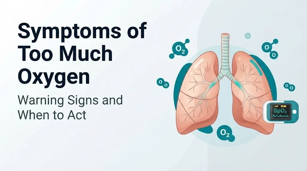
The symptoms of getting too much oxygen range from subtle early warning signs like twitching and visual disturbances to severe, life-threatening presentations including seizures, pulmonary edema, and respiratory failure. This condition, called oxygen toxicity or hyperoxia, is not a theoretical risk: it occurs in real clinical settings every day, affecting COPD patients on high-flow home oxygen, premature infants in neonatal intensive care units, scuba divers breathing enriched air mixtures, and critically ill patients on mechanical ventilators.
The dangers of too much oxygen are well-documented in the clinical literature. According to the Undersea and Hyperbaric Medical Society (UHMS), central nervous system oxygen toxicity can progress from early warning symptoms to a full tonic-clonic seizure within minutes when oxygen partial pressures exceed safe thresholds. For patients in intensive care settings, research published in Intensive Care Medicine has found that liberal oxygen administration is associated with higher mortality compared to conservative oxygen targeting in critically ill adults.
This article covers what happens physiologically when oxygen concentration becomes toxic, what symptoms each affected body system produces, which populations are at the highest risk, and the specific signs that require calling 911 immediately rather than waiting for a scheduled physician appointment.
What Is Oxygen Toxicity and How Does It Happen
Oxygen toxicity occurs when the partial pressure of oxygen in the body’s tissues rises high enough to overwhelm the cell’s antioxidant defense systems, causing direct biochemical damage to cell membranes, proteins, and DNA.
Under normal atmospheric conditions, the air you breathe is approximately 21% oxygen. At sea level, this produces an inspired oxygen partial pressure of roughly 160 mmHg. Your tissues, particularly your mitochondria, are exquisitely tuned to this level. They extract oxygen efficiently while maintaining the balance needed to prevent oxidative harm.
Problems begin when inspired oxygen concentration rises well above 21%, either through supplemental oxygen therapy, breathing compressed gas mixtures during diving, or receiving hyperbaric oxygen treatment. The relevant measure is not the percentage of oxygen being breathed but the partial pressure of oxygen (PO2): the product of total pressure multiplied by oxygen fraction. A scuba diver breathing air at 66 feet of depth experiences a PO2 roughly three times what they would breathe at the surface, even though the oxygen percentage in their tank is unchanged.
Think of the cell’s antioxidant system like a flood drainage canal. Under normal conditions, it manages the steady trickle of reactive oxygen molecules produced by normal metabolism. When PO2 rises pathologically high, the flood of reactive oxygen overwhelms the drainage system completely. The damage that follows is not primarily from the oxygen molecule itself but from the unstable, highly reactive molecules it generates inside cells.
| PO2 Threshold | Form of Toxicity | Primary Affected Organ | Approximate Time to Symptoms |
|---|---|---|---|
| Greater than 1.6 ATA (1216 mmHg) | CNS toxicity (Paul Bert effect) | Brain, spinal cord | Minutes to hours |
| FiO2 greater than 0.6 at 1 ATA for over 24 hours | Pulmonary toxicity (Lorrain Smith effect) | Lung alveoli, tracheobronchial epithelium | Hours to days |
| PaO2 greater than 80 mmHg in premature neonates | Ocular/retinal toxicity | Retinal vasculature | Hours to days with cumulative exposure |
For patients receiving home oxygen therapy, the risk of CNS oxygen toxicity is low because concentrators typically deliver oxygen at 1 to 5 liters per minute at atmospheric pressure, producing PO2 levels that are elevated but generally below CNS toxicity thresholds. Pulmonary toxicity at sustained high flow rates remains the more clinically relevant risk for home oxygen users.
The Biochemistry of Too Much Oxygen: Reactive Oxygen Species and Tissue Damage
The molecular mechanism behind every symptom of oxygen toxicity is the same: an excess of reactive oxygen species (ROS) that overwhelm the cell’s antioxidant defense capacity and destroy cellular structures.
Under normal oxygen partial pressures, the mitochondrial electron transport chain reduces molecular oxygen to water through a series of controlled electron transfers. This process is efficient but imperfect. A small fraction of oxygen molecules accepts electrons at intermediate steps, forming superoxide anion (O2-), the primary ROS produced by mitochondrial metabolism. Normally, the enzyme superoxide dismutase rapidly converts O2- to hydrogen peroxide (H2O2), which catalase and glutathione peroxidase then neutralize to water.
When PO2 is pathologically elevated, the electron transport chain generates superoxide in quantities that exceed the enzymatic capacity of this antioxidant system. The excess O2- is converted to hydrogen peroxide, which reacts with iron ions in a Fenton reaction to produce the hydroxyl radical (OH-): the most reactive and destructive oxidant the body can generate.
The hydroxyl radical attacks the phospholipid bilayer of cell membranes through a process called lipid peroxidation. This is not a reversible insult. Once a phospholipid is oxidized, it releases more reactive molecules that oxidize adjacent phospholipids in a chain reaction, progressively degrading the cell membrane’s structural integrity. The same ROS attack mitochondrial proteins (disrupting energy production), damage cellular enzymes, and cause oxidative breaks in DNA strands.
In the lungs, the type II pneumocytes (the cells responsible for producing surfactant, the detergent-like substance that prevents alveolar collapse) are particularly vulnerable to lipid peroxidation. Their destruction reduces surfactant availability, destabilizes alveoli, and allows fluid to flood into the alveolar space. In the brain, ROS disrupt the inhibitory neurotransmitter system, specifically gamma-aminobutyric acid (GABA) pathways, raising neuronal excitability toward seizure threshold.
Patients with glucose-6-phosphate dehydrogenase (G6PD) deficiency are at elevated risk for ROS-mediated injury from hyperoxia. G6PD generates NADPH, which regenerates glutathione, the primary intracellular ROS scavenger. Without adequate G6PD activity, glutathione cannot be replenished, and the antioxidant defense depletes faster under high-oxygen conditions.
What Are the Symptoms of Getting Too Much Oxygen
The symptoms of getting too much oxygen depend on which organ system is most affected, which is determined by the PO2 level, the duration of exposure, and the individual’s physiological vulnerabilities.
The three recognized clinical forms produce overlapping but distinct symptom sets:
CNS oxygen toxicity symptoms (primarily at high PO2, short exposure):
- Visual disturbances including tunnel vision, flickering, or light sensitivity
- Tinnitus (ringing or buzzing in the ears)
- Nausea with or without vomiting
- Facial muscle twitching or involuntary limb twitching
- Irritability, anxiety, or agitation without clear cause
- Dizziness or vertigo
- Tonic-clonic seizure (the most serious CNS manifestation)
Pulmonary oxygen toxicity symptoms (primarily at sustained high FiO2, longer exposure):
- Substernal chest tightness or burning pain (pleuritic in character)
- Dry, nonproductive cough worsening over hours
- Progressive shortness of breath (dyspnea) at rest or with minimal exertion
- Shallow, rapid breathing (tachypnea)
- Decreased exercise tolerance
Ocular oxygen toxicity symptoms (primarily in premature neonates):
- Not directly symptomatic in the neonate (detected through ophthalmic screening)
- Retinal vascular changes leading to vision impairment or blindness if untreated
For the general reader on home supplemental oxygen, the most practically relevant symptoms to recognize are the pulmonary pattern: a worsening dry cough, increasing chest discomfort, and growing breathlessness at oxygen flow rates previously well tolerated.
Central Nervous System Oxygen Toxicity Symptoms
Central nervous system (CNS) oxygen toxicity produces a recognizable cluster of neurological symptoms that clinicians trained in hyperbaric medicine learn through the mnemonic VENTID: Vision changes, Ears ringing, Nausea, Twitching, Irritability, Dizziness.
The physiological driver of every CNS symptom is the same: ROS-mediated disruption of GABAergic inhibitory neurotransmission. GABA is the brain’s primary inhibitory neurotransmitter, responsible for preventing neurons from firing excessively. When ROS disable the enzymes responsible for GABA synthesis and release, neuronal excitability rises across the cortex and brainstem. The result is the classic progression from subtle sensory disturbances to frank seizure activity.
Vision changes in CNS oxygen toxicity typically present as tunnel vision (progressive narrowing of the visual field), flickering lights at the visual periphery, or sudden monocular brightness changes. These reflect ROS injury to the optic nerve or occipital cortex and are an early warning that CNS toxicity is progressing.
Muscle twitching most commonly begins in the face, particularly around the lips and cheeks, before spreading to the extremities. This is an important early sign: experienced hyperbaric medicine specialists treat any facial twitching during oxygen breathing as a CNS toxicity signal requiring immediate interruption of the oxygen exposure.
According to the UHMS, CNS oxygen toxicity is the primary safety concern in hyperbaric oxygen therapy because the transition from early warning symptoms to a generalized seizure can occur within minutes without any reliable intervening warning. The VENTID symptoms are present in some, but not all, patients before a seizure. Some patients seize without any preceding warning signs.
Patients using prescribed home oxygen at standard flow rates are at low risk for CNS toxicity. The primary populations at CNS toxicity risk are those breathing oxygen at elevated pressure: scuba divers, hyperbaric chamber patients, and occasionally patients receiving high-flow oxygen via non-invasive positive pressure ventilation masks at high FiO2 settings.
Pulmonary Oxygen Toxicity Symptoms
Pulmonary oxygen toxicity produces symptoms through direct injury to the alveolar-capillary membrane and the respiratory epithelium lining the airways, and it develops over hours to days of sustained high-concentration oxygen exposure rather than the minutes-to-hours timeline of CNS toxicity.
The earliest pulmonary symptom is substernal chest tightness: a burning discomfort behind the sternum that worsens with deep inspiration. This reflects injury to the tracheobronchial epithelium, the ciliated mucous membrane lining the trachea and bronchi. ROS attack these cells, triggering inflammatory cytokine release, mucosal edema, and pain with inspiration. The pattern is distinct from cardiac chest pain: it is reproduced by breathing depth, not by exertion.
A dry, persistent cough follows. As type II pneumocyte injury progresses and surfactant levels fall, the alveoli (the tiny air sacs where gas exchange occurs) become unstable. The alveolar-capillary membrane begins to leak fluid into the alveolar space. This is the beginning of pulmonary edema from oxygen toxicity, which presents clinically as progressively worsening dyspnea, declining oxygen saturation paradoxically (the patient is breathing high oxygen but absorbing less of it), and, in severe cases, respiratory failure.
According to research published in the New England Journal of Medicine in studies of mechanically ventilated ICU patients, prolonged FiO2 greater than 0.6 (60% oxygen) at normal atmospheric pressure over 24 to 48 hours is associated with measurable alveolar damage, and FiO2 of 1.0 (100% oxygen) for more than 6 hours produces early pulmonary toxicity detectable on bronchoalveolar lavage.
Patients already receiving treatment for respiratory conditions, including those with pneumonia, acute respiratory distress syndrome (ARDS), or pulmonary fibrosis, are at particular risk because their damaged lung tissue has reduced antioxidant reserve and greater baseline vulnerability to ROS injury.
Key Takeaway: The earliest pulmonary oxygen toxicity symptom, substernal burning chest pain that worsens with deep breathing, is the specific signal that tells you the lining of your airways is being chemically injured by excess oxygen, and it requires same-day evaluation by a pulmonologist or emergency medicine physician.
Ocular Oxygen Toxicity and Vision Changes
Ocular oxygen toxicity produces fundamentally different effects depending on whether it occurs in a premature neonate or in an adult, and this distinction matters enormously for how symptoms are recognized and managed.
In adults receiving supplemental oxygen or undergoing hyperbaric therapy, the most relevant visual symptom is the tunnel vision and peripheral flickering that occurs as part of CNS oxygen toxicity, described above. This reflects central nervous system involvement of the visual cortex and optic pathways rather than direct retinal injury. In adults with normal retinal vasculature, hyperoxia-induced retinal vasoconstriction can reduce retinal blood flow, contributing to visual field changes, but this is typically reversible when oxygen exposure is reduced.
In premature neonates, particularly those born before 32 weeks gestational age, the situation is entirely different and carries consequences that can be permanent. The retina completes its vascular development in the final weeks of gestation, and at premature birth, large areas of the peripheral retina remain avascular (lacking blood vessels). Under normal low-PO2 conditions in the womb, growth factors including vascular endothelial growth factor (VEGF) drive new vessel growth into the avascular peripheral retina.
When a premature neonate receives supplemental oxygen and arterial PaO2 rises above approximately 80 mmHg, this elevated oxygen suppresses VEGF production and arrests retinal vascularization. The avascular retina then responds with disorganized, abnormal vessel growth when oxygen exposure decreases, producing retinopathy of prematurity (ROP). Severe ROP can lead to retinal detachment and permanent blindness.
According to the landmark SUPPORT Trial (published in the New England Journal of Medicine in 2010) and the subsequent NeOProM Collaboration analysis, premature neonates targeted to lower oxygen saturation ranges (85 to 89%) had lower rates of severe ROP but higher mortality rates than those targeted to 91 to 95%, demonstrating the clinical difficulty of oxygen management in this population and the real consequences of both too much and too little oxygen in neonates.
Early Warning Signs of Oxygen Toxicity Before It Becomes Dangerous
Recognizing oxygen toxicity before it escalates to seizure or respiratory failure gives clinicians and patients the opportunity to reduce oxygen exposure and prevent a medical emergency. The early warning signs differ by form of toxicity, but several are consistent enough to learn by name.
For CNS toxicity, the VENTID warning signs are the clinical standard recognized by hyperbaric medicine specialists:
To recognize and respond to early CNS oxygen toxicity warning signs:
- Note any change in visual clarity, peripheral flickering, or tunnel-like narrowing of what you can see, and report it immediately to the supervising clinician or dive buddy.
- Report any ringing or buzzing in the ears that appears during oxygen exposure, as tinnitus from oxygen toxicity tends to be bilateral and appears suddenly.
- Watch for nausea that develops without dietary cause during oxygen breathing, particularly in a diving or hyperbaric context.
- Report any involuntary twitching, particularly of facial muscles (lips, cheeks, eyelids), as this is a pre-seizure warning signal requiring immediate action.
- Note any unexplained irritability, agitation, or anxiety that develops during oxygen exposure, as these reflect limbic system hyperexcitability from GABAergic suppression.
- Report dizziness or any sense of spatial disorientation without positional cause.
For pulmonary toxicity early warning, the key symptoms to monitor are:
- A mild substernal burning sensation that begins with deep inhalation
- A dry tickling cough that appears without infection or allergy cause
- Any reduction in exercise tolerance not explained by the underlying condition
Older adults over 65 receiving supplemental oxygen at home may have diminished awareness of gradual pulmonary symptom onset due to reduced respiratory sensitivity that accompanies aging-related changes in the lung mechanoreceptors. This makes regular check-ins with the prescribing pulmonologist or primary care physician, and adherence to the prescribed flow rate without self-adjustment upward, particularly important for this age group.
How Quickly Does Oxygen Toxicity Develop
The speed at which oxygen toxicity develops depends almost entirely on the PO2 being breathed: the higher the partial pressure, the faster the onset of symptoms and tissue injury.
CNS oxygen toxicity develops on a time scale of minutes to a few hours at the PO2 levels encountered in hyperbaric oxygen therapy (typically 2.0 to 3.0 ATA of pure oxygen) or in scuba diving mishaps. At a PO2 of 1.6 ATA (the UHMS-established upper limit for diving with enriched air nitrox), clinically relevant CNS symptoms can develop within 30 to 60 minutes in susceptible individuals. At higher pressures, the time to seizure shrinks dramatically. This is why hyperbaric therapy protocols include mandatory air breaks, periods of breathing air rather than pure oxygen, to reduce cumulative ROS exposure and prevent toxicity.
Pulmonary oxygen toxicity develops over a longer time scale because it requires cumulative alveolar damage rather than an acute neurological excitability threshold. Research summarized in the American Journal of Respiratory and Critical Care Medicine indicates that breathing 100% oxygen at 1 atmosphere (normal sea level pressure) for six or more hours produces early tracheobronchitis and detectable alveolar injury. Sustained FiO2 greater than 0.6 for 24 hours or more produces measurable pulmonary toxicity in the majority of exposed individuals. Extended exposure beyond 48 hours at high FiO2 can produce diffuse alveolar damage indistinguishable from ARDS on imaging.
According to the American Thoracic Society, the target for mechanically ventilated patients should minimize FiO2 to the lowest level that maintains SpO2 at 92 to 96%, specifically to reduce the risk of time-dependent pulmonary oxygen toxicity while ensuring adequate tissue oxygen delivery.
Individual factors that accelerate toxicity onset include fever (which increases metabolic rate and ROS production), corticosteroid use (which may reduce some antioxidant enzyme expression), and pre-existing inflammatory lung disease that has already depleted local antioxidant reserves.
Key Takeaway: CNS oxygen toxicity can progress from early tingling warning signs to a full seizure in minutes at high PO2 levels, while pulmonary toxicity develops over hours to days at the oxygen concentrations used in hospital mechanical ventilation, making the two forms medically distinct emergencies with different response timelines.
Oxygen Toxicity in COPD Patients on Supplemental Oxygen
For people with chronic obstructive pulmonary disease (COPD) who use home supplemental oxygen, the risk picture is different from either the diving or neonatal context, and it is one that is frequently misunderstood by patients and sometimes by non-specialist clinicians.
The well-established concern is hypercapnia: carbon dioxide retention caused by inappropriately high oxygen flow in COPD patients. Many people with severe COPD have chronically elevated arterial CO2 (PaCO2) and reduced ventilatory drive. When supplemental oxygen raises arterial oxygen saturation above the appropriate target range for COPD (generally 88 to 92% SpO2, per American Thoracic Society and Global Initiative for Chronic Obstructive Lung Disease (GOLD) guidelines), several mechanisms contribute to worsening CO2 retention: release of hypoxic pulmonary vasoconstriction increases dead space ventilation, and the Haldane effect reduces CO2 carrying capacity of hemoglobin. The result is rising PaCO2, which produces a specific symptom pattern:
- Increasing drowsiness or difficulty staying awake
- Confusion or altered mental status
- Morning headache (from nocturnal CO2 retention)
- Flushed, warm skin
- Bounding pulse
This progression toward hypercapnic respiratory failure is a medical emergency. If a COPD patient on home oxygen becomes increasingly drowsy or confused, reduce oxygen flow only if prescribed titration instructions allow it, and call 911 immediately.
Beyond hypercapnia risk, COPD patients on long-term high-flow oxygen also face pulmonary oxygen toxicity risk from the same ROS mechanism described earlier. Their already-damaged lung parenchyma, with depleted local antioxidant reserves from chronic inflammation, is more vulnerable to oxidative injury at lower PO2 levels than healthy lung tissue. A COPD patient who is increasing their home oxygen flow rate above the prescribed level without physician guidance is at real risk of accelerating both hypercapnic and oxidative lung injury.
Any COPD patient whose oxygen prescription was set more than six months ago and who has experienced increasing breathlessness, changed activity tolerance, or started using oxygen at higher flows than prescribed should bring those observations to an appointment with the prescribing pulmonologist, specifically requesting a repeat arterial blood gas to confirm their current CO2 level and oxygen saturation targets.
Oxygen Toxicity in Premature Newborns and NICU Patients
Premature newborns in the neonatal intensive care unit (NICU) represent the population for whom oxygen management is among the most clinically consequential decisions a neonatologist makes, because both too much and too little oxygen carry independent serious risks in this patient group.
Retinopathy of prematurity (ROP) is the most recognized oxygen-related injury in premature neonates, as described in the ocular toxicity section. But hyperoxia in premature infants causes two additional conditions worth understanding: bronchopulmonary dysplasia (BPD) and oxidative brain injury.
BPD is a chronic lung disease that develops in premature infants who require prolonged mechanical ventilation and supplemental oxygen. The incompletely developed premature lung, with fewer alveoli and less surfactant reserve than a term infant, is far more vulnerable to ROS-mediated injury from supplemental oxygen than a mature lung. ROS damage the developing alveolar epithelium and promote inflammatory pathways (via NF-kB activation and IL-6 release) that disrupt alveolar development, producing the characteristic scarring and air trapping of BPD.
According to research published in Pediatrics, the official journal of the American Academy of Pediatrics, very preterm infants (born before 28 weeks gestational age) have substantially lower superoxide dismutase and catalase activity than term infants, explaining their heightened susceptibility to oxygen-mediated oxidative stress at PaO2 levels that would not injure mature lung tissue.
Parents of premature infants in the NICU do not control oxygen management directly, but understanding why neonatologists work to keep oxygen saturations within a narrow target range (typically 91 to 95% for most premature infants) helps make sense of the alarms, adjustments, and monitoring intensity they observe at the bedside. Any questions about their infant’s oxygen targets should be directed to the attending neonatologist, who can explain the specific saturation targets and the reasons for the range chosen for their particular infant.
Oxygen Toxicity in Scuba Divers and Hyperbaric Oxygen Therapy
Scuba divers and patients receiving hyperbaric oxygen therapy (HBO) are the populations most commonly associated with CNS oxygen toxicity in clinical literature, because both groups breathe oxygen at partial pressures well above what is achievable at sea level in open air.
For scuba divers breathing enriched air nitrox (air mixtures with oxygen content above the standard 21%), the risk of CNS oxygen toxicity rises as they descend. The UHMS and Divers Alert Network (DAN) both establish a maximum operating depth (MOD) for any given nitrox mixture, calculated to keep PO2 below 1.4 ATA for routine recreational diving and 1.6 ATA as an absolute ceiling. Divers who descend below their MOD, whether from navigation error, current, or equipment failure, face rapidly escalating CNS toxicity risk.
The clinical danger in diving is that a CNS oxygen toxicity seizure underwater is almost universally fatal without immediate surface assistance, because loss of consciousness during a dive leads to drowning. The VENTID warning symptoms give a brief pre-seizure window if recognized. Experienced divers are trained to signal their buddy and begin ascent immediately if any VENTID symptom appears. The problem is that CNS toxicity can produce seizure without preceding warning symptoms in a minority of cases.
For patients undergoing HBO therapy for conditions such as carbon monoxide poisoning, chronic non-healing wounds, or decompression sickness, therapy protocols at established hyperbaric centers incorporate mandatory air breaks (typically 5 minutes of breathing air per 20 to 30 minutes of oxygen) specifically to interrupt ROS accumulation and reduce CNS toxicity risk. According to the UHMS, the incidence of CNS oxygen toxicity seizures in modern HBO therapy protocols is estimated at approximately 1 in 10,000 treatments when protocols are followed correctly.
Patients with a history of febrile seizures, known epilepsy, or current fever are at elevated risk for CNS oxygen toxicity during HBO therapy. The treating hyperbaric medicine physician should be informed of any seizure history before treatment begins.
Key Takeaway: A scuba diver who notices visual flickering, lip twitching, or ear ringing while underwater on a nitrox mix should immediately signal their buddy and begin a controlled ascent, because these are the pre-seizure warning signs of CNS oxygen toxicity that can progress to loss of consciousness and drowning within minutes.
What Causes and Triggers Oxygen Toxicity
Oxygen toxicity is always caused by the same underlying factor: breathing oxygen at a partial pressure high enough and for a duration long enough to overwhelm cellular antioxidant defenses. The specific triggers that create this condition vary by setting.
| Trigger | Clinical Context | PO2/FiO2 Involved | Risk Level |
|---|---|---|---|
| High-flow supplemental oxygen | Home oxygen, hospital therapy, ICU ventilation | FiO2 greater than 0.6 at 1 ATA | Moderate to high with prolonged exposure |
| Enriched air nitrox diving below MOD | Recreational scuba diving | PO2 greater than 1.6 ATA | High: CNS toxicity within minutes |
| Hyperbaric oxygen therapy without air breaks | HBO treatment for wounds, CO poisoning | PO2 2.0 to 3.0 ATA | High: CNS toxicity without breaks |
| 100% oxygen breathing during anesthesia | Surgical patients under general anesthesia | FiO2 1.0 at 1 ATA | Moderate: pulmonary toxicity with prolonged procedures |
| Oxygen misuse in emergency care | Self-administered pure oxygen by athletes | Variable | Low at atmospheric pressure; higher with pressurized masks |
Several individual factors increase susceptibility to oxygen toxicity at any given PO2 level. These include:
- Fever: increases metabolic rate and ROS production, lowering the effective toxicity threshold
- Physical exertion during oxygen exposure: carbon dioxide produced by exercise is a known CNS oxygen toxicity sensitizer
- Corticosteroid medications: may reduce antioxidant enzyme expression
- G6PD deficiency: reduces glutathione regeneration capacity, increasing ROS vulnerability
- Prior lung disease: depletes local antioxidant reserves in the respiratory epithelium
- Carbon dioxide retention: elevated CO2 enhances CNS excitability under high PO2 conditions
Healthy adults breathing standard supplemental oxygen at 2 to 4 liters per minute through nasal cannula at sea level do not produce PO2 levels that approach CNS toxicity thresholds. Pulmonary toxicity at these flow rates would require sustained high-concentration delivery (typically FiO2 greater than 0.6 via mask) over many hours, a scenario more relevant to hospital care than home oxygen use.
Oxygen Toxicity Versus Hypoxia: Key Differences
Oxygen toxicity and hypoxia sit at opposite ends of the oxygen spectrum, but their symptoms overlap in ways that can create confusion about which condition a person is experiencing.
| Feature | Oxygen Toxicity (Hyperoxia) | Hypoxia (Low Oxygen) |
|---|---|---|
| Cause | PO2 or FiO2 too high | PO2 or FiO2 too low |
| SpO2 reading | High (often above 98 to 99%) | Low (below 90% signals concern) |
| CNS symptoms | Twitching, seizure, visual disturbance | Confusion, drowsiness, impaired judgment |
| Respiratory symptoms | Chest burning, cough, worsening dyspnea | Breathlessness, air hunger, cyanosis |
| Typical onset | Variable: minutes (CNS) to days (pulmonary) | Minutes to hours depending on cause |
| Who is at risk | Divers, HBO patients, ICU patients on high FiO2 | COPD patients, altitude exposure, cardiac disease |
| Emergency response | Reduce oxygen, call 911 for seizure or respiratory failure | Increase oxygen, call 911 for SpO2 below 90% |
The distinction is clinically relevant because the immediate management differs: for oxygen toxicity, the response is to reduce or discontinue oxygen exposure; for hypoxia, the response is to increase oxygen delivery. In a home setting, someone who observes a person becoming confused while receiving supplemental oxygen needs to consider both possibilities. Pulse oximetry provides a quick directional clue: an SpO2 above 98 to 99% in a person with new neurological symptoms suggests oxygen toxicity as a contributor; an SpO2 below 88% in a person with confusion points to hypoxia.
COPD patients occupy a particularly complex position: their appropriate SpO2 target is intentionally kept lower (88 to 92%) than the general adult population (94 to 98%). An SpO2 of 95% in a COPD patient may represent over-oxygenation with hypercapnia risk, while the same reading in a healthy adult is appropriate. Context and physician-set targets are essential for correct interpretation.
How Oxygen Toxicity Is Diagnosed
Oxygen toxicity is diagnosed by a combination of clinical history, symptom pattern, and physiological measurement rather than by a single definitive test.
The diagnostic process typically follows this sequence:
- Clinical history: the physician establishes what oxygen level was being breathed (FiO2, PO2, or liters per minute via which delivery device), for how long, and in what pressure environment. This single piece of information often makes the diagnosis clinically obvious.
- Arterial blood gas (ABG) measurement: an ABG provides arterial PaO2 (confirming hyperoxia if above 120 to 150 mmHg in a non-altitude context), PaCO2 (identifying hypercapnia in COPD patients), and arterial pH (revealing respiratory acidosis from CO2 retention). This is the most informative immediate blood test.
- Pulse oximetry: a noninvasive real-time measure of oxygen saturation (SpO2). It does not measure PO2 directly but provides a rapid screen. SpO2 persistently above 98 to 99% in a symptomatic patient on supplemental oxygen is a signal to reassess the prescribed flow rate.
- Chest imaging: a plain chest radiograph (X-ray) or computed tomography (CT) scan of the chest identifies pulmonary edema, ground-glass opacity patterns, or diffuse alveolar damage from pulmonary oxygen toxicity. These findings are often non-specific in early stages.
- Pulmonary function testing: spirometry showing reduced lung volumes (vital capacity, forced vital capacity) reflects the restrictive pattern that pulmonary oxygen toxicity produces as alveolar damage progresses.
- Electroencephalography (EEG): ordered if CNS oxygen toxicity with seizure occurred, to document post-ictal changes and rule out other seizure causes.
For the neonatal patient population, ophthalmologic screening using indirect ophthalmoscopy to examine the developing retina is the primary diagnostic tool for retinopathy of prematurity. The American Academy of Pediatrics recommends serial ophthalmic screening exams for all premature infants born before 30 weeks gestational age or with birth weight below 1,500 grams, beginning at 31 to 33 weeks corrected gestational age.
When Oxygen Toxicity Becomes a Medical Emergency
Certain manifestations of oxygen toxicity require immediate emergency medical response. Do not wait to see if these resolve on their own.
Call 911 or go to the nearest emergency room immediately if you observe:
- A generalized tonic-clonic seizure in anyone breathing supplemental oxygen or who has recently undergone hyperbaric oxygen therapy: oxygen-induced seizure requires immediate cessation of supplemental oxygen and emergency neurological assessment
- Loss of consciousness in a patient receiving supplemental oxygen, a diver, or a hyperbaric therapy patient: this may represent CNS oxygen toxicity with seizure and requires emergency evaluation
- Severe respiratory distress with declining oxygen saturation despite being on supplemental oxygen: this paradox, getting worse on oxygen, can indicate pulmonary oxygen toxicity with developing pulmonary edema
- Rapidly worsening confusion, inability to be roused, or extreme drowsiness in a COPD patient on home oxygen: this symptom pattern suggests hypercapnic respiratory failure from oxygen-driven CO2 retention
- Facial twitching followed by limb twitching in anyone breathing enriched-air diving gas or undergoing hyperbaric therapy: this is the immediate pre-seizure warning and requires stopping oxygen breathing and surfacing (for divers) or exiting the chamber (for HBO patients) immediately
- Blue discoloration of the lips or fingertips (cyanosis) combined with shortness of breath despite oxygen delivery: suggests severe V/Q mismatch from pulmonary edema secondary to oxygen toxicity
These presentations can indicate oxygen-induced seizure, hypercapnic respiratory failure, or acute pulmonary oxygen toxicity with respiratory failure, all of which require emergency medical assessment with cardiac monitoring, arterial blood gas measurement, and specialist evaluation. None of these presentations is appropriate for an urgent care center; go directly to a hospital emergency department.
How to Prevent Oxygen Toxicity
Preventing oxygen toxicity requires matching oxygen delivery precisely to physiological need, not delivering the maximum available concentration on the assumption that more oxygen is always better.
Evidence-based prevention strategies by setting:
For home oxygen users:
- Use only the prescribed flow rate (in liters per minute). Do not self-adjust upward during breathlessness without physician guidance, as this may worsen rather than relieve the underlying cause.
- Schedule regular arterial blood gas tests as recommended by your pulmonologist to confirm your oxygen saturation targets remain appropriate.
- Report any new chest burning, worsening cough, or increasing breathlessness to your prescribing physician promptly.
For scuba divers using nitrox:
- Calculate and know your maximum operating depth (MOD) before every dive, using the formula: MOD (in meters of seawater) equals (PO2 max / FiO2) minus 10, with PO2 max of 1.4 ATA for recreational diving.
- Never dive solo on nitrox: a buddy trained in recognizing VENTID symptoms and providing emergency ascent assistance is non-negotiable.
- Avoid heavy exercise during enriched-air dives; exercise raises CO2 output, which sensitizes the CNS to oxygen toxicity at any given PO2.
For patients undergoing hyperbaric oxygen therapy:
- Disclose all current medications, prior seizure history, and any fever or illness to the hyperbaric medicine physician before each session, as these factors can lower your CNS toxicity threshold.
- Follow the air break schedule prescribed by your hyperbaric medicine team without shortening it.
- Report any VENTID symptoms during therapy immediately to the treating clinician.
For ICU clinical staff managing ventilated patients:
- Target the minimum FiO2 that maintains SpO2 at 92 to 96%, per American Thoracic Society guidelines, specifically to minimize pulmonary oxygen toxicity risk during prolonged mechanical ventilation.
Key Takeaway: The most effective prevention of oxygen toxicity in every clinical setting, from the home oxygen concentrator to the hyperbaric chamber, is using the minimum oxygen concentration that achieves the clinically appropriate oxygen saturation target, not the maximum available concentration.
Frequently Asked Questions About Symptoms of Getting Too Much Oxygen
What does it feel like to have too much oxygen?
Getting too much oxygen produces sensations that vary by which body system is affected first.
CNS oxygen toxicity typically produces visual flickering or tunnel vision, ringing in the ears, nausea, involuntary muscle twitching (especially in the face), unexplained irritability, and dizziness, progressing potentially to seizure.
Pulmonary oxygen toxicity feels like a worsening burning sensation behind the sternum that increases with deep breathing, accompanied by a dry, progressive cough and increasing breathlessness.
Can you get oxygen toxicity from a home oxygen concentrator?
Home oxygen concentrators operating at standard flow rates of 1 to 5 liters per minute at atmospheric pressure produce oxygen partial pressures that are elevated but generally below the threshold for CNS oxygen toxicity in most adults.
However, prolonged high-flow delivery via mask at FiO2 above 0.6 carries cumulative pulmonary toxicity risk over hours to days, and COPD patients face additional risk of hypercapnic respiratory failure at any flow rate that raises SpO2 above their prescribed target of 88 to 92%.
Any home oxygen user experiencing new chest burning, worsening cough, or confusion while on oxygen should contact their prescribing pulmonologist or primary care physician the same day, or call 911 if symptoms are severe.
How long does it take for too much oxygen to cause damage?
CNS oxygen toxicity can progress from the first warning symptoms to a seizure in minutes when PO2 exceeds 1.6 ATA, as occurs in scuba diving or hyperbaric therapy at therapeutic pressures.
Pulmonary oxygen toxicity requires sustained exposure to FiO2 greater than 0.6 for six or more hours to produce detectable alveolar injury, with more severe damage accumulating over 24 to 48 hours of continuous high-concentration oxygen breathing.
The time course depends entirely on both the oxygen partial pressure being breathed and the individual’s antioxidant reserve capacity.
What is the difference between oxygen toxicity and oxygen overdose?
Oxygen toxicity and oxygen overdose refer to the same clinical condition: tissue injury from breathing oxygen at a partial pressure that overwhelms cellular antioxidant defenses.
“Oxygen overdose” is a colloquial term not used in clinical medicine, while “oxygen toxicity” or “hyperoxia” are the correct clinical terms covering both the CNS (Paul Bert effect) and pulmonary (Lorrain Smith effect) forms.
Both terms describe the same underlying biochemical mechanism of reactive oxygen species overproduction causing lipid peroxidation and cell death.
Is oxygen toxicity reversible?
Early oxygen toxicity is largely reversible if oxygen exposure is reduced promptly before permanent structural damage accumulates.
Pulmonary oxygen toxicity that has not progressed beyond early tracheobronchitis and mild alveolar inflammation typically resolves over days to weeks once FiO2 is reduced, but extensive diffuse alveolar damage may produce persistent lung function impairment.
Retinopathy of prematurity in premature neonates can cause permanent vision loss or blindness if not detected and treated early through ophthalmologic screening and, when indicated, laser photocoagulation or anti-VEGF therapy administered by a pediatric ophthalmologist.
What oxygen level is too high?
An oxygen saturation (SpO2) persistently above 98 to 99% in a patient receiving supplemental oxygen is a signal that the prescribed flow rate may be delivering more oxygen than the body needs.
For COPD patients, an SpO2 above 92% while on supplemental oxygen is generally considered above the appropriate target range and carries risk of CO2 retention and hypercapnic respiratory failure, per American Thoracic Society guidelines.
In terms of partial pressure, the UHMS establishes 1.6 ATA as the absolute PO2 ceiling for recreational scuba diving on nitrox, above which CNS oxygen toxicity risk becomes acutely dangerous.
Closing
The symptoms of getting too much oxygen are not abstract clinical rarities: they occur in living rooms, dive sites, and hospital wards, in patients who assumed that more oxygen meant more safety. The body requires oxygen within a precise physiological range, and exceeding that range triggers a biochemical cascade of reactive oxygen species-mediated damage that affects the lungs, brain, and eyes through mechanisms that are well-understood and entirely preventable.
If you are on home supplemental oxygen, the most important action you can take is using exactly the prescribed flow rate, not higher, and reporting any new chest burning, worsening cough, confusion, or drowsiness to your prescribing physician the same day they appear.
If you dive on nitrox or undergo hyperbaric therapy, know your maximum operating depth, know the VENTID symptoms by name, and know that facial twitching during oxygen breathing requires stopping oxygen immediately. Your awareness of these early warning signs is the only window between a pre-seizure symptom and a full medical emergency.