Broken Foot Symptoms: Signs, Severity and When to Act Now
Broken foot symptoms include localized pain, swelling, bruising, tenderness to touch, and difficulty bearing weight on the affected foot, each produced by the inflammatory cascade that begins at the moment bone continuity is disrupted. These symptoms vary substantially depending on which of the foot’s 26 bones is fractured, how much displacement has occurred, and whether the injury is an acute traumatic fracture or a gradually developing stress fracture.
According to the American Academy of Orthopaedic Surgeons (AAOS), foot fractures are among the most frequently occurring skeletal injuries, with metatarsal fractures alone accounting for approximately 35% of all foot fractures seen in emergency settings. The foot’s complex architecture means that where your pain is located, where bruising appears, and whether you can put weight on it at all are clinically informative details that point to specific bones and specific fracture patterns.
This guide covers every core broken foot symptom with its physiological mechanism, maps pain and bruising locations to the bones most likely fractured, explains the most commonly missed serious foot injury (the Lisfranc fracture-dislocation), and tells you clearly when your symptoms require emergency evaluation today versus an urgent care visit tomorrow.
Broken Foot Symptoms: What You Need to Know First
Broken foot symptoms range from severe immediate pain with inability to stand to gradual aching that builds over weeks, with the exact pattern depending on which bone is fractured, how it fractured, and what forces caused the injury.
The foot is not a simple structure. It contains 26 bones working as an interconnected weight-bearing platform, and a fracture in any one of them disrupts load distribution across the others. A break in the first metatarsal (the thick bone behind the big toe) has very different functional consequences from a fracture of the fifth metatarsal’s base (the bony prominence on the outer edge of the foot) or a calcaneal fracture (the heel bone, which bears the majority of impact forces during walking).
The most important preliminary distinction is between an acute traumatic fracture and a stress fracture. Acute fractures happen suddenly: you drop something on your foot, roll your ankle, or land badly from a height. Symptoms begin immediately and are severe. Stress fractures develop gradually over days to weeks from repetitive loading, and their symptoms are initially subtle enough that many people mistake them for a sore muscle or bruised foot.
What to tell your provider before your appointment:
- The exact mechanism of injury: fall, drop, twist, collision, or gradual onset with no specific trauma
- Where exactly on your foot the pain is most intense: the toes, the ball of the foot, the outer edge, the top (dorsum), the midfoot, or the heel
- Whether bruising has appeared, and where on the foot it is located
- Whether you can put any weight on the foot at all
- Whether you heard or felt a snap, crack, or pop at the time of injury
- Any prior foot fractures, osteoporosis diagnosis, or current steroid medication use
What Is a Broken Foot and Which Bones Are Involved?
A broken foot is a fracture of one or more of the 26 bones that make up the foot, including the 7 tarsal bones, 5 metatarsal bones, and 14 phalangeal bones of the toes.
The foot’s skeletal architecture divides into three functional regions. The hindfoot consists of the calcaneus (heel bone) and the talus (which connects the foot to the ankle). The midfoot includes the navicular, cuboid, and three cuneiform bones (medial, intermediate, and lateral), which form the arch-stabilizing keystone of the foot. The forefoot contains the five metatarsal bones and the 14 phalanges of the toes.
Each region has specific fracture patterns associated with specific injury mechanisms. Hindfoot fractures, particularly calcaneal fractures, typically result from high-energy axial loading such as a fall from height landing on the heel. Midfoot fractures, including Lisfranc injuries, typically result from a twisting force applied to the planted foot or from a crush mechanism. Forefoot fractures, the most common category, result from direct trauma (dropping a heavy object), twisting mechanisms (metatarsal fractures), or repetitive stress (metatarsal stress fractures in runners and military personnel).
According to the National Institute of Arthritis and Musculoskeletal and Skin Diseases (NIAMS), the metatarsal bones are the most commonly fractured bones in the foot, with the fifth metatarsal (the outer metatarsal) and the second metatarsal together accounting for the majority of forefoot fracture presentations.
Older adults over 65 who sustain a foot fracture from a minimal-energy mechanism such as a simple misstep on flat ground should have their bone density considered in clinical assessment. A fracture from a trivial mechanism in this population suggests underlying osteoporosis and warrants bone density evaluation by a primary care physician or endocrinologist in addition to fracture treatment.
Symptoms of a Broken Foot: Pain and Tenderness
The pain of a broken foot is produced by direct activation of periosteal nociceptors, the sensory nerve endings embedded in the periosteum (the fibrous outer membrane of bone), which are among the most sensitive pain receptors in the musculoskeletal system.
The periosteum contains both C-fiber nociceptors (which produce the dull, throbbing background pain) and A-delta fiber nociceptors (which produce the sharp, immediate pain at the moment of fracture and with direct pressure on the fracture site). When bone cortex is disrupted, the periosteum tears at the fracture site, releasing bradykinin from the plasma kinin cascade and substance P from the damaged nerve endings themselves. These mediators sensitize surrounding nociceptors, lowering their firing threshold and producing the exquisite tenderness to touch that characterizes a fracture site.
The pain of a broken foot is typically highly localized in acute fractures. Pressing directly on the fracture site (point tenderness) produces a sharp increase in pain that is disproportionate to the gentle pressure applied. This is different from the diffuse tenderness of a sprain, where the entire ligament attachment area and surrounding soft tissue ache, but no single point produces a dramatically sharper pain response than adjacent areas.
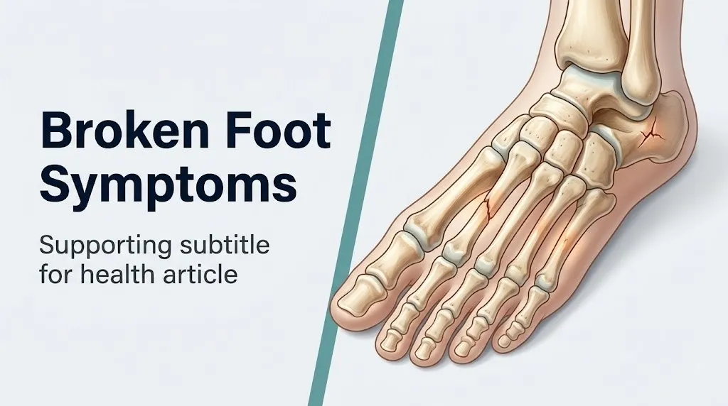
The American Academy of Orthopaedic Surgeons notes that point tenderness directly over a bone is one of the most clinically reliable physical examination findings for identifying a fracture, and it forms part of the basis for the Ottawa Foot Rules, the validated clinical decision instrument used in emergency settings to determine whether foot X-rays are needed.
People taking nonsteroidal anti-inflammatory drugs (NSAIDs) regularly for arthritis or other chronic pain conditions may have a blunted pain response at the fracture site. Their periosteal nociceptors are partially suppressed by prostaglandin inhibition, meaning a fracture may feel less sharp than expected. This population should not use reduced pain as reassurance that a fracture is unlikely.
Broken Foot Swelling and Bruising Symptoms
Swelling and bruising after a broken foot occur through distinct but related physiological mechanisms, and their pattern, location, and timing provide clinically useful information about the type and severity of the underlying fracture.
Swelling begins within minutes of fracture. Bone fracture ruptures the periosteal blood vessels and the small vessels within the marrow cavity, producing an acute hematoma at the fracture site. Simultaneously, the release of histamine from mast cells and damaged endothelial cells, combined with prostaglandin E2 from the COX-2 pathway in injured tissue, increases the permeability of the capillary walls in the surrounding soft tissue. Fluid and plasma proteins pour out of the capillaries into the interstitial space, producing the visible swelling (edema) around the fracture site. Swelling typically reaches its peak between 24 and 72 hours after the injury, which is why the foot often looks significantly worse the morning after a fracture than it did the night of the injury.
Bruising (ecchymosis) appears because extravasated blood from ruptured vessels tracks along fascial planes toward the skin. This tracking follows gravity and the anatomical pathways of least resistance. A fracture on the dorsum (top) of the foot frequently produces bruising that appears on the sole of the foot, the heel, and the ankle, because blood tracks downward through the fascial layers. This is not a sign of additional injury in those locations. It is simply blood migrating through the tissue.
Think of it like a leak in a water pipe inside a wall. The water doesn’t stay at the leak site. It travels through cavities and channels until it emerges somewhere visible. The visible water stain on the ceiling two rooms away doesn’t mean the ceiling is damaged. It just means water traveled the path of least resistance from the original leak.
Bruising that appears specifically on the sole of the foot (plantar surface) after a midfoot injury is a red flag for a Lisfranc injury rather than a simple sprain or metatarsal fracture. This symptom is addressed in detail in the Lisfranc section below.
Older adults and people taking anticoagulant medications such as warfarin, apixaban, or rivaroxaban develop more extensive bruising from the same injury due to impaired clotting, and their bruising may spread across a larger area of the foot, ankle, and lower leg than expected.
Key Takeaway: Swelling peaks at 24 to 72 hours after a foot fracture, so a foot that looks manageable the evening of injury may look alarmingly swollen by the next morning. That progression does not necessarily mean the injury is worsening. But bruising that appears on the sole of the foot after a midfoot twisting injury is a specific warning sign for a Lisfranc injury and warrants same-day imaging.
Can You Walk on a Broken Foot?
Whether you can walk on a broken foot depends entirely on which bone is fractured, whether it is displaced, and how much pain you can tolerate, making weight-bearing ability an unreliable indicator of fracture severity on its own.
This is one of the most common misconceptions about broken feet: that if you can walk on it, it is not broken. That is not clinically accurate. Some fractures that require surgical fixation still allow painful limping, because the fractured bone retains enough mechanical continuity to transmit limited weight without the fracture segments collapsing. Conversely, some undisplaced fractures produce such severe periosteal pain that any contact with the ground feels impossible, even though the fracture alignment is stable and will heal without surgery.
Fracture types that often still allow some weight bearing:
- Nondisplaced fifth metatarsal base fractures (pseudo-Jones avulsion fractures)
- Nondisplaced lateral metatarsal shaft fractures
- Toe fractures of the lesser toes (second through fifth)
- Early-stage stress fractures before structural failure occurs
Fracture types that typically produce complete inability to bear weight:
- Displaced calcaneal fractures
- Jones fractures (zone 2 fifth metatarsal fractures) with instability
- Lisfranc injuries with tarsometatarsal disruption
- Fractures of the first metatarsal (weight bearing produces immediate collapse of the medial arch)
- Open fractures with visible bone or skin disruption
The Ottawa Foot Rules, validated by randomized controlled trial data and endorsed by the American College of Emergency Physicians, recommend foot X-rays for anyone who cannot bear weight for four steps at the time of injury or in the emergency department, or who has point tenderness over the base of the fifth metatarsal or navicular bone. These rules have a sensitivity exceeding 99% for identifying clinically important fractures and allow many radiographically negative (unbroken) feet to avoid unnecessary imaging.
Children under 12 years old are an important exception. Growth plate fractures (Salter-Harris fractures) at the physis of the metatarsals and phalanges may allow partial weight bearing because the physis (growth plate cartilage) absorbs energy before structural failure of the bone cortex. A child who is walking with a limp after a foot injury should be evaluated by a pediatric emergency medicine physician or pediatric orthopedic surgeon, because unrecognized growth plate injury can affect bone growth.
Broken Foot Symptoms by Location: Top, Side, and Heel
The location of your pain and swelling on the foot is the most useful piece of information for predicting which bone is fractured before imaging is performed.
The following framework maps symptom location to the most probable fractured bone based on clinical anatomy and injury mechanism patterns.
| Pain Location | Most Probable Bone(s) | Typical Mechanism | Clinical Priority |
|---|---|---|---|
| Top of the foot (midfoot) | Metatarsal shafts, navicular, cuneiform bones | Dropping heavy object, crush, twisting | Urgent: rule out Lisfranc injury |
| Outer edge (lateral border) | Base of fifth metatarsal, fifth metatarsal shaft | Ankle inversion twist | Urgent: distinguish Jones from pseudo-Jones |
| Inner edge (medial border) | First metatarsal, medial cuneiform, navicular | Direct blow, twisting | Urgent: first metatarsal fracture affects weight-bearing axis |
| Ball of the foot (forefoot) | Second, third, fourth metatarsal heads and shafts | Repetitive impact (stress), drop injury | Routine-urgent: stress fracture vs acute |
| Heel | Calcaneus | Fall from height landing on heel | Emergency if high-energy: calcaneal fracture |
| Across the full dorsum with plantar bruising | Tarsometatarsal joint (Lisfranc complex) | Planting and twisting, low-speed MVA | Emergency: do not bear weight; imaging required immediately |
| Toes | Phalanges (proximal, middle, distal) | Stubbing, dropping object on toes | Routine: most heal without surgery |
This location-to-bone mapping is not a diagnosis. A licensed emergency medicine physician, urgent care physician, or board-certified orthopedic surgeon must perform physical examination and order appropriate imaging to confirm the fracture type and displacement. But knowing where your pain is located helps you communicate accurately to the provider and helps you understand which fractures are urgency-level concerns.
Metatarsal Fracture Symptoms
Metatarsal fracture symptoms include sharp pain across the top of the forefoot or midfoot, localized swelling over the affected metatarsal shaft or base, bruising that may extend across the dorsum of the foot, and pain that worsens with any weight bearing but may allow a painful limp in nondisplaced fractures.
The metatarsal bones are five long bones running from the midfoot tarsal bones to the bases of the toes. Each metatarsal has three anatomical segments: the base (where it connects to the tarsal bones), the shaft, and the head (where it meets the proximal phalanx of the toe). The most common metatarsal fracture locations are the shaft (often from a direct blow or crush) and the base of the fifth metatarsal (from an ankle inversion injury where the peroneus brevis tendon avulses a fragment of bone from its attachment at the fifth metatarsal base).
Weight bearing loads the metatarsal shafts in a specific pattern. The first metatarsal bears approximately 40% of the forefoot load during the push-off phase of gait, because it is wider and has two sesamoid bones beneath its head that distribute force. The second through fourth metatarsals share the remaining forefoot load. A fracture of the first metatarsal therefore produces more profound gait disruption than a fracture of a lesser metatarsal, because no alternative load distribution path is available for the medial forefoot.
The second metatarsal is the most frequently affected site for stress fractures in runners, military recruits, and ballet dancers, a specific pattern called a march fracture. Stress fracture symptoms of the second metatarsal begin as a diffuse aching across the forefoot that is present during activity and relieved by rest. Over days to weeks, the pain intensifies and begins to occur earlier in activity, eventually being present at rest. Point tenderness directly over the second metatarsal shaft is a reliable clinical indicator of a march fracture.
Research published in Foot and Ankle International found that metatarsal stress fractures account for approximately 9% of all stress fractures across body sites in military training populations, with female recruits having a measurably higher rate than male recruits, attributable in part to differences in average bone density and biomechanical loading patterns during running.
Broken Fifth Metatarsal Symptoms and Jones Fracture
Fifth metatarsal fractures produce pain and tenderness at the bony prominence on the outer edge of the foot, and the distinction between a Jones fracture and a pseudo-Jones fracture determines whether surgery is needed.
The base of the fifth metatarsal is the most easily palpable bony prominence on the outer (lateral) border of the foot, approximately where the foot begins to curve toward the ankle. Fractures here are among the most frequently occurring foot fractures and also among the most frequently mismanaged, because two distinct fracture patterns at this location have completely different treatment requirements and outcomes.
A pseudo-Jones fracture (also called a zone 1 or styloid process avulsion fracture) occurs at the very tip of the fifth metatarsal base, where the peroneus brevis tendon attaches. During an ankle inversion injury, the peroneus brevis contracts reflexively and the force of contraction literally pulls a flake of bone away from the base of the fifth metatarsal. Most avulsion fractures heal reliably with conservative management (a walking boot or hard-soled shoe) within 4 to 8 weeks.
A Jones fracture (zone 2 fracture) occurs approximately 1.5 to 2 centimeters distal to the styloid process, at the junction of the fifth metatarsal base and shaft. This specific location has a relatively poor blood supply because it lies between the nutrient artery feeding the metatarsal shaft and the periosteal vessels feeding the base. The combination of poor vascular supply and the mechanical stresses at this junction (it is a pivot point for forefoot bending) gives Jones fractures a high rate of delayed healing, nonunion, and refracture.
According to the AAOS OrthoInfo patient education resource, Jones fractures in athletes are often treated surgically with intramedullary screw fixation to reduce the nonunion risk and allow faster return to sport, whereas the same injury in a sedentary adult may be treated conservatively with non-weight-bearing casting.
Symptoms alone cannot reliably distinguish a pseudo-Jones from a Jones fracture. Both produce lateral foot pain and tenderness at the base of the fifth metatarsal. Only a weight-bearing X-ray with proper oblique views can confirm the fracture location and type. Any person with pain at the outer edge of the foot after an ankle-twisting injury should have imaging performed, because treating a Jones fracture as an ankle sprain and allowing weight bearing risks fracture displacement and nonunion.
Key Takeaway: Pain at the bony bump on the outer edge of your foot after twisting your ankle is not necessarily just a sprained ankle. It needs an X-ray to rule out a fifth metatarsal fracture, and the specific fracture type (pseudo-Jones vs. Jones) completely changes the treatment required.
Stress Fracture Foot Symptoms
Stress fracture foot symptoms develop gradually over days to weeks without a specific acute injury, beginning as activity-related aching in a specific area of the foot that initially resolves with rest and progressively worsens until it is present even at rest.
A stress fracture is not a crack from a single force. It is a fatigue failure of bone that occurs when repetitive submaximal loading accumulates micro-damage faster than the normal bone remodeling cycle can repair it. The osteoblasts (bone-building cells) and osteoclasts (bone-resorbing cells) work in coordinated cycles called bone remodeling units. Under excessive repetitive loading, osteoclastic resorption at the stressed site outpaces osteoblastic repair temporarily, creating a zone of bone weakness. If loading continues, this zone can progress from micro-damage to a complete fracture.
The earliest symptom of a stress fracture is activity-related pain that stops within minutes of ceasing the activity. This is the stage at which imaging with plain X-rays is often normal, because the periosteal callus that would be visible on X-ray has not yet formed. MRI is the preferred imaging modality for early stress fracture detection because it identifies the bone marrow edema pattern that precedes visible cortical changes.
Stress fracture symptoms by location in the foot:
- Second metatarsal shaft: pain across the ball of the foot during running; point tenderness over the metatarsal shaft on the dorsum
- Navicular: deep, vague midfoot pain during activity, often described as a cramp; point tenderness at the “N-spot” (the dorsal proximal navicular, palpated by pressing along the top of the midfoot just distal to the ankle)
- Calcaneus: heel pain that is worst with the first few steps after rest and diminishes with walking; tenderness when the heel is squeezed from the sides (positive calcaneal squeeze test)
- Base of the second metatarsal: pain at the root of the second toe; associated with high-heel shoe wear and abnormal forefoot loading patterns
Female athletes in endurance sports who have irregular menstrual cycles or amenorrhea are at substantially elevated risk of stress fractures due to the female athlete triad: the combination of low energy availability (inadequate calorie intake for training load), functional hypothalamic amenorrhea (reduced estrogen from hypothalamic suppression), and low bone mineral density. Low estrogen reduces the activity of osteoblasts, slowing bone remodeling repair and predisposing the athlete to stress injury under training loads that would be tolerable for a normally menstruating athlete.
Lisfranc Injury Symptoms
A Lisfranc injury produces midfoot pain, swelling across the dorsum of the foot, and characteristically bruising on the sole of the foot (plantar ecchymosis), and it is the most frequently missed serious foot injury in emergency settings.
The Lisfranc joint complex consists of the articulations between the five metatarsal bases and the three cuneiform bones and cuboid, held together by a network of ligaments. The most important stabilizing structure is the Lisfranc ligament itself, the interosseous ligament running between the medial cuneiform and the base of the second metatarsal. This ligament’s strength means that when the forces acting on the tarsometatarsal joint exceed the ligament’s capacity, something must give: either the ligament ruptures, or the bone on either side of it fractures, or both.
The classic Lisfranc injury mechanism is a low-energy twist: stepping off a curb, catching a foot in a hole, or falling with the foot plantarflexed (pointed downward). High-energy mechanisms, such as motor vehicle accidents, produce more severe injuries with greater displacement. The distinguishing feature of a Lisfranc injury compared to a simple midfoot sprain is the involvement of the bony or ligamentous architecture of the tarsometatarsal joint, rather than just the capsular soft tissue.
Research published in the Journal of the American Academy of Orthopaedic Surgeons found that Lisfranc injuries are missed in the emergency setting in up to 20% of cases when weight-bearing radiographs are not obtained, because a non-weight-bearing X-ray may appear nearly normal in a subtle Lisfranc injury. The tarsometatarsal joint only demonstrates its instability under the compressive load of body weight.
Specific symptoms that suggest a Lisfranc injury rather than a midfoot sprain:
- Plantar ecchymosis: bruising on the sole of the foot, visible within 12 to 24 hours of injury; this occurs because blood from the disrupted Lisfranc ligament and surrounding vessels tracks plantarward through the medial plantar compartment of the foot
- Pain when standing on the tiptoes of the affected foot
- Pain with passive abduction of the forefoot (the examiner holds the heel steady and pushes the front of the foot outward)
- Visible widening between the first and second metatarsals on a weight-bearing X-ray
Any person with midfoot pain and bruising on the sole of their foot after a twisting injury should be evaluated by an emergency medicine physician with weight-bearing X-rays of the foot. Do not continue walking on a suspected Lisfranc injury while awaiting an appointment. Continued loading of an unstable tarsometatarsal joint can displace the injury from a ligamentous sprain into a fracture-dislocation requiring surgical reduction.
Calcaneal Fracture Symptoms
Calcaneal fracture (broken heel bone) symptoms include severe heel pain immediately after a fall from height, an inability to stand, dramatic swelling and bruising around the heel and ankle, and a widened or flattened appearance of the heel compared to the other side.
The calcaneus is the largest tarsal bone and serves as the foundation of the posterior foot, transmitting the entire body weight from the talus to the ground during heel-strike. Calcaneal fractures are high-energy injuries typically caused by axial loading (a person falling from a height and landing on their heels), making them the most frequently fractured tarsal bone and one of the most complex foot fractures in terms of management.
The subtalar joint (the joint between the calcaneus and the talus above it) is frequently involved in calcaneal fractures, because the force of impact drives the talus downward like a wedge, splitting the calcaneus. The degree of subtalar joint involvement determines the fracture’s complexity and prognosis: extra-articular fractures (not involving the subtalar joint) have a better prognosis than intra-articular fractures that disrupt the joint surface.
Observable calcaneal fracture symptoms:
- Immediate, severe heel pain that makes standing impossible
- Rapid swelling of the entire heel region, producing a characteristic “Mondor sign” (wrinkling of the skin over the lateral heel in the first few hours before maximum swelling develops)
- Ecchymosis that develops within hours and typically spreads to both the medial and lateral aspects of the heel and ankle
- A heel that appears wider than the opposite heel (because the calcaneus splays laterally when the fracture compresses the bone)
- Flattening of the arch due to loss of calcaneal height
Calcaneal fractures from fall injuries are associated with spinal compression fractures (particularly at the T12 and L1 vertebrae) in approximately 10% of cases, because the axial loading force travels from the heel upward through the skeleton. Any person who falls from height and sustains a calcaneal fracture should have their lumbar and thoracic spine assessed for pain and tenderness, and spinal imaging should be considered based on clinical examination findings by an emergency medicine physician.
Broken Foot Symptoms vs Sprained Foot Symptoms
Distinguishing broken foot symptoms from a sprained foot by symptoms alone is not reliably possible without imaging, but specific clinical features make fracture more or less likely before an X-ray is performed.
A sprain is a ligament injury. When the ankle or foot is twisted, the ligaments on the outer ankle (most often the anterior talofibular ligament and calcaneofibular ligament) are stretched or torn, producing local swelling and bruising around the ankle and outer foot. A sprain does not involve bone disruption, so the periosteal nociceptors over bone are not activated. The differentiating principle: sprains produce soft tissue pain and tenderness, while fractures produce point tenderness directly over bone.
| Feature | Broken Foot (Fracture) | Sprained Foot (Ligament Injury) |
|---|---|---|
| Mechanism | Twist, crush, direct blow, fall | Twist, inversion (rolling ankle) |
| Tenderness location | Directly over bone | Over ligament attachment or joint capsule |
| Point tenderness on bone | Yes: sharp when bone pressed | No: bone pressing not disproportionately painful |
| Plantar bruising | Possible (Lisfranc, calcaneal) | Absent |
| Snap or crack heard | Sometimes present | Usually absent |
| Weight bearing | Variable: partial to none | Often possible with pain |
| Swelling onset | Immediate, peaks 24 to 72 hours | Variable: can be delayed |
| Deformity | Possible in displaced fractures | Absent unless dislocation |
| Ottawa Foot Rules positive | More likely | Less likely |
The Ottawa Foot Rules are positive (recommending imaging) if the patient has point tenderness over the base of the fifth metatarsal or the navicular, or if they cannot bear weight for four steps. A negative Ottawa Foot Rule finding (no bone point tenderness at these specific sites and ability to walk four steps) reduces the probability of a clinically significant fracture substantially, though it does not eliminate it entirely.
People with peripheral neuropathy from diabetes mellitus present particular diagnostic challenges. Their reduced pain sensation may mean a fracture produces minimal discomfort and limited swelling, while the underlying injury is substantial. Any diabetic patient with a foot injury, even one that seems minor, should have imaging performed and should be evaluated by a primary care physician or podiatrist, because unrecognized fractures in this population can progress to Charcot neuroarthropathy (a destructive process of joint and bone disintegration from repeated unrecognized trauma to an insensate foot).
Key Takeaway: Point tenderness directly over a bone, rather than over a ligament or joint, is the most reliable clinical indicator that a fracture rather than a sprain is present, and it is what the Ottawa Foot Rules are based on. If pressing on a specific bone produces a sharp spike in pain disproportionate to gentle pressure, that bone needs imaging.
Broken Foot Symptoms in Older Adults and People with Osteoporosis
Older adults and people with osteoporosis fracture foot bones from lower-energy mechanisms than younger healthy adults, and their symptoms may be less severe than the fracture’s radiographic appearance would suggest, due to blunted inflammatory and pain responses.
Osteoporosis is defined by the World Health Organization as a bone mineral density more than 2.5 standard deviations below the young adult mean (T-score below -2.5). In osteoporotic bone, the trabecular bone architecture is thinned and the cortical bone is reduced in thickness, making bones structurally weaker under loading. A fall from standing height, which typically produces only a sprain in a young adult, can produce a calcaneal fracture, metatarsal fracture, or midfoot fracture in a person with osteoporosis.
The reduced inflammatory response in older adults means that swelling and bruising may be less dramatic than expected for the injury severity. An older adult who has modest foot swelling and is able to limp may have a displaced metatarsal or calcaneal fracture that in a younger person would produce obvious deformity and inability to walk. Clinicians should have a lower threshold for imaging in older adults with foot pain after any fall, regardless of how mild the symptoms appear.
Postmenopausal women are at the highest risk within the osteoporotic population due to the accelerated bone loss that follows estrogen withdrawal after menopause. Estrogen maintains osteoblast activity and inhibits osteoclast differentiation; its loss after menopause shifts the bone remodeling balance toward net bone resorption, reducing bone mineral density by approximately 1 to 3% per year in the early postmenopausal years according to data from the National Institute of Arthritis and Musculoskeletal and Skin Diseases (NIAMS).
People taking long-term corticosteroids (prednisone, dexamethasone, or other systemic steroids) for conditions such as rheumatoid arthritis, inflammatory bowel disease, or asthma are also at elevated fracture risk, because corticosteroids inhibit osteoblast differentiation and reduce calcium absorption from the gut, producing corticosteroid-induced osteoporosis. Any foot injury in a person on long-term steroids should be evaluated with imaging at the first clinical encounter.
Broken Foot Symptoms in Athletes and Children
Athletes and children present with foot fracture symptoms that differ from the general adult population in mechanism, fracture type, and the clinical significance of normal-appearing initial imaging.
In competitive athletes and military personnel, the dominant foot fracture concern is the stress fracture. Athletes in endurance sports (running, cycling, triathlons), jumping sports (basketball, volleyball), and military training are exposed to the high-repetition mechanical loading that accumulates fatigue damage in bone. The specific pattern of stress fracture in athletes depends on the sport’s biomechanical loading: runners predominantly develop second metatarsal and navicular stress fractures; ballet dancers predominantly develop second metatarsal and tibial shaft stress fractures; military recruits develop stress fractures across the metatarsals, navicular, and tibia.
Navicular stress fractures deserve specific attention because they are notoriously difficult to diagnose. The navicular bone sits at the apex of the medial longitudinal arch and is subjected to high compressive stress during push-off. Pain from a navicular stress fracture is often described as a vague midfoot ache or cramping that the athlete attributes to shoe fit or general foot fatigue. The “N-spot” (the dorsal proximal navicular) produces point tenderness when pressed, but this finding is only apparent to an examiner who specifically tests for it. According to a clinical review in Foot and Ankle International, navicular stress fractures are among the most frequently missed stress fracture diagnoses, and delayed diagnosis significantly worsens long-term outcomes.
In children, the key difference from adults is the presence of the physis (growth plate). The growth plate is a layer of cartilage at the ends of long bones where longitudinal growth occurs. Because cartilage is weaker than bone, fractures in children often occur through the physis rather than through the bone shaft or cortex, producing Salter-Harris fractures. A child with foot pain after an injury may have a growth plate fracture that allows limited walking but produces point tenderness at the metatarsal growth plate location. Plain X-rays may underestimate growth plate injuries because the cartilaginous physis is not visible on standard radiographs. Any child with persistent foot pain after an injury should be evaluated by a pediatric emergency medicine physician or a pediatric orthopedic surgeon rather than assumed to have a sprain.
Emergency Symptoms: When to Call 911 or Go to the ER for a Broken Foot
Certain broken foot symptoms represent limb-threatening or life-threatening emergencies that require immediate evaluation and cannot wait for an urgent care appointment the next day.
Call 911 or go to the nearest emergency room immediately if you have any of the following:
- An open fracture: bone is visible through a wound in the skin, or there is a wound near the fracture site that communicates with the bone; open fractures require emergency surgical debridement within hours to prevent osteomyelitis (bone infection)
- Visible deformity of the foot: the foot is angulated, shortened, or rotated in a way that was not present before the injury, indicating significant displacement requiring urgent reduction
- Loss of sensation in the toes: numbness or absence of feeling in one or more toes indicates nerve damage at the fracture site or developing compartment syndrome
- Toes that are white, blue, or pulseless: vascular compromise of the forefoot from a displaced fracture pressing on the dorsalis pedis artery or posterior tibial artery is a surgical emergency; loss of arterial supply to the toes produces irreversible tissue death within hours
- Severe pain that is worsening despite elevation and ice, with a hard, tense feeling to the foot: these are the hallmark symptoms of compartment syndrome of the foot, a limb-threatening emergency requiring surgical fasciotomy (incision to release compartment pressure) within hours; compartment syndrome can follow crush injuries, calcaneal fractures, and Lisfranc injuries
- Pain with passive extension of the toes (someone gently pulling your toes upward produces severe pain in the foot): a highly specific clinical sign of compartment syndrome, because passive stretch of muscles in a pressurized compartment produces ischemic pain
- A high-energy injury mechanism (fall from more than twice standing height, motor vehicle accident, industrial crush): these mechanisms produce fracture patterns associated with calcaneal fractures and Lisfranc injuries that require emergency imaging and often urgent surgical evaluation
These presentations can indicate open fracture, vascular injury, or compartment syndrome of the foot, all of which require emergency room evaluation, not a phone call to your primary care office in the morning.
How Is a Broken Foot Diagnosed?
A broken foot is diagnosed through a combination of clinical history, physical examination applying the Ottawa Foot Rules, and imaging beginning with plain weight-bearing X-rays, with advanced imaging (CT or MRI) ordered when initial X-rays are insufficient to characterize the injury.
The diagnostic process a clinician follows:
- History: mechanism of injury, location of maximum pain, ability to bear weight, presence of deformity, hearing or feeling a crack, history of prior fractures or osteoporosis, current medications including steroids or anticoagulants
- Physical examination: inspection for deformity, swelling, bruising, skin integrity (to exclude open fracture); palpation for point tenderness directly over specific bones (fifth metatarsal base, navicular, calcaneus, metatarsal shafts); assessment of neurovascular status (sensation in toes, capillary refill, pulse in dorsalis pedis and posterior tibial arteries); assessment of plantar bruising as a Lisfranc injury indicator
- Ottawa Foot Rules application: the clinician determines whether imaging is indicated based on point tenderness over the fifth metatarsal base or navicular, and weight-bearing ability
- Plain X-rays (weight-bearing when possible): three views are standard: anteroposterior (AP), lateral, and oblique; weight-bearing films are specifically required for Lisfranc injury assessment
- CT scan: ordered for calcaneal fractures (to characterize subtalar joint involvement and plan surgery), Lisfranc injuries with subtle findings on plain films, and complex midfoot fractures where surgical planning is needed
- MRI: ordered for suspected stress fractures with normal plain X-rays; MRI identifies bone marrow edema and periosteal reaction before cortical stress fracture lines become visible on plain films; also used to assess Lisfranc ligament integrity
- Bone scan: technetium-99m scintigraphy is an older imaging modality still used for stress fracture detection when MRI is unavailable or contraindicated (pacemakers, certain metallic implants)
| Imaging Test | What It Shows | When Ordered |
|---|---|---|
| Weight-bearing X-ray (3 views) | Bone alignment, cortical breaks, joint relationships | First-line for all suspected acute fractures |
| CT scan | 3D bone architecture, subtalar joint, surgical planning | Calcaneal fractures, Lisfranc injuries, complex midfoot |
| MRI | Bone marrow edema, stress fracture, ligament integrity | Stress fractures with normal X-ray, Lisfranc ligament assessment |
| Bone scan | Increased metabolic activity at fracture site | Stress fracture when MRI unavailable |
A board-certified orthopedic surgeon with foot and ankle subspecialty training should evaluate any displaced fracture, Jones fracture, calcaneal fracture, or Lisfranc injury, because these injuries have specific management requirements and high complication rates when mismanaged.
Key Takeaway: Never assume a foot injury is “just a sprain” because the foot is not visibly deformed and some weight bearing is possible. The Ottawa Foot Rules, applied by an emergency medicine physician with appropriate X-rays, are the only reliable way to determine whether a fracture is present.
Frequently Asked Questions About Broken Foot Symptoms
Can you walk on a broken foot?
You can sometimes walk with a painful limp on a broken foot, particularly with nondisplaced metatarsal shaft fractures or toe fractures of the lesser toes.
However, the ability to walk does not exclude a fracture, and some fractures that require surgery (including Jones fractures and Lisfranc injuries) may initially allow limited weight bearing before they displace further.
Any foot injury that produces point tenderness over a bone, plantar bruising, or significant swelling should be evaluated by an emergency medicine physician or urgent care physician within 24 hours, regardless of weight-bearing ability.
How do I know if my foot is broken or just sprained?
The most reliable clinical indicator of fracture rather than sprain is point tenderness directly over a bone when pressed gently, which produces a sharper pain response than pressing on the surrounding soft tissue.
Plantar bruising (bruising on the sole of the foot) after a midfoot twisting injury is a specific sign of Lisfranc injury, not a simple sprain, and requires imaging.
Only a physical examination applying the Ottawa Foot Rules and appropriate X-rays can reliably distinguish a fracture from a sprain, because symptoms alone overlap substantially between the two injuries.
What does a broken foot feel like?
A broken foot from an acute traumatic fracture typically feels like an immediate sharp pain at the injury site, followed by a throbbing ache that worsens with any attempt to bear weight.
A stress fracture feels like a gradually worsening ache in a specific area of the foot that is present during activity and initially relieves with rest, before becoming persistent even at rest as the stress injury progresses.
Both types of fracture produce point tenderness: pressing directly on the fractured bone produces a sharp pain response that is disproportionate to the gentle pressure applied.
How long does a broken foot take to heal?
Healing time depends on which bone is fractured and how severe the fracture is: toe fractures typically heal in 3 to 6 weeks, metatarsal shaft fractures in 4 to 8 weeks, and calcaneal or Lisfranc fractures requiring surgery in 3 to 6 months or longer.
Stress fractures of the navicular and Jones fractures are known for prolonged healing due to their poor blood supply, and they may require 6 to 8 weeks of non-weight-bearing in a boot or cast, with surgical fixation considered for competitive athletes.
Older adults with osteoporosis and people on corticosteroids typically have longer healing times because of impaired osteoblast activity and reduced inflammatory repair capacity.
Should I go to the ER or urgent care for a broken foot?
Go to the emergency room immediately for open fractures, foot deformity, loss of sensation or pulses in the toes, or any suspected compartment syndrome (severe worsening pain with a hard, tense foot).
Urgent care is appropriate for most acute foot injuries without deformity, neurovascular compromise, or open wounds, where plain X-rays are available and the injury occurred within 24 to 48 hours.
A primary care physician or sports medicine physician is appropriate for gradually developing foot pain without acute trauma that suggests a stress fracture, where same-day evaluation is not required but imaging within several days is needed.
Can a broken foot heal on its own without a cast?
Some broken feet, specifically nondisplaced lesser toe fractures (second through fifth toes) and nondisplaced fifth metatarsal base avulsion fractures, heal reliably without casting using a hard-soled shoe, buddy-taping of the toe, or a walking boot for protection.
However, no foot fracture should be allowed to heal without at minimum a clinical examination and imaging to confirm the fracture type and alignment, because a fracture that appears stable may be displaced or involve a joint surface that requires surgical intervention.
Jones fractures, calcaneal fractures, Lisfranc injuries, and first metatarsal fractures should never be managed without orthopedic surgeon input, because attempting conservative management of these injuries without appropriate expertise risks malunion, nonunion, and long-term disability.
Closing
The two most consequential decisions after a foot injury are whether to keep walking on it and whether to get imaging promptly. Walking on an unstable fracture can displace bone that would otherwise have healed in alignment, and delayed imaging of a Lisfranc injury or Jones fracture can convert a manageable injury into one requiring complex surgery.
If your foot has point tenderness directly over a bone, bruising on the sole of your foot, or you cannot take four steps, go to an urgent care or emergency room today for weight-bearing X-rays. If you have any of the emergency signs described in this article (deformity, absent pulse or sensation in the toes, worsening pain that is disproportionate to the injury), go to the emergency room now.
You now understand which symptoms map to which bones, why swelling peaks at 24 to 72 hours, why Lisfranc plantar bruising is a red flag, and exactly which clinical thresholds separate a fracture that can wait from one that cannot. That understanding gives you what you need to make the right call tonight.