What Causes Yellow Teeth and Why Your Teeth Discolor
What causes yellow teeth is one of two entirely different biological processes: either colored compounds from food, drink, or tobacco are staining the protein film on the tooth surface, or the tooth structure itself has changed color from within due to aging, medication effects, genetic factors, or systemic illness. The cause determines whether the discoloration is reversible with cleaning and bleaching, or whether it is permanent and requires dental restoration.
According to the National Institute of Dental and Craniofacial Research (NIDCR), oral health affects the entire body, and tooth discoloration is sometimes the first visible sign of an underlying nutritional deficiency, medication history, or systemic condition that deserves clinical attention beyond cosmetic concern. Understanding which biological process is responsible for your specific discoloration pattern is the starting point for any meaningful intervention.
This article explains the specific anatomical and biochemical mechanisms behind every major cause of yellow and discolored teeth, the critical distinction between surface staining and structural discoloration, which causes are reversible and which are not, and when discolored teeth warrant evaluation by a dentist or a physician.
What Causes Yellow Teeth: Extrinsic Versus Intrinsic Discoloration
The fundamental distinction in understanding what causes yellow teeth is whether the discoloration is extrinsic (on the tooth surface) or intrinsic (within the tooth structure), because these two categories have entirely different causes, different appearances, and entirely different reversibility.
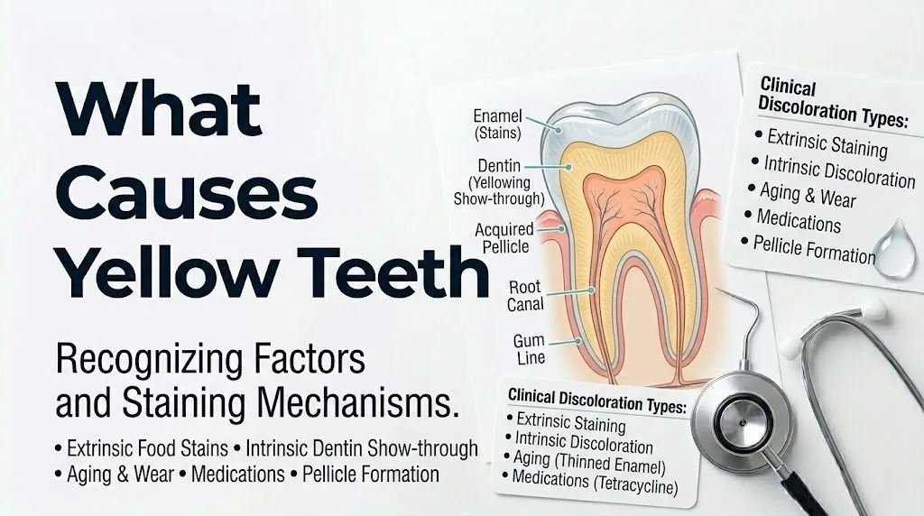
Extrinsic tooth staining occurs when colored compounds from food, beverages, tobacco, or medications are deposited on the acquired enamel pellicle (AEP), the thin protein film that forms on the tooth surface within minutes of cleaning. The staining sits on or just within this surface film and does not penetrate the intact enamel crystal structure beneath. This type of staining is potentially removable.
Intrinsic tooth discoloration involves color change within the enamel or dentin itself, from structural damage, chemical incorporation during development, or pigment diffusion through dentin. Intrinsic discoloration cannot be removed by cleaning or standard bleaching in most cases. It requires more intensive dental treatment to address aesthetically.
The two-pathway framework for tooth discoloration:
| Category | Location | Reversibility | Primary Causes | Clinical Appearance |
|---|---|---|---|---|
| Extrinsic staining | Acquired enamel pellicle surface | Often reversible with prophylaxis and bleaching | Coffee, tea, red wine, tobacco, poor hygiene | Yellow-brown, diffuse, uniform distribution |
| Intrinsic discoloration | Within enamel or dentin structure | Generally not reversible without dental restoration | Aging, tetracycline, fluorosis, trauma, genetics, systemic illness | Yellow, gray-brown, banding, white spots, single tooth darkening |
According to the American Dental Association (ADA), the clinical distinction between extrinsic and intrinsic discoloration is the first assessment a dentist makes when evaluating tooth color change, because this classification directly determines which interventions are appropriate.
Older adults often present with a combination of both: a lifetime of dietary chromogen exposure producing surface staining layered over the structural yellowing from enamel thinning and dentin darkening that develops with age. Attributing the color change to a single cause in this population is rarely accurate.
The Anatomy of Tooth Color: Enamel, Dentin, and Why Teeth Yellow
Teeth appear white or off-white because of the optical properties of the two main hard tissues: dental enamel on the outside and dentin underneath. Understanding how each contributes to tooth color explains why so many different factors can produce yellowing.
Dental enamel is the hardest tissue in the human body, composed of approximately 96% calcium hydroxyapatite mineral (Ca10(PO4)6(OH)2) organized into tightly packed enamel prisms (enamel rods) approximately 4 to 8 micrometers in diameter, formed by ameloblasts during tooth development. Enamel is semi-translucent: it allows light to pass through partially, reflecting some light back from the surface. Its natural color is off-white to slightly gray, and its thickness over different parts of the tooth determines how much light reaches the dentin beneath.
Dentin is the bulk of the tooth structure beneath the enamel, composed of approximately 70% hydroxyapatite and 30% organic matrix (primarily type I collagen), with a network of microscopic dentin tubules running from the pulp to the dentinoenamel junction. Dentin’s natural color is inherently yellow. The white appearance of healthy teeth is largely an optical effect: the relatively opaque enamel layer reflects white light from its surface while the yellow dentin provides warmth and depth to the overall tooth color.
Think of a tooth like a wine glass made of frosted white glass over a yellow inner lining. The glass looks white from a distance, but as the frosted coating wears thinner, the yellow lining becomes more visible through it. That is exactly what happens in aging-related tooth yellowing: enamel thins from decades of dietary acid exposure and mechanical wear, and the yellow dentin becomes progressively more visible through the increasingly translucent outer layer.
According to the Journal of Dental Research, the optical properties of enamel and dentin together determine perceived tooth color far more than the enamel surface staining alone, which is why intrinsic changes in dentin color from aging or medication produce a fundamentally different and more resistant type of yellowing than surface chromogen deposits.
Children’s primary teeth (baby teeth) often appear whiter than permanent teeth because primary enamel has slightly different optical properties and the dentin beneath is thinner. This is a normal anatomical difference, not a sign that primary teeth are healthier than permanent teeth.
Coffee, Tea, and Red Wine as Causes of Yellow Teeth
Coffee, tea, and red wine cause yellow teeth through a specific biochemical mechanism involving the acquired enamel pellicle and the polyphenolic compounds (chromogens) present in these beverages.
The acquired enamel pellicle is a thin protein film that reforms on the tooth surface within minutes of cleaning. It is composed of salivary proteins including mucins, proline-rich proteins, statherins, and histatins, deposited through electrostatic and hydrophobic interactions with the enamel surface. This pellicle is the substrate on which dietary chromogens bind.
Coffee contains chlorogenic acid, a polyphenolic chromogen that adsorbs strongly onto pellicle proteins. Tea contains tannic acid and other tannins, which do two things simultaneously: they bind directly to pellicle proteins as chromogens themselves, and they catalyze oxidative cross-linking reactions between other chromogen molecules and pellicle protein side chains, creating stable, colored complexes that are progressively harder to remove as they oxidize and polymerize over time. Red wine contains both tannins and anthocyanins (deep red-blue flavonoid pigments), making it one of the most efficient staining beverages because the tannins act as binding agents for the anthocyanin chromogens.
According to research published in the Journal of Dental Research, tannin concentration in the beverage is a stronger predictor of extrinsic staining potential than chromogen concentration alone, which explains why green tea (lower tannin) stains less than black tea (higher tannin) despite both containing similar chromogens.
Key chromogen-containing foods and beverages ranked by relative staining potential:
- Black tea: very high tannin content; highest staining potential among beverages tested in controlled studies
- Coffee: high chlorogenic acid content; high staining potential, particularly espresso and cold brew
- Red wine: combined tannins and anthocyanins; very high staining potential
- Berries (blueberries, blackberries, pomegranate): high anthocyanin content; moderate-to-high staining potential
- Tomato-based sauces: moderate chromogen content with acid that increases enamel surface roughness
- Green tea: lower tannin content than black tea; lower staining potential
- White wine: low chromogen content but high acid; contributes to enamel erosion that increases staining susceptibility
Individuals with xerostomia (dry mouth) from any cause are at substantially higher risk for beverage-related staining because saliva normally dilutes, buffers, and washes chromogens from the tooth surface. Reduced saliva flow means chromogens have longer contact time with the pellicle before clearance.
Key Takeaway: Coffee, tea, and red wine cause yellow teeth by binding tannin-chromogen complexes to the acquired enamel pellicle protein film, not by penetrating enamel itself, which means this staining is potentially reversible with professional dental prophylaxis, but reducing contact time by drinking through a straw or rinsing with water immediately after consumption reduces accumulation.
Tobacco and Smoking as Causes of Yellow Teeth Staining
Tobacco causes yellow and brown tooth staining through two distinct chemical mechanisms that together produce some of the most resistant and deeply penetrating extrinsic staining seen in clinical dentistry.
Nicotine, while colorless in its pure form, oxidizes on contact with air and oral fluids to form cotinine and other nitrogen-containing metabolites that are pale yellow to yellow-brown in color. These compounds bind strongly to the acquired enamel pellicle and the organic matrix within surface enamel defects and microcracks, producing the characteristic yellow-brown staining associated with tobacco use.
Tar is the far more significant contributor to tobacco staining. Tar is a complex mixture of combustion products generated during tobacco burning, including polycyclic aromatic hydrocarbons, phenols, and catechols. These compounds are intensely colored brown-to-black and adsorb onto both the pellicle surface and the exposed organic components within subsurface enamel defects. Tar compounds are chemically resistant to removal by standard oral hygiene products and require professional abrasive polishing for effective reduction.
Smokeless tobacco (chewing tobacco, snuff) produces a different but equally severe pattern. The nicotine and tar compounds in smokeless products are maintained in prolonged direct contact with the teeth and gingiva, producing intense staining concentrated particularly on the lower anterior teeth and in gingival sulcus areas where the tobacco is habitually placed.
According to the American Dental Association, tobacco use is one of the primary risk factors for periodontal disease, and the combination of tobacco staining, calculus buildup, and gingival recession in long-term tobacco users produces a complex staining picture that professional cleaning can partially address but rarely fully resolve without cessation of tobacco use.
Electronic cigarettes (vaping) produce less tar-related staining than combustible tobacco because vaping does not involve combustion. However, nicotine oxidation and certain propylene glycol-based flavor compounds still contribute to pellicle staining in regular vapers. The long-term dental effects of vaping remain an area of ongoing clinical research, with available evidence based primarily on observational data rather than long-term controlled trials.
Poor Oral Hygiene and Plaque as Causes of Yellow Teeth
Poor oral hygiene causes tooth yellowing through two related but distinct mechanisms: the accumulation of dental plaque and the mineralization of that plaque into calculus (tartar).
Dental plaque is a structured biofilm composed primarily of bacteria (principally Streptococcus mutans, Streptococcus sanguinis, and Lactobacillus species), embedded in an extracellular matrix of bacterial polysaccharides, salivary proteins, and lipids. Plaque itself has a pale yellow to cream color from its bacterial and protein content. When plaque accumulates on tooth surfaces without adequate mechanical removal through brushing and interdental cleaning, this pale yellow film becomes visible as a diffuse, soft, scrapable coating, particularly along the gingival margin.
Importantly, un-removed plaque also serves as an enhanced substrate for chromogen binding. The rough, porous surface of mature plaque biofilm binds dietary chromogens far more effectively than clean enamel, because chromogen molecules can penetrate into the biofilm matrix where they are protected from salivary dilution and mechanical removal.
Calculus (tartar) is mineralized plaque: when plaque is not removed, salivary calcium and phosphate ions gradually precipitate into the plaque matrix, converting the soft biofilm into a hard, rough, yellow-to-brown mineral deposit that cannot be removed by brushing. Calculus is porous and provides an ideal surface for further chromogen adsorption and additional plaque accumulation. Its rough surface and yellow-brown color contribute directly to the yellow appearance of teeth, particularly visible near the gumline where salivary mineral concentrations are highest and calculus formation is fastest.
According to the American Academy of Periodontology (AAP), supragingival calculus (calculus above the gumline, visible to the patient) forms fastest on the lingual surfaces of the lower front teeth and the buccal surfaces of the upper molars, corresponding precisely to the sites nearest the openings of the major salivary glands (submandibular and sublingual glands for lower front teeth, parotid gland for upper molars). This anatomical relationship explains why those specific tooth surfaces are consistently the first to show calculus-related yellowing.
Older adults who have reduced manual dexterity from arthritis or neurological conditions may have difficulty maintaining adequate plaque removal through brushing and interdental cleaning, making them more susceptible to plaque and calculus-related discoloration even with consistent effort.
Aging as a Cause of Yellow Teeth: Enamel Thinning and Dentin Exposure
Aging causes progressive tooth yellowing through two simultaneous structural changes in the tooth: the thinning of the enamel layer from the outside and the thickening of the dentin from the inside, both of which increase the visual contribution of the inherently yellow dentin to the overall tooth color.
Enamel thinning occurs through cumulative acid erosion and mechanical wear over decades of use. Every exposure to dietary acids (from carbonated beverages, fruit juices, citrus fruits, and wine) temporarily softens the surface hydroxyapatite of the enamel in a process called acid demineralization. While saliva partially remineralizes the softened surface between exposures, the rate of remineralization does not fully compensate for the rate of acid exposure in most adults, resulting in a net thinning of the enamel layer over years. Mechanical wear from tooth-to-tooth contact during chewing, particularly in people with bruxism (habitual grinding), compounds this thinning. As the enamel thins, it becomes more translucent, allowing the yellow dentin beneath to show through with increasing prominence.
Simultaneously, odontoblasts (the dentin-forming cells lining the pulp chamber) continue to deposit secondary dentin throughout life. This secondary dentin is a form of reparative response to ongoing mechanical and chemical stimulation from the oral environment. As secondary dentin accumulates on the pulp-facing surface of the existing dentin layer, the dentin mass increases and the pulp chamber narrows. Thicker dentin means more yellow material beneath whatever enamel remains, deepening the yellow appearance visible through the progressively thinning enamel.
According to the American Dental Association, this dual mechanism of enamel thinning and secondary dentin deposition is the primary reason teeth naturally yellow with age, independent of dietary staining or tobacco use.
Postmenopausal women experience accelerated enamel demineralization in some studies, associated with declining estrogen levels that may affect calcium homeostasis in oral tissues. While the available evidence for this is based primarily on observational data rather than controlled trials, this population warrants attention to dietary acid minimization and adequate calcium intake to support enamel remineralization.
Key Takeaway: Aging yellows teeth through a combination of enamel thinning from acid erosion (exposing yellow dentin) and secondary dentin growth from the inside (adding yellow bulk beneath), and this type of intrinsic discoloration does not respond fully to surface bleaching because the yellow color is within the tooth structure itself.
Tetracycline Antibiotics as a Cause of Intrinsic Tooth Discoloration
Tetracycline antibiotics cause one of the most clinically distinct and well-characterized forms of intrinsic tooth discoloration, and understanding the mechanism explains precisely why this discoloration is permanent and why it affects only specific populations.
Tetracycline belongs to a class of antibiotics that chelate divalent metal cations, particularly calcium ions (Ca2+). During the mineralization phase of tooth development, when ameloblasts and odontoblasts are depositing calcium hydroxyapatite crystals into the developing enamel and dentin matrix, circulating tetracycline molecules form stable chelates with the calcium ions at active mineralization sites. These tetracycline-calcium chelate complexes are incorporated directly into the growing hydroxyapatite crystal lattice as a structural component of the forming dentin and enamel.
The incorporated tetracycline-calcium chelate is initially fluorescent yellow, which is why tetracycline-stained teeth initially appear yellow. Upon prolonged light exposure (ultraviolet and visible light), the incorporated tetracycline undergoes oxidation to form stable quinone derivatives that are gray-brown in color. This is why tetracycline-stained teeth typically appear yellow in young patients and progressively darken to gray-brown in adults, a color shift driven entirely by photochemical oxidation of the incorporated drug.
The clinical consequence is that tetracycline staining produces characteristic horizontal yellow-gray-brown banding across affected teeth, corresponding to the specific periods of tooth development during which the drug was administered. Different teeth calcify at different times during development, so the banding pattern reflects the developmental chronology of the affected teeth.
According to the American Academy of Pediatric Dentistry (AAPD), tetracycline antibiotics are contraindicated in children under 8 years of age and in pregnant women after the 4th month of pregnancy for this specific reason: permanent teeth begin calcifying during fetal development and continue through approximately age 7 to 8, making this the critical window for tetracycline-induced intrinsic discoloration. Administration after this developmental window does not cause tooth discoloration.
Minocycline and doxycycline (semi-synthetic tetracycline derivatives) used in adults for acne treatment or periodontal disease can also cause tooth discoloration in adults, but through a different mechanism: these drugs bind to iron ions in the saliva and gingival crevicular fluid, forming iron-minocycline complexes that deposit in dentin as a gray-green or blue-gray color in erupted teeth. This is a distinct and less severe pattern than developmental tetracycline banding.
Dental Fluorosis as a Cause of Yellow and Brown Tooth Discoloration
Dental fluorosis causes tooth discoloration through disruption of the enamel maturation process during tooth development, producing enamel that is structurally altered in ways visible as white spots, yellow-brown patches, or deep pitting depending on severity.
The mechanism begins with excessive fluoride exposure during the enamel maturation phase, which is the post-secretory stage when ameloblasts remove proteins from the enamel matrix while increasing its mineral density to near-final hardness. At elevated fluoride concentrations, fluoride ions interfere with the activity of matrix metalloproteinase 20 (MMP20) and kallikrein-related peptidase 4 (KLK4), the enzymes responsible for degrading and removing amelogenin and other enamel matrix proteins during maturation. With impaired protein removal, the enamel retains more protein than normal and mineralizes incompletely.
The retained protein and impaired mineralization produce enamel that is more porous and less optically uniform than normal enamel. In mild fluorosis, this appears as opaque white spots or streaks (the classic “snow-capping” on cusp tips). In moderate fluorosis, the porous, protein-retaining enamel takes up extrinsic chromogens from the diet more readily than normal enamel, producing the characteristic yellow-to-brown mottled discoloration. In severe fluorosis, the structural disruption produces pitting, chipping, and brown-black surface staining from chromogen penetration into the porous enamel body.
According to the Centers for Disease Control and Prevention (CDC), approximately 23% of Americans aged 6 to 49 have some degree of dental fluorosis, with the majority falling in the mild category (white spots and faint streaking) that has minimal aesthetic impact. Moderate and severe fluorosis, producing the brown mottling most people associate with fluorosis, is significantly less prevalent.
Children who grew up in areas with naturally high fluoride in well water (above 2.0 mg/L), or who swallowed fluoride toothpaste regularly during the ages of approximately 6 months to 8 years when permanent teeth are developing, are at highest risk for clinically visible fluorosis affecting permanent teeth.
Tooth Trauma and Pulp Necrosis as Causes of Grey and Brown Discoloration
A tooth that turns dark gray, brown, or black following physical trauma is not yellowing from extrinsic staining but from a completely different process: the breakdown of blood and tissue inside the tooth pulp following disruption of the blood supply.
When a tooth receives a traumatic blow (from a sports injury, fall, or direct impact), the periodontal ligament and the apical blood vessels that enter the tooth through the root apex can be disrupted or severed. If blood flow to the pulp is reduced enough to cause pulp tissue death (pulp necrosis), the hemoglobin in the red blood cells that were trapped in the pulp chamber and dentin tubules at the time of injury begins to degrade.
Hemoglobin degradation products, including hemosiderin, methemoglobin, and ultimately iron sulfide (a dark brown-black compound formed when iron from hemoglobin reacts with hydrogen sulfide produced by anaerobic bacteria in the necrotic pulp), diffuse outward from the pulp space through the dentin tubules into the surrounding dentin. This diffusion of dark pigments throughout the dentin network produces the characteristic dark grayish discoloration that appears weeks to months after the traumatic event.
According to the American Dental Association, a tooth that changes color after trauma requires prompt dental evaluation regardless of the presence or absence of pain, because pulp necrosis can progress to periapical abscess formation even in the absence of symptoms. Pulp vitality testing (electric pulp test or cold test) and periapical radiographic examination are the standard initial assessments.
Key clinical features distinguishing trauma-related discoloration from other causes:
- Affects a single tooth or a small group of teeth corresponding to the injury site
- Appears weeks to months after the traumatic event
- Color change is gray, brown, or black rather than yellow
- May be painless despite indicating dead pulp tissue
- May have associated periapical pathology visible on dental X-ray
Children involved in contact sports or playground activities who experience dental trauma should have any subsequently darkening tooth evaluated by a pediatric dentist promptly, even if the tooth appeared unaffected immediately after the injury. Early intervention with root canal treatment in a non-vital tooth can sometimes prevent the darkest stage of discoloration from developing fully.
Key Takeaway: A single tooth that turns dark gray or brown weeks to months after an injury, even without pain, almost certainly has a dead pulp and needs same-day dental evaluation for pulp vitality testing and X-ray to check for abscess formation.
Medications That Cause Tooth Staining: Chlorhexidine, Iron, and Chemotherapy
Several medications cause tooth staining through mechanisms distinct from dietary chromogen deposition, and clinicians often underrecognize these causes because the connection between the medication and the dental change is not always obvious to patients.
Chlorhexidine gluconate, widely used as a prescription antibacterial mouthwash for periodontal disease management and plaque control, is one of the most potent causes of extrinsic tooth staining among medications. Chlorhexidine is a cationic antiseptic that binds strongly to the anionic proteins of the acquired enamel pellicle through electrostatic attraction. Once bound in the pellicle, chlorhexidine molecules then complex with dietary chromogens (particularly tannins from tea, coffee, and wine) through a secondary chemical reaction, producing the characteristic dark brown-black staining on the lingual (tongue-facing) surfaces of the lower anterior teeth that is the hallmark of chlorhexidine use. This staining is extrinsic (removable by professional prophylaxis) but accumulates rapidly with concurrent dietary chromogen consumption.
Ferrous sulfate and other iron supplements cause a distinctive dark staining on teeth when liquid preparations are allowed to contact tooth surfaces directly or when iron reacts with hydrogen sulfide produced by oral bacteria, forming iron sulfide as a dark brown-black deposit in the pellicle. Liquid iron supplements should be taken through a straw and followed immediately by rinsing to minimize tooth contact.
Chemotherapy agents, particularly those used in childhood cancer treatment, can cause intrinsic enamel defects and discoloration through direct toxicity to ameloblasts and odontoblasts during active tooth development. Cyclophosphamide and doxorubicin (anthracycline chemotherapy) administered during critical periods of tooth calcification can produce enamel hypoplasia and intrinsic yellow-brown discoloration similar in appearance to fluorosis or systemic illness-related enamel defects.
According to a clinical review published in the Journal of the American Dental Association, pediatric cancer survivors who received chemotherapy before age 8 have substantially higher rates of dental developmental abnormalities including enamel hypoplasia, microdontia (abnormally small teeth), and intrinsic discoloration compared to age-matched controls, making dental surveillance an important component of long-term pediatric oncology follow-up care.
What Causes Teeth to Turn Brown: Severe Staining and Decay
Teeth that have turned brown rather than yellow represent either the extreme end of extrinsic chromogen accumulation or the beginning of a structurally destructive process that goes beyond cosmetic staining.
The progression from yellow to brown in extrinsic staining follows the natural course of chromogen oxidation and polymerization in the acquired enamel pellicle. As tannin-chromogen complexes in the pellicle layer continue to oxidize and cross-link over months to years, the initially yellow deposits progressively darken to orange-brown, then brown, then deep brown-black. Tobacco tar deposits follow a similar oxidative darkening pathway. The resulting brown staining is chemically resistant because the oxidized, polymerized chromogen complexes have higher molecular weights, stronger binding affinity for pellicle proteins, and greater resistance to mechanical and chemical removal than the freshly deposited yellow precursors.
Dental caries (tooth decay) can produce brown discoloration at a distinctly different level: not at the enamel surface but within the enamel and dentin structure itself. When cariogenic bacteria, principally Streptococcus mutans, produce lactic acid from fermentable carbohydrates, the localized acid demineralizes the hydroxyapatite of enamel and dentin, creating a porous, protein-rich zone. Bacterial metabolites, iron compounds, and degraded dentin matrix proteins accumulate in this carious lesion, producing the characteristic brown-to-black discoloration visible in an active or arrested cavity.
According to the National Institute of Dental and Craniofacial Research (NIDCR), dental caries remains the most prevalent chronic disease in children and adults in the United States, affecting approximately 90% of adults and 42% of children aged 2 to 11. Brown or black discoloration of a tooth surface that is localized to a specific point, pit, or fissure rather than distributed across the entire tooth is more likely to represent carious demineralization than extrinsic staining.
People with a history of eating disorders, particularly bulimia nervosa, experience a specific pattern of erosive brown discoloration on the palatal (tongue-facing) surfaces of the upper front teeth from repeated acid exposure during purging episodes. This lingual erosion pattern is clinically distinct from dietary acid erosion or staining and should prompt a sensitive discussion with both a dentist and a physician about the broader health context.
Systemic Diseases and Conditions That Cause Yellow or Discolored Teeth
Several systemic medical conditions produce tooth discoloration as a clinically recognizable manifestation of their underlying pathophysiology, and recognizing these patterns can sometimes lead to earlier diagnosis of conditions that the patient was not aware of.
Celiac disease produces enamel hypoplasia (incomplete enamel formation) in approximately 20 to 40% of affected individuals, according to research published in the Journal of Pediatric Gastroenterology and Nutrition. The mechanism involves malabsorption of calcium, phosphate, and vitamin D during critical periods of tooth development, impairing the mineralization of developing enamel. The resulting enamel defects appear as opaque white, yellow, or brown patches and grooves in a symmetrical, chronological pattern across multiple teeth. These enamel changes may precede gastrointestinal symptoms in some patients, making the dental presentation a potential early clinical indicator for celiac disease evaluation in children with unexplained enamel defects.
Neonatal hyperbilirubinemia (jaundice in newborns) can cause intrinsic staining of primary teeth if bilirubin levels are severely elevated during the period when primary tooth enamel and dentin are forming. Bilirubin (yellow) and its reduced form biliverdin (green) can be deposited within the forming dentin of primary teeth, producing the characteristic yellow-green discoloration of primary teeth in children who had severe neonatal jaundice. The permanent teeth are generally not affected because their calcification occurs primarily after the neonatal period.
Erythropoietic porphyria (EP), a rare inherited disorder of heme biosynthesis, causes pink-red discoloration of teeth from porphyrin deposition in dentin and enamel, with characteristic pink-red fluorescence of the teeth under ultraviolet light (a finding called erythrodontia). This is one of the most distinctive intrinsic tooth discolorations and is essentially pathognomonic for the condition in the clinical setting.
People undergoing radiation therapy to the head and neck for cancer treatment develop radiation-related xerostomia (dry mouth) from damage to salivary gland tissue, which then increases susceptibility to both dental caries and extrinsic chromogen staining. The combination of reduced saliva and radiation-weakened enamel produces a rapidly progressive pattern of discoloration and decay that requires intensive preventive dental management alongside cancer treatment.
Genetic Causes of Yellow Teeth: Amelogenesis Imperfecta and Enamel Hypoplasia
Inherited genetic conditions affecting enamel formation produce tooth discoloration that is present from tooth eruption, affecting all or most teeth, and is fundamentally unrelated to dietary or lifestyle factors.
Amelogenesis imperfecta (AI) is a group of genetic conditions caused by mutations in genes encoding enamel matrix proteins or enzymes required for enamel development, including AMELX (encoding amelogenin), ENAM (encoding enamelin), and MMP20 (encoding matrix metalloproteinase 20). These mutations disrupt the normal secretion and maturation of enamel, producing enamel that is either insufficient in quantity (hypoplastic type), insufficiently mineralized (hypomineralized type), or improperly matured (hypomaturation type).
The hypomineralized and hypomaturation types of AI produce yellow-to-brown teeth with enamel that chips and fractures easily because of its reduced hardness and increased porosity. The enamel may appear yellow-brown or creamy-brown rather than the white of normal enamel, and the color is intrinsic and diffuse across all affected teeth rather than the localized patterning seen in fluorosis or tetracycline staining.
Enamel hypoplasia is a related but broader category of developmental enamel defects caused not by genetic mutations but by environmental insults during the period of enamel formation. These insults include high fever during infancy or early childhood, premature birth, nutritional deficiencies, and certain infections. The deficient enamel in hypoplasia appears as horizontal grooves, pits, or discolored bands at the specific tooth zones that were developing at the time of the insult, producing a chronological record of the illness in the tooth structure.
According to the American Academy of Pediatric Dentistry, enamel developmental defects including hypoplasia and hypomineralization are found in approximately 10 to 25% of children, with rates substantially higher in premature infants. Any child with yellow, brown, or spotted enamel affecting multiple teeth from eruption should be evaluated by a pediatric dentist to distinguish developmental enamel defects from staining and to implement protective dental management before the structurally weakened enamel suffers further damage.
Key Takeaway: Yellow or brown teeth that are present from the time of tooth eruption, affect multiple teeth symmetrically, and cannot be explained by dietary or tobacco exposure should be evaluated by a pediatric dentist or dentist for possible amelogenesis imperfecta, enamel hypoplasia, or other developmental conditions, because these require protective management rather than just aesthetic treatment.
What Causes Yellow Teeth in Children and During Pregnancy
Yellow teeth in children and tooth color changes during pregnancy have specific causes and mechanisms that differ from the general adult population, and understanding these differences prevents both misdiagnosis and unnecessary parental anxiety.
In children, the most frequently confused distinction is between the normal appearance of newly erupted permanent teeth alongside retained primary teeth and pathological tooth discoloration. Permanent teeth are naturally slightly more yellow than primary teeth because their enamel is thicker and their dentin is more densely structured. When a child has mixed dentition (some baby teeth and some permanent teeth at the same time, typically ages 6 to 12), the contrast between the whiter primary teeth and the naturally yellower permanent teeth can look alarming to parents who are unfamiliar with this normal developmental difference.
Genuinely pathological tooth color in children includes the enamel hypoplasia patterns from prematurity or childhood illness described above, fluorosis from excessive fluoride exposure, and tetracycline staining from antibiotic administration under age 8. According to the American Academy of Pediatric Dentistry, any child with visible enamel defects should have a dental examination by a pediatric dentist, both to assess the structural integrity of the affected teeth and to determine whether a systemic condition may be the underlying cause.
Pregnancy-related changes in tooth color are primarily driven by indirect mechanisms rather than direct hormonal effects on tooth structure. Morning sickness with frequent vomiting exposes tooth enamel to gastric acid (pH approximately 2.0), which is strongly erosive. Repeated acid exposure over weeks to months of first-trimester nausea can thin the enamel on the palatal surfaces of the upper front teeth, increasing dentin visibility and producing yellow discoloration specifically in this location.
According to the American College of Obstetricians and Gynecologists (ACOG), dental care during pregnancy is safe and recommended, and pregnant women experiencing morning sickness should rinse with water or a fluoride mouthwash after vomiting rather than brushing immediately, because brushing acid-softened enamel within 30 minutes of acid exposure can accelerate erosion.
Dry Mouth (Xerostomia) as a Cause of Yellow Teeth
Xerostomia (dry mouth, from the Greek meaning “dry mouth”) causes tooth yellowing and accelerated discoloration through the loss of saliva’s multiple protective functions that normally limit chromogen accumulation and enamel demineralization.
Saliva performs four functions that protect tooth color. First, it mechanically washes dietary chromogens and staining compounds from the tooth surface within minutes of consumption, reducing their pellicle contact time. Second, saliva buffers dietary acids through its bicarbonate and phosphate content, limiting acid demineralization of the enamel surface that would otherwise increase enamel surface roughness and chromogen penetration. Third, salivary calcium and phosphate ions drive the remineralization of partially demineralized enamel surface zones, maintaining the smoothness and relative impermeability of the enamel surface. Fourth, salivary proteins including mucins and proline-rich proteins compete with chromogens for binding sites on the pellicle, occupying the same binding sites that dietary tannin-chromogen complexes would otherwise occupy.
When salivary flow is reduced by any mechanism, all four of these protective functions are diminished simultaneously, allowing chromogens more contact time, less acid buffering, slower remineralization, and less competitive pellicle protein binding. The result is accelerated and more severe extrinsic staining alongside increased susceptibility to caries.
According to the Mayo Clinic, xerostomia affects an estimated 10% of the general population and up to 30% of older adults, with the most frequent causes being anticholinergic medications (including first-generation antihistamines, tricyclic antidepressants, and bladder medications), radiation therapy to the head and neck, and Sjogren’s syndrome (an autoimmune condition targeting the salivary and lacrimal glands).
Common medication classes that cause xerostomia and secondary tooth discoloration:
- First-generation antihistamines (diphenhydramine, hydroxyzine): muscarinic receptor blockade at salivary glands
- Tricyclic antidepressants (amitriptyline, nortriptyline): anticholinergic mechanism
- Bladder anticholinergics (oxybutynin, solifenacin): muscarinic blockade
- Diuretics (furosemide, hydrochlorothiazide): systemic dehydration reducing salivary output
- Decongestants (pseudoephedrine): alpha-adrenergic reduction of salivary secretion
Individuals taking multiple xerogenic medications simultaneously, which is common in older adults, face compounded salivary suppression and significantly amplified tooth discoloration risk compared to single-medication users.
How Tooth Discoloration Is Assessed and When to See a Dentist
The professional assessment of tooth discoloration follows a structured clinical process that distinguishes extrinsic from intrinsic causes, identifies reversible from permanent discoloration, and determines whether any associated systemic condition requires medical attention alongside dental management.
A dentist evaluating tooth discoloration will typically follow this approach:
- Clinical history: dietary habits (coffee, tea, wine consumption frequency), tobacco use, medication history (particularly tetracycline in childhood, chlorhexidine use, iron supplements, chemotherapy), developmental history (premature birth, childhood illnesses, fluoride exposure history), trauma history for any single dark tooth, and family history (for possible amelogenesis imperfecta or other genetic conditions)
- Visual examination: distribution of discoloration (single tooth versus multiple teeth; localized versus diffuse; surface versus structural); color character (yellow versus gray-brown versus banded versus white spots); association with gingival margin (supragingival calculus), pit and fissure anatomy (possible caries), or specific developmental regions (chronological enamel defects)
- Shade measurement: clinical shade assessment using the VITA shade guide (a standardized system using tabs from A1 to D4 across four hue ranges) or objective digital spectrophotometry for more precise color documentation
- Radiographic examination: periapical X-rays to assess for carious lesions, periapical pathology in traumatized teeth, and root or pulp chamber changes suggesting pulp necrosis
- Pulp vitality testing for single discolored teeth: electric pulp test or cold test to assess whether the pulp of a darkened tooth is still vital or has undergone necrosis
When to make an appointment with a board-certified dentist for tooth discoloration:
- Any single tooth that has darkened following an injury, even without pain
- Yellow-brown banding or patterned discoloration on multiple teeth that appeared from eruption
- Tooth discoloration in a child that is not explained by dietary factors and has been present since tooth eruption
- White or yellow-brown spots with associated chipping or surface fragility
- Any discoloration accompanied by tooth sensitivity, pain on biting, or visible holes or dark pits in the tooth surface
- Discoloration that has changed noticeably in the past several months
According to the American Dental Association, patients should inform their dentist about all current medications at every dental examination, specifically because many medication-related tooth color changes are only recognizable in the context of a complete medication history and cannot be identified from visual examination alone.
Frequently Asked Questions About What Causes Yellow Teeth
What is the most common cause of yellow teeth?
The most common cause of yellow teeth in the general adult population is extrinsic staining from dietary chromogens in coffee, tea, and red wine binding to the acquired enamel pellicle, combined with the progressive natural yellowing from enamel thinning and dentin darkening that occurs with aging.
These two causes frequently coexist in adults over 30, making most cases of adult tooth yellowing a combination of surface staining and structural change rather than a single identifiable cause.
A dentist can distinguish the relative contribution of each through clinical examination, with surface staining appearing as removable deposits concentrated near the gumline and extrinsic surfaces, while structural yellowing appears as a uniform change visible through the translucent enamel.
Does aging always cause teeth to turn yellow?
Aging causes a progressive yellowing in nearly all adults through the dual mechanism of enamel thinning and secondary dentin deposition, which together increase the visual prominence of the inherently yellow dentin beneath progressively thinner enamel.
The rate and severity of this yellowing varies based on individual dietary acid exposure, tooth grinding habits, and the initial enamel thickness at tooth eruption, meaning some people yellow faster than others despite similar ages.
This type of age-related yellowing is intrinsic and does not respond fully to surface bleaching because the yellow color originates within the dentin structure rather than on the enamel surface.
Can yellow teeth be a sign of a health problem?
Yellow teeth can occasionally be the first visible indicator of a systemic condition, particularly when the discoloration is present from tooth eruption, affects multiple teeth in a symmetrical or chronological pattern, or occurs in a child with no obvious dietary or lifestyle explanation.
Enamel hypoplasia from celiac disease, bilirubin staining from neonatal hyperbilirubinemia, and enamel defects from chemotherapy during tooth development all produce yellow or brown intrinsic discoloration that warrants medical evaluation alongside dental assessment.
Any child with unexplained enamel defects affecting multiple teeth should be evaluated by a pediatric dentist and their primary care physician, who can assess for underlying systemic causes including celiac disease or nutritional deficiencies.
Why do some people have yellow teeth even with good oral hygiene?
Some people have inherently yellower teeth from the start because of natural variation in enamel thickness and translucency, dentin color, or mild fluorosis that produced slightly porous enamel that traps chromogens more readily than normal.
Genetic factors including variations in enamel matrix protein genes can produce enamel that is slightly thinner or more translucent than average, making the underlying dentin more visible regardless of how well the teeth are cleaned.
Additionally, enamel surface roughness from dietary acid erosion (even in healthy eaters who consume citrus fruits and sparkling water daily) increases chromogen retention even with excellent brushing habits.
At what age does tetracycline cause permanent tooth discoloration?
Tetracycline causes permanent intrinsic tooth discoloration only when administered during the calcification phase of tooth development, which for permanent teeth extends from approximately the 4th month of fetal development through approximately age 7 to 8 years.
The American Academy of Pediatric Dentistry states this is why tetracycline antibiotics are contraindicated in children under 8 years of age and in pregnant women after the 4th month of pregnancy.
Tetracycline administered to adults after tooth development is complete does not produce the banded intrinsic discoloration, though the related antibiotic minocycline can cause a different type of gray-green discoloration in erupted adult teeth through a separate iron-chelation mechanism.
Is yellow tooth discoloration always reversible with whitening?
Extrinsic staining from dietary chromogens, tobacco, and plaque is potentially reversible with professional dental prophylaxis and bleaching (hydrogen peroxide or carbamide peroxide-based treatments), though results vary based on the depth and duration of staining.
Intrinsic discoloration from aging, tetracycline, fluorosis, trauma, or genetic enamel defects is generally not reversible with bleaching because the color change is within the tooth structure rather than on the surface; these cases typically require dental veneers, bonding, or crowns for aesthetic management.
A dentist can determine whether a patient’s discoloration pattern is likely to respond to bleaching after a clinical examination, and attempting bleaching on intrinsic discoloration without professional guidance often produces disappointingly limited results.
Making Sense of Your Tooth Color
Yellow teeth have a specific cause in every person, and that cause is either on the tooth surface or within the tooth structure. The distinction matters because surface staining responds to professional cleaning and bleaching, while structural yellowing from aging, medications, or development requires different dental approaches entirely.
The first practical step for anyone concerned about tooth color is a dental examination. Bring your complete medication list (including over-the-counter supplements and any antibiotics taken in childhood if that history is available), be prepared to describe your dietary habits and any relevant health history, and ask your dentist specifically whether your discoloration is extrinsic, intrinsic, or both. That answer determines what interventions are realistic.
If your dentist identifies an unusual pattern suggesting a possible systemic cause (symmetrical enamel defects, chronological banding not explained by tetracycline history, or discoloration in multiple teeth from eruption), they will guide you toward the appropriate medical evaluation alongside dental management. Yellow teeth are usually benign and cosmetic, but they occasionally tell you something worth knowing about your health.