L5-S1 Nerve Root Compression Symptoms 2026: Every Warning Sign Explained
L5-S1 nerve root compression symptoms include lower back pain, leg pain radiating from the buttock to the foot, numbness and tingling in the leg or foot, muscle weakness in specific leg muscle groups, and in severe cases, loss of bladder or bowel control. The specific symptoms depend critically on whether the L5 nerve root, the S1 nerve root, or both are compressed at the lumbosacral junction, as each root controls a distinct territory of skin sensation and muscle function.
According to the North American Spine Society (NASS), the L5-S1 disc level is one of the two most frequently affected sites in lumbar disc herniation, with L4-L5 being the other. The lumbosacral junction bears the highest mechanical load of any spinal segment in the upright human body, which is a primary reason disc degeneration and nerve compression concentrate here so reliably across all adult age groups.
This article covers every established symptom of L5-S1 nerve root compression with the anatomical and neurochemical mechanism behind each one, how L5 and S1 root symptoms differ from each other, how population-specific factors change the clinical picture, and exactly which symptoms require you to stop reading and go to an emergency room.
L5-S1 Nerve Root Compression Symptoms: The Full Clinical Picture
L5-S1 nerve root compression symptoms encompass lower back and buttock pain, leg pain in a dermatomal distribution, paresthesia (tingling and numbness), motor weakness in specific myotomal muscle groups, reflex changes, and in severe compression, autonomic dysfunction affecting bladder and bowel control.
The symptom cluster varies by which of the two nerve roots is primarily compressed. L5 and S1 roots exit the lumbosacral spinal canal through adjacent but distinct neural foramina. Each root then divides into anterior and posterior branches, with the anterior ramus contributing to the sciatic nerve and eventually innervating specific leg muscle groups and skin territories.
The complete symptom inventory at L5-S1 includes:
- Lower back pain localized to the lumbosacral junction, often described as deep, aching, and poorly localized
- Buttock pain, arising from referred pain from the posterior ramus and from sciatic nerve root irritation
- Radicular leg pain traveling from the buttock down the posterior or lateral thigh, into the leg and foot in a dermatomal pattern specific to L5 or S1
- Paresthesia (tingling, burning, pins-and-needles) in the leg or foot, following the same dermatomal territory as the pain
- Hypoesthesia or anesthesia (reduced or absent skin sensation) in the dermatomal territory of the compressed root
- Motor weakness in the myotome supplied by the compressed root
- Deep tendon reflex changes, most reliably the Achilles reflex in S1 compression
- Pain worsening with specific positions including prolonged sitting or forward bending
- Gait disturbance, ranging from a subtle Trendelenburg gait in L5 weakness to a frank foot drop
Symptoms are typically unilateral in the early and moderate stages. Bilateral symptoms at L5-S1 warrant urgent evaluation for cauda equina syndrome, which is discussed in a dedicated section below.
What Is L5-S1 Nerve Root Compression
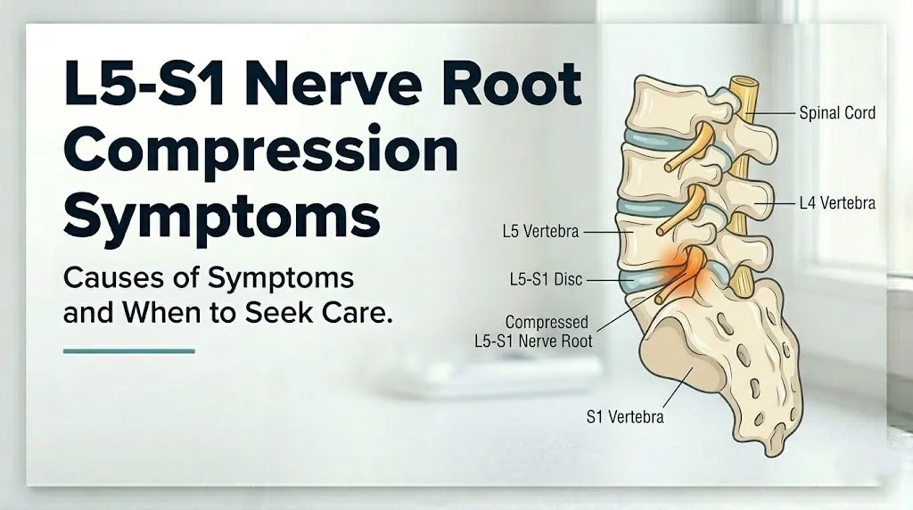
L5-S1 nerve root compression is a condition in which the L5 nerve root, the S1 nerve root, or both are mechanically compressed or chemically irritated at the level of the fifth lumbar and first sacral vertebrae, most commonly by a herniated intervertebral disc, bony foraminal narrowing, or thickened ligamentum flavum.
The L5-S1 intervertebral disc is a fibrocartilaginous structure composed of an outer annulus fibrosus (concentric lamellae of type I collagen) and an inner nucleus pulposus (a gelatinous matrix of type II collagen, proteoglycans, and water). The nucleus pulposus maintains disc height and distributes compressive load across the disc. When the annulus fibrosus develops radial tears, the nucleus pulposus can protrude into the spinal canal or neural foramen, compressing the adjacent nerve root.
What competitors don’t explain is that the compression is only part of the pain mechanism. The nucleus pulposus of a herniated disc contains and releases phospholipase A2, an enzyme that initiates the arachidonic acid cascade. This cascade produces prostaglandin E2 and other inflammatory mediators that directly sensitize the dorsal root ganglion of the compressed nerve root. Tumor necrosis factor-alpha (TNF-alpha) from the disc and infiltrating macrophages amplifies this sensitization. The result is that even a relatively small disc herniation that barely contacts a nerve root can produce severe, burning radicular pain, because the chemical inflammation does much of the work that physical pressure alone cannot explain.
Think of it like a garden hose getting both kinked and sprayed with acid at the same time. The kink reduces the signal flow. The acid inflames the hose itself. Both together produce a pain response far more severe than either alone would.
According to the National Institute of Neurological Disorders and Stroke (NINDS), lumbar disc herniation most commonly occurs in adults between 30 and 50 years old, though degenerative foraminal narrowing causing nerve root compression is more common in adults over 50.
L5 Nerve Root Compression Symptoms
L5 nerve root compression produces pain, numbness, and tingling along the lateral leg, the dorsum (top) of the foot, and the first web space between the great toe and second toe, with weakness primarily in toe dorsiflexion and foot eversion.
The L5 dermatomal territory runs from the lateral hip, down the lateral thigh and knee, along the outer shin, across the dorsum of the foot, and into the first web space. A patient with L5 compression who places their hand on the outer shin and traces a line to the top of the foot toward the big toe is tracing the exact territory where numbness and tingling will concentrate.
The myotomal territory of L5 includes the tibialis anterior (which lifts the forefoot, tested by heel walking), the extensor hallucis longus (which extends the great toe upward, a highly specific L5 indicator), and the peroneus longus and brevis (foot eversion). Weakness in these muscles produces the clinical picture of foot drop in severe L5 compression: the patient cannot adequately lift the forefoot during the swing phase of gait, producing a characteristic steppage gait where they lift the knee higher than normal to clear the foot.
A critical anatomical fact that competitor pages consistently omit: the L5 nerve root does not have a reliable associated deep tendon reflex. The patellar reflex tests L4, and the Achilles reflex tests S1. Clinical examination of L5 compression relies on motor testing and sensory testing, not reflex testing.
| L5 Root: Key Clinical Features | Finding | Clinical Significance |
|---|---|---|
| Dermatomal pain territory | Lateral leg, dorsum of foot, first web space | Distinguishes L5 from S1 (posterior calf, heel) |
| Weakness: toe dorsiflexion | Cannot raise great toe | Highly specific for L5 myotomal involvement |
| Weakness: heel walking | Shuffling or impossible heel walk | Tibialis anterior and toe extensor weakness |
| Reflex change | No reliable reflex; L5 has no specific DTR | Cannot use reflex alone to confirm L5 |
| Sensory deficit | Numbness dorsum of foot, first web space | Maps to L5 dermatome |
Older adults with concurrent lumbar spinal stenosis may experience L5 symptoms that worsen with walking (neurogenic claudication) rather than with sitting, a positional pattern that differs from the typical disc herniation presentation and complicates clinical differentiation.
S1 Nerve Root Compression Symptoms
S1 nerve root compression produces pain, numbness, and tingling along the posterior calf, the lateral border of the foot, the heel, and the plantar surface of the foot, with weakness primarily in plantarflexion (pushing down through the foot) and an absent or diminished Achilles tendon reflex.
The S1 dermatomal territory runs from the posterior buttock and posterior thigh, down the posterior calf, around the lateral ankle, along the outer edge of the foot, and into the heel and sole. Patients with S1 compression often describe a deep ache in the posterior calf that many initially mistake for a calf muscle strain or a deep vein thrombosis. The distinguishing feature is the path: the pain traces from the buttock downward rather than starting in the calf itself.
The S1 myotome includes the gastrocnemius and soleus (the primary plantarflexors, tested by toe walking and single-leg heel raises), the gluteus maximus (hip extension), and the small intrinsic muscles of the foot via the tibial nerve’s continuation. Weakness in S1-innervated muscles produces difficulty pushing off during the propulsive phase of gait, difficulty climbing stairs, and in severe cases, an inability to perform a single-leg toe raise on the affected side.
The Achilles tendon reflex (ankle jerk) is the most clinically reliable reflex test for S1 integrity. An absent or diminished Achilles reflex on the affected side, compared with the contralateral side, is a consistent finding in S1 nerve root compression. According to the American Academy of Orthopaedic Surgeons (AAOS), loss of the Achilles reflex is one of the most reproducible physical examination findings in confirmed S1 radiculopathy.
Patients taking medications that affect neuromuscular junction function, including certain antibiotic classes and neuromuscular blocking agents, may have altered reflex responses that complicate this examination finding. Any patient on such medications should disclose them to their examining physician before reflex testing is interpreted.
Key Takeaway: L5 compression weakens toe lifting and causes numbness on the top of the foot; S1 compression weakens toe pushing and causes numbness on the outer foot and heel, and only S1 reliably loses the Achilles reflex.
L5-S1 Radiculopathy and Sciatica Symptoms
Radiculopathy at L5-S1 refers to a constellation of symptoms produced by dysfunction of the L5 or S1 nerve root, including pain, sensory changes, motor weakness, and reflex changes, while sciatica describes specifically the radiating leg pain component that travels along the course of the sciatic nerve.
Sciatica from L5-S1 is one of the most recognizable presentations in clinical spine medicine. The pain typically starts in the lower back or buttock and radiates down one leg, following either the L5 dermatomal path (lateral leg and foot) or the S1 dermatomal path (posterior leg and heel). The quality of the pain is specific: it is not a dull muscle ache. Radicular pain from nerve root compression tends to be sharp, shooting, burning, or electric, because it is generated by ectopic discharge from sensitized A-delta and C-fiber axons in the compressed nerve root rather than from standard nociceptor activation in tissue.
According to research published in the European Spine Journal, approximately 90 percent of cases of acute sciatica from lumbar disc herniation resolve with conservative management within 6 to 12 weeks, though the timeline varies considerably by the degree of disc herniation and the amount of nerve root involvement. This evidence supports a conservative-first approach in the absence of red flags, while making it equally clear that persistent or progressive symptoms warrant specialist evaluation.
The straight leg raise test (also called the Lasegue test) is the primary clinical examination tool for confirming radiculopathy at L5-S1. The examiner lifts the patient’s straight leg while the patient lies flat. Reproduction of the patient’s radicular leg pain (not just back pain or hamstring tightness) between 30 and 70 degrees of elevation is a positive test, indicating nerve root tension consistent with L5-S1 disc herniation. Research published in the Journal of Bone and Joint Surgery found that a positive straight leg raise has a sensitivity of approximately 80 percent for lumbar disc herniation, making it one of the most useful bedside diagnostic maneuvers in spinal medicine.
Athletes and physically active adults may have particularly tight hamstrings that produce hamstring pain during the straight leg raise, which can be confused with a positive radiculopathy test. A clinician differentiates these by noting the location and quality of the reproduced pain: hamstring tightness produces posterior thigh discomfort at the limits of the raise, while radiculopathy produces shooting or burning pain below the knee, often into the foot.
L5-S1 Numbness and Tingling Symptoms
Numbness and tingling from L5-S1 nerve root compression are produced by impaired sensory axon conduction in the compressed nerve root, specifically in the large-diameter A-beta fibers (which carry touch and vibration) and the smaller A-delta fibers (which carry pain and temperature), and they follow precise dermatomal boundaries that map directly to which root is compressed.
The paresthesia (tingling, pins-and-needles, buzzing) is often the first symptom patients notice, appearing before frank numbness or pain in many cases. This is because A-beta sensory fibers are relatively large, myelinated, and susceptible to early compression-related conduction changes. As compression deepens or persists, the sensation progresses from intermittent paresthesia to persistent hypoesthesia (reduced sensation) and eventually to frank anesthesia (absent sensation) in the most severe cases.
The dermatomal distribution of numbness and tingling provides the most precise anatomical clue available without imaging:
- L5 numbness: Lateral shin, dorsum of the foot, first toe and first web space. Patients often describe it as the top of the foot feeling “like a sock is on it” when no sock is present.
- S1 numbness: Posterior calf, outer ankle, lateral border of the foot, heel, and sole. Patients often report the heel feels “dead” or “wooden” when walking barefoot.
- L5 and S1 both compressed (central large disc herniation): Numbness spanning the dorsum and lateral foot simultaneously, sometimes extending toward the sole.
Intermittent numbness that comes and goes, particularly with position changes, indicates a dynamic compression pattern where the nerve root is periodically pinched more severely in certain positions. Constant numbness that does not vary with position suggests a more established compression with ongoing axonal conduction failure.
Patients with pre-existing type 2 diabetes mellitus may experience a superimposed diabetic peripheral neuropathy, which causes stocking-and-glove pattern numbness and tingling in the feet bilaterally. This overlap can obscure the dermatomal pattern of L5-S1 compression and make clinical localization more difficult. A board-certified neurologist can separate these contributions using nerve conduction velocity (NCV) testing and electromyography (EMG).
L5-S1 Muscle Weakness Symptoms
Muscle weakness from L5-S1 nerve root compression reflects specific motor axon injury in the compressed root’s myotomal territory, with L5 compression producing weakness in the muscles that lift and extend the foot and toes, while S1 compression produces weakness in the muscles that push down through the foot and extend the hip.
Motor axon injury from nerve root compression occurs through two mechanisms. Acute severe compression can produce axonal disruption (axonotmesis), resulting in rapid-onset weakness within days. More commonly, chronic or moderate compression produces progressive axonal demyelination, resulting in gradual weakness over weeks to months. According to clinical observation reported in the Spine journal, patients with chronic L5-S1 compression often underestimate the degree of their motor weakness because they compensate unconsciously with hip flexion strategies during walking.
To assess L5-S1 motor function at home before a physician appointment:
- Heel walk test for L5: Stand and walk on your heels with your toes raised off the ground for 5 to 10 steps. Inability to lift the forefoot, or a foot that slaps down despite effort, suggests tibialis anterior and extensor hallucis longus weakness consistent with L5 myotome involvement.
- Toe walk test for S1: Stand and walk on the balls of your feet with your heels raised for 5 to 10 steps. Inability to rise onto the toes on one side, or rapid fatigue and drop of the heel on one side, suggests gastrocnemius-soleus weakness consistent with S1 myotome involvement.
- Single-leg heel raise for S1: Stand on one foot and raise up onto the toes 10 times consecutively. Compare left and right sides. Fewer than 10 repetitions on one side compared to the other, or pain and weakness preventing the full test, is a meaningful asymmetry to report to your physician.
- Great toe extension for L5: Sit and attempt to extend the great toe upward against light resistance from your own finger. Compare both sides. Asymmetric weakness of great toe extension is one of the most specific physical signs of L5 nerve root involvement.
These tests do not constitute a diagnosis. They provide meaningful clinical information to share with your primary care physician or with a board-certified orthopedic spine surgeon or physiatrist at your initial evaluation.
Weakness that progresses rapidly over hours or days, particularly if accompanied by increasing pain or new sensory symptoms, requires urgent evaluation rather than home monitoring.
Key Takeaway: Inability to walk on your heels points toward L5 nerve root weakness; inability to walk on your toes points toward S1 nerve root weakness; either finding deserves evaluation by a physician within 24 to 72 hours.
L5-S1 Symptoms Worse Sitting or Standing
L5-S1 nerve root compression symptoms that worsen with sitting are typically caused by disc herniation, because sitting increases intradiscal pressure and posteriorly shifts the nucleus pulposus toward the neural foramen. Symptoms that worsen primarily with standing and walking suggest foraminal stenosis or lumbar spinal stenosis rather than an isolated disc herniation.
The mechanism behind positional variation is grounded in spinal biomechanics. Intradiscal pressure at L5-S1 is lowest when lying flat (approximately 25 kg of load), rises substantially in the upright standing position (approximately 100 kg), and rises further in the seated forward-flexed position (approximately 175 kg), based on landmark biomechanical measurements published by Nachemson in clinical research that continues to inform spinal medicine today. Forward bending while seated, such as leaning toward a desk, further increases posterior disc pressure and can provoke or intensify radicular symptoms within minutes.
This is why many patients with disc-mediated L5-S1 nerve root compression find that lying on their side with the knees slightly flexed toward the chest (the fetal position) provides the most reliable temporary relief. This position reduces intradiscal pressure and opens the posterior neural foramen slightly, reducing mechanical pressure on the nerve root.
By contrast, patients whose L5-S1 symptoms worsen with standing or walking and improve with sitting or leaning forward are more likely experiencing neurogenic claudication from lumbar spinal stenosis, in which the spinal canal narrows in the upright position and becomes relatively more spacious in flexion. This positional pattern is a clinically important differentiator because these two conditions require different diagnostic approaches and have different treatment pathways.
Patients over 60 years old are more likely to experience both conditions simultaneously, a disc herniation superimposed on pre-existing spinal stenosis, which produces a mixed positional symptom pattern that can be particularly difficult to sort out without lumbar magnetic resonance imaging (MRI).
L5-S1 Nerve Root Compression Symptoms in Females
L5-S1 nerve root compression symptoms in females follow the same dermatomal, myotomal, and reflex patterns as in males, but several female-specific biological and anatomical factors influence when compression occurs, how severely it presents, and how quickly it progresses.
During pregnancy, the hormone relaxin, produced by the corpus luteum and later the placenta, causes systemic ligament laxity throughout the musculoskeletal system. At the lumbosacral junction, relaxin-mediated laxity in the posterior longitudinal ligament and facet joint capsules reduces spinal stability, allowing greater segmental motion that increases annular stress on the L5-S1 disc. The simultaneous shift in center of gravity from fetal growth increases lumbar lordosis and posterior loading on the L5-S1 disc. These combined effects mean that L5-S1 disc herniation and nerve root compression are not uncommon in the second and third trimesters of pregnancy.
Pregnant women who develop new radicular leg pain, foot numbness, or leg weakness should contact their obstetrician-gynecologist immediately. MRI of the lumbar spine is considered safe during pregnancy (without gadolinium contrast), and it is the preferred imaging study in pregnant women with suspected nerve root compression, as it avoids ionizing radiation. CT scanning and plain X-ray should be avoided unless no alternative exists.
Postmenopausal women experience accelerated lumbar disc degeneration related to estrogen-withdrawal effects on disc proteoglycan and collagen metabolism. Research published in the European Spine Journal has found an association between estrogen decline and reduced disc hydration and height, which accelerates foraminal narrowing and increases the risk of L5-S1 nerve root compression from degenerative rather than traumatic causes. The symptom presentation in postmenopausal women is therefore more likely to involve gradual-onset pain with positional variation than the acute severe onset more typical of a disc herniation in younger adults.
Women who work in prolonged standing occupations (healthcare, retail, food service) and those with a history of prior lumbar disc problems face compounded mechanical risk at L5-S1 that warrants earlier referral to a board-certified orthopedic spine surgeon or physiatrist if symptoms persist beyond four weeks.
L5-S1 Disc Herniation Symptoms
L5-S1 disc herniation symptoms are produced when the nucleus pulposus protrudes through a tear in the annulus fibrosus and contacts or compresses the L5 or S1 nerve root, generating a combination of mechanical compression and intense local chemical inflammation that produces the characteristic radicular symptom pattern.
The direction of the herniation matters clinically. A posterocentral (central) herniation protrudes into the middle of the spinal canal and can affect both the L5 and S1 roots bilaterally, or can compress the cauda equina directly. A posterolateral herniation (the most common type, occurring at approximately the 4 to 5 o’clock position on an axial disc view) typically compresses the exiting nerve root on one side only. A foraminal herniation compresses the nerve root within the neural foramen itself, often producing more severe root pain than a posterolateral herniation because there is less available space for the nerve root to accommodate the displaced disc material.
Disc herniation-related symptoms tend to have a characteristic onset pattern: a sudden increase in back pain followed within hours to days by the onset of radicular leg pain as the herniated material contacts and inflames the adjacent nerve root. Many patients recall a specific lifting, twisting, or straining event that precipitated symptoms, though in some cases there is no identifiable inciting event.
According to the North American Spine Society (NASS) clinical guidelines for lumbar disc herniation, the natural history of acute lumbar disc herniation with radiculopathy is generally favorable, with the majority of patients experiencing substantial improvement within 3 months with conservative care. However, NASS guidelines specifically identify progressive motor weakness, bilateral symptoms, and bowel or bladder dysfunction as indicators for expedited specialist evaluation regardless of symptom duration.
Patients who are obese (body mass index over 30) experience higher baseline intradiscal pressure at L5-S1, which increases both the risk of herniation and the severity of compression. A board-certified physiatrist or orthopedic spine surgeon can provide guidance on activity modification and targeted rehabilitation strategies appropriate for this population.
Key Takeaway: A sudden onset of severe lower back pain followed within hours by shooting leg pain following a dermatomal path is the textbook onset pattern of L5-S1 disc herniation with nerve root compression, and it warrants a physician evaluation within days, not weeks.
What Are the Symptoms of L5 Nerve Damage
The symptoms of L5 nerve damage include pain and paresthesia in the lateral leg and dorsum of the foot, weakness of the muscles that dorsiflex the foot and extend the great toe, a characteristic steppage gait pattern when the weakness is severe, and sensory loss concentrated in the first web space and top of the foot, without a reliable reflex change.
The distinction between L5 nerve root damage from disc compression and L5 nerve damage from other causes is clinically important. Foot drop (inability to dorsiflex the foot against gravity) can arise from L5 nerve root compression, from common peroneal nerve injury at the fibular head (from prolonged leg crossing or direct trauma), or from upper motor neuron lesions. The dermatomal sensory loss, lumbar pain, and radicular onset pattern help localize the damage to the L5 nerve root rather than to the peripheral common peroneal nerve.
Chronic L5 nerve damage from longstanding compression produces changes beyond the initial pain and paresthesia. Prolonged motor axon injury causes denervation of the L5 myotome muscles, resulting in visible muscle wasting (atrophy) of the tibialis anterior and extensor hallucis longus. This atrophy is observable as a visible thinning or concavity along the anterior shin compared to the opposite leg.
Severe or long-standing L5 nerve damage can also cause neuropathic skin changes in the L5 dermatomal territory, including dryness, reduced sweating, and altered temperature regulation in the lateral foot and great toe area. These autonomic fiber changes reflect damage to small-fiber populations within the L5 root alongside the large sensory and motor fibers.
Anyone experiencing foot drop (the foot dragging or slapping during normal walking, or inability to clear the foot during gait) should be evaluated by a board-certified orthopedic spine surgeon or neurosurgeon urgently, as prolonged denervation reduces the likelihood of full motor recovery after surgical or interventional treatment.
L2 Nerve Root Impingement Symptoms
L2 nerve root impingement symptoms are distinct from L5-S1 compression and include anterior thigh pain and numbness, groin pain, and hip flexor weakness, because the L2 nerve root exits the spinal canal at a much higher lumbar level and supplies a completely different anatomical territory.
The L2 nerve root exits between the first and second lumbar vertebrae. Its dermatomal territory covers the anterior and medial upper thigh, and it contributes to the femoral nerve, which innervates the iliopsoas (hip flexion) and contributes to quadriceps innervation alongside L3 and L4. Patients with L2 impingement typically describe pain in the front of the thigh and groin rather than the buttock and posterior leg pattern typical of L5-S1 involvement.
This comparison is included here because patients with multi-level lumbar disease may have both upper lumbar (L2-L3) and lower lumbar (L5-S1) nerve root involvement simultaneously. When a patient reports both groin and anterior thigh pain alongside classic sciatica, multi-level radiculopathy should be considered.
| Feature | L2 Nerve Root Impingement | L5-S1 Nerve Root Compression |
|---|---|---|
| Pain territory | Anterior thigh, groin, medial knee | Buttock, posterior or lateral leg, foot |
| Numbness location | Anterior and medial thigh | Lateral leg, dorsum of foot, heel |
| Primary muscle weakness | Hip flexion (iliopsoas), knee extension contribution | Foot dorsiflexion (L5), plantarflexion (S1) |
| Reflex change | Contribution to patellar reflex (with L3, L4) | Achilles reflex (S1 only) |
| Common cause | Upper lumbar disc herniation, lumbar stenosis L1-L2 | Lower lumbar disc herniation, L5-S1 foraminal stenosis |
| Typical age pattern | More common in adults over 50 with degenerative stenosis | Broad adult range, peak 30-50 for disc herniation |
L2 nerve root impingement is less common than L4-L5 or L5-S1 compression, and it is more commonly associated with degenerative lumbar stenosis in older adults than with isolated disc herniation. A board-certified neurologist or orthopedic spine surgeon can differentiate upper from lower lumbar radiculopathy with physical examination and lumbar MRI.
Cauda Equina Syndrome Symptoms at L5-S1
Cauda equina syndrome is a rare but surgical emergency that occurs when a large central disc herniation at L5-S1 (or another lumbar level) compresses the cauda equina, the bundle of nerve roots that descend below the conus medullaris and supply the bladder, bowel, genitalia, and lower limbs, producing a constellation of symptoms that requires emergency decompression surgery, typically within 24 to 48 hours.
The cauda equina descends from the conus medullaris (which ends at approximately the L1-L2 vertebral level) through the spinal canal. A massive central L5-S1 disc herniation can compress multiple cauda equina nerve roots simultaneously, affecting S2, S3, and S4 roots that control the external urethral sphincter, external anal sphincter, and perineal sensation.
Emergency Symptoms: When to Call 911 or Go to the ER
Certain symptoms associated with L5-S1 nerve root compression require immediate emergency evaluation. Do not wait to see if these resolve on their own.
Call 911 or go to the nearest emergency room immediately if you experience:
- Sudden loss of bladder control (urinary incontinence) or inability to urinate despite a full bladder (urinary retention): both indicate cauda equina nerve root compression affecting the external urethral sphincter via S2-S4 nerve roots
- Sudden loss of bowel control (fecal incontinence) or inability to feel the urge to defecate: indicates sacral nerve root compression of the external anal sphincter
- Saddle anesthesia: numbness or loss of sensation in the inner thighs, perineum, genitalia, and perianal area, the exact area that would contact a saddle when riding; this is the signature sensory deficit of cauda equina compression
- Sudden bilateral leg weakness: weakness in both legs simultaneously at L5-S1 level indicates bilateral nerve root involvement, not the typical unilateral radiculopathy pattern
- Rapidly progressive motor weakness: new foot drop, rapidly worsening leg weakness, or leg giving out during walking that has developed within hours to days
- Severe back pain with any of the above: severe central low back pain accompanied by any of the symptoms above increases the likelihood of a large central disc herniation
These presentations indicate cauda equina syndrome, a condition requiring emergency neurosurgical or orthopedic spine surgery evaluation, not an urgent care appointment or a scheduled physician visit.
The clinical evidence supporting rapid surgical intervention for cauda equina syndrome is well-established. Research published in the Journal of Bone and Joint Surgery has found that patients who undergo surgical decompression within 24 to 48 hours of symptom onset have substantially better outcomes for bladder and bowel recovery than those who present later. Every hour of delay reduces the likelihood of full neurological recovery.
Acute vs Chronic L5-S1 Nerve Compression Symptoms
Acute L5-S1 nerve root compression symptoms appear suddenly, often following a specific mechanical event, and produce severe pain and rapid neurological change, while chronic L5-S1 compression develops gradually over months to years and produces a more insidious pattern of worsening leg pain, slowly progressive numbness, and subtle motor weakness that many patients initially attribute to aging or fatigue.
Acute presentations, typically from a disc herniation, produce sharp, severe radicular pain that can be incapacitating within hours of onset. The acute phase often involves intense inflammation around the nerve root as phospholipase A2 and TNF-alpha from the newly herniated disc material trigger an immediate inflammatory response. This acute inflammatory phase peaks within the first several days and then partially subsides, which is why many patients report that the most severe pain of an acute disc herniation gradually improves over two to four weeks even without specific treatment.
Chronic presentations, typically from progressive foraminal stenosis or degenerative disc disease, develop so gradually that patients often cannot name when the symptoms began. The distinguishing features of chronic versus acute compression are:
| Feature | Acute L5-S1 Compression | Chronic L5-S1 Compression |
|---|---|---|
| Onset pattern | Sudden, often linked to specific event | Gradual, no identifiable inciting event |
| Pain severity | Typically severe, incapacitating acutely | Moderate, progressive, variable |
| Motor changes | Rapid if present | Slow, often underrecognized |
| Inflammatory component | High (TNF-alpha, prostaglandin E2) | Lower, fibrotic changes predominate |
| Natural history | Majority improve within 6-12 weeks | Slower to resolve, may require earlier intervention |
| Imaging findings | Disc herniation with nerve root contact | Foraminal stenosis, disc space narrowing, facet hypertrophy |
| Response to anti-inflammatory treatment | Often rapid and noticeable | More variable, slower response |
Patients with chronic L5-S1 compression and pre-existing lumbar spondylosis (degenerative osteoarthritis of the lumbar facet joints) may experience episodic flares of acute-on-chronic symptoms, where a sudden increase in disc or joint inflammation superimposed on existing chronic compression produces a clinical picture that resembles acute herniation. Distinguishing these from new acute events requires lumbar MRI comparison with any prior imaging studies.
Key Takeaway: Acute L5-S1 nerve compression produces severe pain rapidly and is often self-limiting over 6 to 12 weeks; chronic compression builds silently over months and may not improve without targeted intervention because the underlying structural change is fibrotic rather than inflammatory.
How Is L5-S1 Nerve Root Compression Diagnosed
L5-S1 nerve root compression is diagnosed through a combination of clinical history, physical examination findings (including dermatomal sensory testing, myotomal motor strength testing, deep tendon reflex assessment, and the straight leg raise test), and confirmatory imaging, most commonly lumbar magnetic resonance imaging (MRI).
The clinical examination is the foundation of diagnosis. A physician assessing possible L5-S1 nerve root compression will specifically test heel walking and toe walking to assess L5 and S1 motor function, assess great toe extension strength against resistance for L5 specificity, test the Achilles tendon reflex bilaterally for S1 integrity, perform the straight leg raise test at both 30 and 70 degrees elevation, and map the patient’s sensory deficit to a dermatomal territory.
The diagnostic process from first visit to confirmed diagnosis typically unfolds as follows:
- Primary care physician or emergency physician evaluation: History taking, neurological examination, and initial clinical diagnosis of likely lumbar radiculopathy. Plain X-ray may be ordered to assess bony alignment and rule out fracture or spondylolisthesis, but plain X-ray does not visualize disc material or nerve roots.
- Lumbar MRI (without contrast): The definitive imaging study for L5-S1 disc herniation and nerve root compression. MRI visualizes the disc, the nerve roots, the spinal canal, and the neural foramina without radiation. It identifies the specific location, direction, and degree of disc herniation and nerve root contact. The American Academy of Orthopaedic Surgeons (AAOS) recommends MRI as the first-line imaging study for lumbar radiculopathy that does not improve with conservative care within 4 to 6 weeks, or immediately in the presence of red flag symptoms.
- Electromyography (EMG) and nerve conduction velocity (NCV) testing: Performed by a board-certified neurologist or physiatrist when the diagnosis remains uncertain or when multiple nerve root levels may be involved. EMG detects denervation changes in specific muscles and can confirm which nerve root is functionally compromised. NCV testing assesses large-fiber sensory and motor conduction and helps differentiate radiculopathy from peripheral neuropathy.
- CT myelogram: Used when MRI is contraindicated (patients with certain implanted devices), or when MRI findings are inconclusive. Involves injecting contrast dye into the spinal fluid and then performing CT imaging; this outlines the nerve roots within the spinal canal and foramina.
Patients with obesity may have technically limited MRI studies due to magnet bore size constraints. Open MRI systems with lower field strength are an option but provide reduced image resolution compared to standard closed-bore 1.5 or 3 Tesla systems. Discussing this with the ordering physician before the imaging appointment allows appropriate scanner selection.
When to See a Doctor for L5-S1 Nerve Root Compression
The appropriate urgency for seeking medical evaluation for L5-S1 nerve root compression depends on the nature and severity of symptoms: mild radicular pain without motor or autonomic involvement warrants evaluation within days to weeks, while progressive weakness, bilateral symptoms, or any bowel or bladder change warrants same-day or emergency evaluation.
Knowing which type of provider to see matters as much as knowing when to go. The pathway is not always straight to a spine surgeon.
- New, mild-to-moderate radicular leg pain without weakness or bowel/bladder symptoms: Contact your primary care physician within one to two weeks. Bring a written description of the pain’s location, quality, onset, what makes it better or worse, and any associated symptoms. Request a lumbar MRI referral if symptoms persist beyond four weeks of conservative management.
- Moderate radiculopathy with dermatomal numbness and mild motor changes, persisting beyond six weeks without improvement: Ask your primary care physician for referral to a board-certified physiatrist (physical medicine and rehabilitation specialist) or a board-certified orthopedic spine surgeon for evaluation, imaging review, and discussion of epidural steroid injection or other interventional options.
- Progressive motor weakness: worsening foot drop, worsening inability to heel walk or toe walk, increasing leg weakness: Contact a board-certified orthopedic spine surgeon or board-certified neurosurgeon urgently, within 24 to 72 hours. Bring any prior lumbar MRI results. Progressive denervation from ongoing motor axon compression reduces the probability of full functional recovery with each passing day.
- Any bowel or bladder symptom, saddle anesthesia, or bilateral leg weakness: Go to the emergency room immediately. Do not wait for a primary care appointment.
What to bring to your first physician appointment:
- Written timeline of symptom onset and progression
- Description of the exact location and radiation pattern of the pain
- List of movements, positions, or activities that provoke or relieve symptoms
- Current medications, including over-the-counter analgesics and any anticoagulants (relevant to procedural planning)
- Any prior lumbar imaging results, including MRI or X-ray reports
- Description of any bowel, bladder, or sexual function changes, even if you think they may be unrelated
Patients with a history of lumbar surgery at L5-S1 who develop new radicular symptoms require imaging evaluation more promptly than first-presentation patients, because post-surgical scar tissue (epidural fibrosis) and adjacent level disease produce a more complex diagnostic picture that benefits from earlier specialist involvement.
Frequently Asked Questions About L5-S1 Nerve Root Compression Symptoms
What does L5-S1 nerve root compression feel like?
L5-S1 nerve root compression typically feels like deep lower back or buttock pain combined with a sharp, shooting, or burning sensation traveling down one leg into the foot.
The leg pain often has an electric or radiating quality that distinguishes it from a muscle ache, because it is generated by ectopic nerve fiber discharge rather than standard tissue nociceptor activation.
Numbness, tingling, and a “pins-and-needles” sensation in the foot are common accompanying sensations, following a dermatomal path specific to the L5 or S1 nerve root involved.
What is the difference between L5 and S1 nerve root compression symptoms?
L5 nerve root compression causes pain and numbness in the lateral shin, dorsum of the foot, and first web space, with weakness in foot dorsiflexion and great toe extension, and no reliable deep tendon reflex change.
S1 nerve root compression causes pain and numbness in the posterior calf, outer ankle, lateral foot, and heel, with weakness in plantarflexion, and a characteristically diminished or absent Achilles tendon reflex on the affected side.
These two roots have distinct dermatomal territories and distinct muscle groups they control, which is why precise symptom localization helps a physician identify which root is compressed before imaging is performed.
Can L5-S1 nerve compression cause bladder or bowel problems?
Yes, but only in the serious condition called cauda equina syndrome, where a large central disc herniation at L5-S1 compresses multiple sacral nerve roots (S2-S4) that control the external urethral sphincter and external anal sphincter.
Bladder or bowel symptoms from L5-S1 compression, including inability to urinate, urinary incontinence, fecal incontinence, or loss of the urge to defecate, constitute a medical emergency requiring immediate emergency room evaluation and likely emergency surgery.
These symptoms should never be monitored at home or assigned to a scheduled appointment; go to the emergency room immediately if they occur alongside back or leg pain.
How long does L5-S1 nerve root compression last?
According to research published in the European Spine Journal, approximately 90 percent of patients with acute lumbar disc herniation and radiculopathy experience substantial improvement within 6 to 12 weeks with conservative management.
Chronic L5-S1 nerve root compression from degenerative foraminal stenosis tends to follow a slower recovery timeline and may not improve spontaneously without interventional or surgical treatment.
Symptoms that persist beyond 6 to 12 weeks, or that involve progressive motor weakness or sensory loss, warrant evaluation by a board-certified orthopedic spine surgeon or physiatrist to assess whether conservative management should continue or whether procedural options are appropriate.
What makes L5-S1 nerve root compression symptoms worse?
Prolonged sitting, forward bending, lifting, twisting, and coughing or sneezing typically worsen L5-S1 nerve root compression symptoms because these positions increase intradiscal pressure at the lumbosacral junction, pushing the herniated disc material more firmly against the nerve root.
Symptoms from lumbar spinal stenosis at L5-S1 behave differently, worsening with standing and walking (from spinal canal narrowing in the upright position) and improving with sitting or forward leaning.
Tracking whether your symptoms are worse with sitting versus walking is one of the most practically useful observations to share with your physician, as it helps differentiate disc herniation from stenosis as the primary driver of nerve root compression.
Do I need surgery for L5-S1 nerve root compression?
Most people with L5-S1 nerve root compression do not require surgery, as research published in the European Spine Journal found that approximately 90 percent of acute lumbar radiculopathy cases improve substantially with conservative management within 6 to 12 weeks.
Surgical evaluation is warranted when symptoms persist beyond 6 to 12 weeks of conservative care without meaningful improvement, when progressive motor weakness is present, or when cauda equina syndrome symptoms develop.
A board-certified orthopedic spine surgeon or neurosurgeon can review lumbar MRI findings alongside clinical examination findings to determine whether microdiscectomy, epidural steroid injection, or continued conservative management is the most appropriate path for a specific patient’s presentation.
Closing
L5-S1 nerve root compression is a condition that announces itself with a specific, recognizable language: shooting leg pain in a dermatomal path, numbness on the top or outer edge of the foot, weakness in the muscles that lift or push through the foot, and a diminished ankle reflex when the S1 root is involved. Understanding which root is compressed, and why each symptom feels the way it does, puts you in a far better position to describe your symptoms accurately to a physician and understand what imaging and testing are intended to confirm.
The most important action you can take right now depends on where your symptoms fall on this spectrum. Mild to moderate radicular pain without weakness warrants a primary care physician appointment within one to two weeks. Progressive weakness, worsening numbness, or symptoms persisting beyond six weeks warrant referral to a board-certified orthopedic spine surgeon or physiatrist. Any bowel or bladder change, saddle anesthesia, or bilateral leg weakness means you go to the emergency room immediately, without waiting to see if it resolves.
Your anatomy is specific. Your symptoms are specific. The provider you see and the question you ask at that appointment should be equally specific: request lumbar MRI review, ask whether both the L5 and S1 nerve roots are affected, and ask what the imaging shows about the direction and degree of disc material contact with the nerve root.