TMJ Nerve Damage Symptoms: Signs, Nerves & When to Act
TMJ nerve damage symptoms include facial numbness, tingling, persistent ear pain, jaw and tooth numbness, taste changes, tinnitus, and in severe cases, facial muscle weakness, all produced when dysfunction of the temporomandibular joint (TMJ) compresses or chemically irritates the network of nerves that pass through and around the joint space. These symptoms go well beyond the jaw clicking and muscle soreness most people associate with TMJ disorder, and they are often misattributed to dental problems, ear infections, or neurological conditions for months or years before the jaw joint is identified as the source.
According to the National Institute of Dental and Craniofacial Research (NIDCR), temporomandibular disorders (TMD) affect an estimated 10 million Americans, and neurological symptoms including orofacial pain, numbness, and ear-related complaints are among the most frequently reported but least understood presentations. Research published in the Journal of Orofacial Pain consistently identifies nerve sensitization as a core mechanism in moderate to severe TMD.
This article covers every clinically recognized neurological symptom of TMJ nerve involvement, explains exactly which nerve is responsible for each one, distinguishes reversible nerve irritation from lasting neuropathic damage, and tells you precisely when your symptoms require emergency evaluation rather than a scheduled appointment.
TMJ Nerve Damage Symptoms
TMJ nerve damage symptoms are the neurological manifestations that occur when temporomandibular joint dysfunction mechanically compresses, chemically inflames, or chronically sensitizes the nerve branches that run through the infratemporal fossa and retrodiscal tissue surrounding the joint.
The symptom list is wider than most people expect. Pain is the most recognized complaint, but the neurological picture extends to sensory changes, autonomic effects, and even motor function in advanced cases. Symptoms may be unilateral (one side, corresponding to the affected joint) or, in bilateral TMD or chronic central sensitization, can spread to involve both sides of the face.
Core TMJ nerve damage symptoms, ranked by clinical frequency:
- Preauricular pain (pain in front of the ear), produced by auriculotemporal nerve compression
- Facial numbness or altered sensation along the jaw, chin, or lower lip
- Tingling in the tongue, lower teeth, or gum tissue
- Persistent ear pain (otalgia) without infection, from referred auriculotemporal nerve irritation
- Tinnitus (ringing, buzzing, or pulsing sound in the ear) from nerve and vascular disruption near the joint
- Temporal headache radiating from the preauricular region into the scalp
- Altered taste, from chorda tympani nerve involvement near the petrotympanic fissure
- Burning mouth sensation or burning tongue, from sensitized lingual nerve fibers
- Facial weakness or drooping in severe cases, from facial nerve branch compression
- Scalp hypersensitivity or tingling in the temporal region, from auriculotemporal nerve distribution
Women aged 20 to 45 experience TMD and its nerve-related symptoms at substantially higher rates than men. The NIDCR reports that women seek care for TMD at rates approximately 3 to 9 times higher than men, a disparity linked in part to estrogen receptor beta expression in synovial tissue, which may amplify the inflammatory response within the joint.
What Is TMJ Nerve Damage and How Does It Happen
TMJ nerve damage occurs through three distinct mechanisms: direct mechanical compression of nerve tissue by a displaced mandibular condyle, chemical sensitization of peripheral nerve fibers by inflammatory mediators released from inflamed retrodiscal tissue, and chronic central sensitization that produces a self-sustaining neuropathic pain state in the brainstem and cortex.
The temporomandibular joint sits at the junction of the mandibular condyle (the rounded upper portion of the lower jaw) and the glenoid fossa of the temporal bone, with an articular disc separating the two bony surfaces. Behind the disc lies the retrodiscal tissue (also called the bilaminar zone), a richly innervated and vascularized pad of connective tissue. This tissue is not designed to bear compressive load. When the disc displaces anteriorly (a pattern called anterior disc displacement), the condyle bears directly on the retrodiscal tissue during jaw movements, compressing the nerve fibers within it.
Think of the retrodiscal tissue like the padding inside a shoe heel. It’s built to cushion, not to be crushed directly under the heel bone. When the shoe’s inner structure collapses (like a displaced disc), the bone grinds directly into the padding. Every jaw movement becomes a compression event against tissue packed with nerve fibers.
The inflammatory cascade that follows involves release of substance P, calcitonin gene-related peptide (CGRP), prostaglandins E2 (PGE2), and cytokines including interleukin-1 beta (IL-1β) and tumor necrosis factor-alpha (TNF-alpha). These mediators lower the firing threshold of adjacent nerve fibers, producing spontaneous pain and abnormal sensory responses from stimuli that would not normally be painful.
Patients with pre-existing fibromyalgia or chronic widespread pain conditions are particularly vulnerable to rapid progression from peripheral nerve sensitization to full central sensitization, because their central pain-processing systems are already altered. A board-certified orofacial pain specialist should be involved in the care of any TMD patient with known fibromyalgia who develops new neurological symptoms.
Which Nerves Are Affected by TMJ Disorders
The primary nerves affected by TMJ disorders are branches of the trigeminal nerve (cranial nerve V), specifically the mandibular division (V3), along with the chorda tympani branch of the facial nerve (cranial nerve VII) and, in severe cases, motor branches of the facial nerve itself.
The trigeminal nerve is the main sensory nerve of the face and the motor nerve of the muscles of mastication. Its three divisions supply the forehead (V1, ophthalmic), the cheek and upper jaw (V2, maxillary), and the lower jaw, tongue, and ear (V3, mandibular). The mandibular division travels through the infratemporal fossa immediately adjacent to the TMJ, making its branches directly vulnerable to any pathological process within or around the joint.
| Nerve | Division | TMJ Vulnerability | Symptom Territory |
|---|---|---|---|
| Auriculotemporal nerve | V3 branch | Passes posterior to mandibular condyle | Preauricular pain, ear, temporal scalp |
| Inferior alveolar nerve | V3 branch | Travels through mandibular foramen near condyle | Lower teeth, chin, lower lip numbness |
| Lingual nerve | V3 branch | Runs medial to pterygomandibular space | Tongue numbness, burning, altered sensation |
| Chorda tympani | CN VII branch joining lingual | Passes through petrotympanic fissure near joint | Taste changes, anterior tongue |
| Facial nerve (motor branches) | CN VII | Compressed in parotid region near joint in severe cases | Facial muscle weakness, drooping |
| Long buccal nerve | V3 branch | Passes through buccinator area near joint | Cheek and gum numbness |
The American Academy of Orofacial Pain (AAOP) recognizes that TMD can produce a range of neuropathic pain presentations through both peripheral sensitization of these nerve branches and central sensitization of the trigeminal nucleus caudalis in the brainstem.
Key Takeaway: TMJ nerve damage produces symptoms far beyond jaw pain because the mandibular division of the trigeminal nerve and its branches run directly through and around the joint space, making them vulnerable to both mechanical compression and inflammatory chemical sensitization from retrodiscal tissue injury.
Auriculotemporal Nerve and TMJ Pain
The auriculotemporal nerve is the most consistently affected nerve in TMJ disorders, and understanding its anatomical path explains why so many TMD patients experience ear pain, temporal headache, and scalp tingling with no ear pathology found on examination.
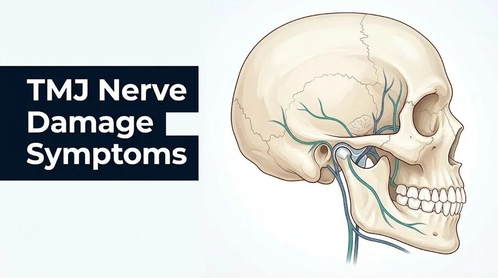
This nerve is a branch of V3. It wraps around the posterior and medial surface of the mandibular condyle before ascending through the parotid gland to supply the skin in front of the ear, the external auditory canal, the outer surface of the tympanic membrane (eardrum), the TMJ capsule itself, and the skin of the temporal scalp. Its path around the condyle neck is the key anatomical vulnerability: when the condyle displaces posteriorly (backward), the auriculotemporal nerve is pinched between the condyle and the surrounding tissue.
A 2019 study published in the Journal of Orofacial Pain found that preauricular pain (pain directly in front of the ear) and referred ear pain in TMD patients corresponded anatomically to the sensory distribution of the auriculotemporal nerve in the majority of cases studied, supporting direct nerve involvement rather than purely muscular referred pain.
The auriculotemporal nerve also carries sympathetic nerve fibers that supply the parotid gland. Compression can occasionally produce autonomic symptoms including increased salivation or changes in salivary quality, though this is a less frequently reported clinical observation.
Symptoms specifically produced by auriculotemporal nerve involvement:
- Deep aching pain in front of and just below the ear
- Sensation of ear fullness or pressure without middle ear fluid on otoscopy
- Burning or stabbing pain in the external ear canal
- Tingling or hypersensitivity of the skin in the temporal scalp region above the ear
- Scalp tenderness when combing hair or touching the temple area
- Preauricular swelling or a sense of fullness in the parotid region
Older adults with arthritic changes in the TMJ, particularly those with condylar osteophytes (bony spurs on the condyle), face higher risk of persistent auriculotemporal nerve compression because the altered condylar shape maintains abnormal pressure on the nerve even during rest position.
Trigeminal Nerve Symptoms from TMJ Dysfunction
Trigeminal nerve symptoms from TMJ dysfunction arise when chronic joint inflammation and mechanical dysfunction sensitize the peripheral branches of the trigeminal nerve (cranial nerve V) and, over time, upregulate pain-signaling pathways in the trigeminal nucleus caudalis in the brainstem.
The trigeminal nucleus caudalis is the brainstem structure where first-order (peripheral) trigeminal nerve fibers synapse onto second-order neurons that relay signals to the thalamus and then to the somatosensory cortex. In acute TMD, sensitization stays peripheral. In chronic TMD, the continuous barrage of pain signals from the joint causes synaptic changes in the nucleus caudalis itself, a process clinicians and researchers call central sensitization. Once central sensitization is established, pain can be experienced across a wider facial territory than the actual joint can explain.
The trigeminal nerve overlap with migraine pathways is clinically important. The same CGRP pathway implicated in migraine pathogenesis is activated in trigeminal sensitization from TMD. Research published in the Journal of Dental Research has found that patients with TMD have significantly higher rates of migraine comorbidity than the general population, and that the trigeminal sensitization from TMD can lower the threshold for migraine attacks.
Trigeminal symptoms in TMD that indicate nerve sensitization rather than simple muscle pain:
- Allodynia: pain from normally non-painful stimuli, such as light touch on the jaw or face causing burning or sharp pain
- Hyperalgesia: amplified pain response from typically mildly painful stimuli (biting down, cold air)
- Spreading facial pain that extends beyond the jaw into the cheek, eye region, or forehead (following V2 or V1 distribution despite the joint being a V3 structure)
- Spontaneous electric-shock-like jabs in the face, occasionally resembling trigeminal neuralgia
Distinguishing TMD-related trigeminal sensitization from classical trigeminal neuralgia (idiopathic trigeminal neuralgia, ICD-10: G50.0) requires specialist evaluation. Classical trigeminal neuralgia produces paroxysmal electric-shock pain triggered by a specific trigger zone and typically has no relationship to jaw mechanics. TMD-related trigeminal symptoms are more directly tied to jaw use, jaw position, and palpation of the joint.
Adolescents with TMD who also have anxiety disorders appear to develop trigeminal sensitization more rapidly than those without anxiety, based on observational data from specialty orofacial pain clinics.
Inferior Alveolar Nerve and Lingual Nerve Involvement in TMJ
The inferior alveolar nerve and lingual nerve, both branches of the mandibular division (V3), can be affected in TMJ disorders when inflammation or mechanical changes in the pterygomandibular space alter the tissue environment through which these nerves travel.
The inferior alveolar nerve enters the mandible through the mandibular foramen, a small opening on the medial surface of the mandibular ramus just below the condyle. It travels through the inferior alveolar canal within the mandibular bone, supplying the mandibular teeth (lower molars, premolars, canines, and incisors) and exiting at the mental foramen as the mental nerve to supply the chin and lower lip. When severe TMD produces inflammatory changes in the pterygomandibular space (the tissue envelope lateral to the pharynx and medial to the mandibular ramus), the proximal inferior alveolar nerve can be chemically irritated, producing tooth numbness, lower lip numbness, or a persistent “dental anesthesia” sensation in the lower jaw.
Many patients with this pattern present to a dentist first, assuming a tooth problem is responsible. When no dental pathology is found and the numbness persists, the TMJ should be formally evaluated.
The lingual nerve runs just medial to the inferior alveolar nerve, passing close to the lower third molar region before spreading across the floor of the mouth to the anterior two-thirds of the tongue. Inflammatory changes from TMD rarely affect the lingual nerve directly, but myofascial hypertonicity of the medial pterygoid muscle (one of the muscles of mastication that originates near the pterygomandibular space) can indirectly compress or irritate the lingual nerve, producing tongue numbness, burning, or altered sensation.
The chorda tympani, a branch of the facial nerve (cranial nerve VII) that joins the lingual nerve as it passes near the petrotympanic fissure (immediately adjacent to the posterior TMJ capsule), deserves specific mention. Retrodiscal tissue inflammation that extends to the petrotympanic fissure can irritate the chorda tympani, altering taste sensation from the anterior two-thirds of the tongue. This is an underrecognized TMJ-related symptom: patients may notice that food tastes different, metallic, or blunted on the affected side.
According to the Mayo Clinic, taste alterations in the context of jaw pain and clicking should prompt evaluation of both the TMJ and the petrotympanic fissure region, as chorda tympani involvement near the joint can be confirmed on high-resolution MRI.
Key Takeaway: Numbness or tingling in the lower teeth, chin, lower lip, or tongue in the context of jaw symptoms is not a dental problem until proven otherwise; it may reflect inferior alveolar, lingual, or chorda tympani nerve irritation from TMJ inflammation in the pterygomandibular and petrotympanic regions.
Facial Nerve Damage from TMJ Disorders
Facial nerve (cranial nerve VII) involvement in TMJ disorders is uncommon but represents the most serious neurological complication of this condition, because the facial nerve controls the muscles of facial expression and its damage produces visible motor deficits.
The facial nerve exits the skull through the stylomastoid foramen just posterior and inferior to the TMJ, then enters the parotid gland, which wraps around the TMJ capsule laterally. Within the parotid, the nerve divides into five main branches: temporal, zygomatic, buccal, marginal mandibular, and cervical. The proximity of the parotid gland and facial nerve to the joint capsule means that severe retrodiscal inflammation, a large parotid cyst or tumor mimicking TMD, or postoperative TMJ changes can all potentially involve facial nerve branches.
Pure facial nerve involvement from conservative TMD alone (without surgery or tumor) is rare. When facial nerve motor symptoms (drooping eyelid, asymmetric smile, inability to close the eye completely) appear alongside jaw pain, clinicians must rule out other diagnoses before attributing motor weakness to TMD. These diagnoses include Bell’s palsy (idiopathic cranial nerve VII palsy), Ramsay Hunt syndrome (varicella-zoster reactivation in the geniculate ganglion), parotid gland tumor, acoustic neuroma, and brainstem lesion.
The American Association of Oral and Maxillofacial Surgeons (AAOMS) notes that iatrogenic (procedure-related) facial nerve injury is a recognized risk of open TMJ surgery, and any patient who develops facial nerve symptoms following TMJ procedures should be evaluated urgently.
Facial nerve symptoms that require same-day emergency evaluation regardless of TMJ history:
- Sudden inability to move one side of the face
- Inability to close one eye completely
- Drooping of one side of the mouth at rest
- Inability to raise one eyebrow
- Ear pain with facial weakness (possible Ramsay Hunt syndrome)
These symptoms should not be assumed to be TMJ-related without ruling out stroke, Bell’s palsy, and other causes first. Go to the nearest emergency room immediately if any of these appear.
TMJ-Related Numbness and Tingling
TMJ-related numbness and tingling are the sensory changes produced by nerve fiber dysfunction along the distribution of any nerve branch affected by joint inflammation, compression, or chemical sensitization.
Numbness in the neurological sense, called hypoesthesia, means reduced sensitivity to touch, temperature, or pain stimuli. Tingling, called paresthesia, refers to spontaneous abnormal sensations (pins and needles, buzzing, crawling) arising from a nerve fiber that is partially damaged or chemically altered rather than completely interrupted. Both can occur in TMD without any actual severing or structural destruction of the nerve, because inflammation alone is sufficient to alter nerve fiber membrane excitability.
The distribution of numbness or tingling tells a clinician which nerve is most likely involved. Lower lip and chin numbness (the mental nerve territory) suggests inferior alveolar nerve involvement. Tingling in the anterior tongue suggests lingual nerve irritation. Scalp tingling above and in front of the ear suggests auriculotemporal nerve involvement. Cheek or gum tingling may reflect the long buccal nerve.
Acute vs. chronic TMJ numbness patterns:
| Feature | Acute Nerve Irritation | Chronic Neuropathic Change |
|---|---|---|
| Onset | Days to weeks into TMD flare | Months to years of untreated TMD |
| Character | Intermittent tingling, comes and goes | Persistent, may include burning |
| Relationship to jaw use | Worsens with chewing, yawning | May be constant, less jaw-related |
| Response to joint rest | Often improves with reduced jaw loading | Less responsive to simple rest |
| Reversibility | Usually reversible with treatment | Less predictable, may persist |
| Distribution | Follows one nerve territory | May spread across multiple nerve territories |
Patients taking anticoagulant medications should inform their treating dentist or orofacial pain specialist before any TMJ procedures, because these medications affect tissue healing and can alter the presentation and recovery pattern of nerve-related symptoms.
TMJ Nerve Pain in the Ear and Temple
TMJ nerve pain felt in the ear and temple is one of the most diagnostically confusing patterns in all of craniofacial medicine, because it reliably mimics otitis media (middle ear infection), mastoiditis, or temporalis muscle tension headache in its symptom quality and location.
The mechanism is direct: the auriculotemporal nerve supplies both the TMJ capsule and the external ear canal and the tympanic membrane (eardrum) with sensory fibers from the same nerve trunk. When the joint is inflamed and the auriculotemporal nerve is irritated, pain signals travel centrally but the brain interprets them as arising from the ear itself. This is a classic example of referred pain through a shared nerve: the same nerve trunk serving two anatomical regions causes the brain to localize pain to the wrong source.
Research published in the Journal of Orofacial Pain found that in patients with confirmed TMD, ear pain of non-otological origin (no middle ear infection or drum pathology) was present in approximately 42% of cases, and in most of these, the pain disappeared or substantially reduced with successful TMJ treatment, confirming the auriculotemporal nerve as the mechanism.
Temporal pain from TMJ nerve involvement follows the auriculotemporal nerve’s distribution into the temporal scalp. This presents as a unilateral headache that is worst at the temple, often described as pressure or aching rather than pulsating (distinguishing it from the classic throbbing of migraine, though both can coexist).
What to tell your primary care physician if you have TMJ-related ear and temple pain:
- Note the exact location of the pain: in front of the ear, inside the ear canal, or above the ear on the scalp
- Document whether the pain changes with jaw position (opening, closing, chewing, yawning)
- Note whether an ear examination by a clinician has shown normal tympanic membranes with no fluid, infection, or perforation
- Record any recent dental work, jaw injury, or change in bite that preceded the ear pain
- Bring a record of how long the ear pain has been present and whether antibiotics prescribed for assumed ear infection failed to help
In children, recurrent “ear pain” not responding to antibiotics should prompt evaluation for TMD by a pediatric dentist or orofacial pain specialist, as TMJ dysfunction in children is underdiagnosed and frequently misattributed to chronic otitis media.
Key Takeaway: Ear pain and temporal headache without ear infection found on physical examination are the most common nerve-referred symptoms of TMJ disorder, produced by the auriculotemporal nerve supplying both the joint capsule and the ear with sensory fibers from the same nerve trunk.
Tinnitus and Hearing Changes from TMJ Nerve Involvement
Tinnitus related to TMJ nerve involvement is a real, clinically documented phenomenon, though the precise mechanisms are still being studied, and it represents one of the more distressing neurological symptoms of TMD because it can be constant and affect sleep and concentration.
Tinnitus in TMD is thought to arise through several overlapping pathways. The auriculotemporal nerve irritation produces abnormal afferent (incoming) signaling in the auditory pathway. The retrodiscal tissue of the TMJ sits immediately adjacent to the anterior wall of the external auditory canal and the middle ear, separated by a thin layer of bone. When this tissue is inflamed or thickened, mechanical transmission of joint sounds and pressure changes into the middle ear system can occur. The chorda tympani nerve, which crosses through the petrotympanic fissure near the posterior TMJ capsule before entering the middle ear cavity, may also play a role: irritation of this nerve near the joint could create abnormal neural signals perceived as sound.
Aural fullness, the sensation that the ear is “plugged” or full, is a closely related symptom. It is produced by the same auriculotemporal nerve pathway and, less commonly, by spasm of the tensor tympani muscle (which is innervated by a branch of V3) in response to chronic TMJ-related muscle hypertonicity in the pterygoid muscles.
The Cleveland Clinic notes that TMJ-associated tinnitus is typically unilateral, corresponds to the side of greater joint involvement, and is often described as low-pitched buzzing, clicking, or pulsing rather than the high-pitched ringing more characteristic of sensorineural hearing loss.
Postmenopausal women with TMD appear to have higher rates of tinnitus as a symptom, according to clinical observations reported in specialty orofacial pain literature, possibly related to the interaction between declining estrogen levels, trigeminal sensitization, and auditory pathway sensitization.
Any new tinnitus should be evaluated by an otolaryngologist (ENT specialist) or neurotologist to rule out acoustic neuroma, Meniere’s disease, and sensorineural hearing loss before attributing it to TMJ involvement, even in a patient with confirmed TMD.
Headaches and Neurological Symptoms of TMJ Nerve Damage
TMJ-related headaches arise from both peripheral nerve sensitization and central sensitization of the trigeminal pain pathway, and they represent one of the most clinically important neurological symptoms of TMJ nerve involvement because they are frequently misdiagnosed as primary headache disorders.
The temporalis muscle (one of the four muscles of mastication, innervated by the deep temporal branches of V3) is attached to the temporal fossa of the skull and inserts on the coronoid process of the mandible. Chronic hypertonicity or myofascial trigger points in the temporalis muscle produce referred pain into the temple and forehead, mimicking tension-type headache. This is not direct nerve damage, but it is a neurological symptom: myofascial trigger points develop when peripheral sensitization alters the excitability of motor and sensory neurons supplying the muscle.
Beyond muscle-referred headache, the trigeminal sensitization described in earlier sections can produce headaches that feel indistinguishable from migraine. The CGRP pathway is the shared mechanism. Trigeminal terminals in the joint and surrounding tissues release CGRP when sensitized, and CGRP is the same vasodilatory neuropeptide implicated in migraine pathogenesis. Research published in Pain (the journal of the International Association for the Study of Pain, IASP) found that patients with TMD had a significantly higher prevalence of migraine than matched controls, and that successful TMD treatment reduced headache frequency in a proportion of these patients.
Neurological headache symptoms that suggest TMJ nerve involvement rather than primary headache disorder:
- Unilateral temporal headache that worsens specifically with chewing, yawning, or wide mouth opening
- Headache triggered or worsened by jaw clenching (bruxism-related)
- Headache accompanied by ipsilateral (same-side) ear fullness, preauricular pain, or jaw clicking
- Headache that preceded or coincided with the onset of jaw symptoms
- Headache partially relieved by jaw rest or a dental splint
Patients with pre-existing migraine disorder who develop TMD face a particularly challenging clinical picture, because the two conditions share trigeminal pathways and amplify each other. A board-certified neurologist with expertise in headache medicine, working in coordination with an orofacial pain specialist, provides the most effective evaluation for this overlap presentation.
Central Sensitization and Chronic TMJ Neuropathic Pain
Central sensitization in TMJ disorders is the neuroplastic state that develops when chronic peripheral nerve sensitization from the joint permanently alters pain-processing pathways in the trigeminal nucleus caudalis and higher brain structures, producing pain that outlasts and exceeds what the peripheral joint pathology can explain.
Central sensitization is not a psychological phenomenon. It is a measurable neurobiological change: synaptic strengthening in the trigeminal nucleus caudalis, upregulation of NMDA (N-methyl-D-aspartate) glutamate receptors in second-order neurons, and increased activation of descending pain facilitation pathways from the brainstem. Quantitative sensory testing (QST), a validated research and clinical tool that measures sensory thresholds at multiple body sites, has been used in TMD research to document central sensitization. Studies using QST have shown that patients with chronic TMD display lowered pain thresholds not just in the jaw but at distant body sites (forearm, shin), indicating that the sensitization has generalized beyond the trigeminal system.
A 2021 study published in the Journal of Dental Research found that approximately 30 to 40% of patients with chronic TMD (defined as symptoms lasting more than 6 months) showed QST profiles consistent with central sensitization, compared to essentially zero in age- and sex-matched healthy controls.
Once central sensitization is established, treatment of the peripheral joint alone is often insufficient to fully resolve pain. Management requires a multidisciplinary approach including the peripheral joint (TMJ), the central pain processing system, and comorbid psychological contributors (anxiety and depression both amplify central sensitization through shared HPA axis and serotonergic pathways).
Patients who have had TMD symptoms for more than 6 months without adequate treatment, who report widespread pain beyond the jaw and face, or who have comorbid fibromyalgia should specifically ask their orofacial pain specialist about central sensitization evaluation using QST and about whether a multidisciplinary pain program is appropriate for their care.
Key Takeaway: Central sensitization transforms TMJ pain from a local joint problem into a brain-level pain amplification state, which is why patients with chronic, longstanding TMD often report pain that seems out of proportion to their imaging findings and why joint treatment alone may not resolve all neurological symptoms.
Can TMJ Nerve Damage Be Permanent
TMJ nerve damage can be permanent in a subset of patients, particularly those with longstanding untreated disc displacement, severe condylar degeneration, or established central sensitization, though many patients with early or acute nerve irritation experience full or substantial recovery with appropriate treatment.
The reversibility of TMJ-related nerve symptoms depends on what type of nerve injury is present. In clinical nerve injury classification (Sunderland classification, applied here to the context of compression neuropathy rather than traumatic nerve transaction), mild nerve compression produces neuropraxia: temporary conduction block without structural nerve damage, fully reversible with decompression. Prolonged compression with some internal nerve architecture disruption produces axonotmesis: recovery is possible but slower and incomplete in some fibers. Severe or longstanding injury with complete architectural disruption (neurotmesis) has the worst recovery potential.
In TMD, the majority of nerve symptoms fall into the neuropraxia category: they resolve or substantially improve when the joint is decompressed, inflammation is reduced, and jaw loading is decreased. The AAOP notes that most patients with TMD-associated numbness or tingling who undergo appropriate non-surgical management (occlusal splint therapy, physical therapy, and anti-inflammatory management) show symptom improvement within 3 to 12 months.
However, three scenarios carry higher risk of permanent or long-lasting neurological change:
- Central sensitization (as described in the previous section) may persist even after peripheral joint treatment
- Severe condylar degeneration with bony ankylosis (fusion) that has chronically deformed the path of the auriculotemporal nerve
- Post-surgical nerve injury from open TMJ procedures, where direct intraoperative nerve trauma has occurred
Older patients with osteoarthritic TMJ changes and years of untreated symptoms are less likely to experience complete neurological recovery than younger patients with early-stage disc displacement and recent onset of nerve symptoms.
Risk Factors for Developing TMJ Nerve Damage Symptoms
Several factors increase the likelihood that a person with TMJ disorder will develop nerve involvement beyond simple jaw clicking and muscle pain, and identifying these factors helps prioritize which patients need earlier and more aggressive clinical evaluation.
Biological sex is the strongest demographic risk factor. Women are affected by TMD at rates 3 to 9 times higher than men, per NIDCR data, and estrogen receptor beta in TMJ synovial tissue may intensify the inflammatory response in the retrodiscal tissue. Women who are pregnant or perimenopausal experience hormonal fluctuations that can worsen joint laxity and synovial inflammation, potentially accelerating nerve irritation.
Bruxism (sleep or awake grinding and clenching) dramatically increases the compressive load on the retrodiscal tissue during sleep. A person who clenches at night applies forces to the retrodiscal tissue for hours at a time, producing sustained compression of the auriculotemporal nerve and sustained chemical irritation from inflammatory mediators.
Key risk factors for TMJ nerve damage symptoms:
- Moderate to severe bruxism, particularly nocturnal (sleep) bruxism
- Female sex, especially ages 20 to 45
- Perimenopausal or postmenopausal hormonal status
- Prior jaw trauma (condylar fracture, mandibular fracture, whiplash affecting the TMJ)
- Long-standing untreated disc displacement without reduction (closed lock)
- Comorbid fibromyalgia, migraine, or chronic widespread pain (central sensitization risk)
- Anxiety disorder or major depressive disorder (both amplify HPA axis cortisol output and lower pain thresholds)
- Highly stressful life circumstances with parafunctional habits (clenching during stress)
- Previous TMJ surgery with possible intraoperative nerve proximity involvement
- Hypermobility syndrome or Ehlers-Danlos syndrome (joint laxity increases condylar instability and retrodiscal loading)
Patients with Ehlers-Danlos syndrome (hypermobile type) deserve specific mention. Joint hypermobility allows the condyle to translate excessively, repeatedly stressing the retrodiscal tissue and auriculotemporal nerve. This population often develops TMD-related nerve symptoms earlier and more severely than the general TMD population, and requires specialized care from an orofacial pain specialist familiar with hypermobility management.
How TMJ Nerve Damage Is Diagnosed
TMJ nerve damage is diagnosed through a structured clinical evaluation that combines patient history, physical examination, targeted imaging of the joint, and, in appropriate cases, neurophysiological testing to quantify the degree and distribution of nerve dysfunction.
The evaluation begins with a detailed history: when symptoms started, what makes them worse (jaw use, clenching, wide opening, cold, touch), where exactly the numbness or tingling is located (mapped against known nerve distributions), whether symptoms are constant or intermittent, and whether any triggering event (dental procedure, trauma, jaw locking episode) preceded the onset of neurological symptoms.
Physical examination by a board-certified orofacial pain specialist includes palpation of the TMJ capsule and retrodiscal tissue with the mouth open and closed, assessment of mouth opening range (normal is approximately 40 to 55mm interincisal distance in adults), assessment of lateral and protrusive jaw movements, palpation of all muscles of mastication for tenderness and trigger points, and basic sensory testing of the face and tongue to map any areas of reduced sensation.
Diagnostic pathway for suspected TMJ nerve damage:
- Complete oral and craniofacial history with symptom mapping by nerve distribution territory
- Physical examination of TMJ, muscles of mastication, and cervical spine
- Basic sensory screening of V1, V2, V3 territories and tongue (anterior two-thirds) on both sides
- Panoramic radiograph (OPG) as initial screening for condylar bony changes
- MRI of the TMJ (bilateral, with mouth open and closed protocol) to assess disc position, retrodiscal tissue condition, and joint effusion
- Cone-beam CT (CBCT) if bony condylar pathology (erosion, osteophyte, ankylosis) is suspected
- Quantitative sensory testing (QST) if central sensitization is suspected based on widespread pain or disproportionate symptom severity
- Nerve conduction velocity (NCV) study or electromyography (EMG) if motor nerve involvement (facial nerve) is suspected
- Audiology evaluation and otolaryngology consultation if tinnitus or hearing change is present
- Neurology consultation if presentation overlaps with trigeminal neuralgia, multiple sclerosis, or other central neurological diagnoses
The DC/TMD (Diagnostic Criteria for Temporomandibular Disorders), published by the International Network for Orofacial Pain and Related Disorders Methodology (INfORM) group and widely adopted by the AAOP, provides standardized clinical criteria for classifying TMD subtypes. Using DC/TMD criteria ensures that nerve involvement is appropriately categorized as a pain-related or non-pain-related neurosensory disorder within the TMD spectrum.
Key Takeaway: Diagnosing TMJ nerve damage requires MRI of the joint combined with a nerve distribution sensory examination, because imaging alone cannot capture sensory symptoms and physical examination alone cannot visualize disc displacement and retrodiscal tissue inflammation.
When TMJ Nerve Symptoms Require Urgent Medical Evaluation
Most TMJ nerve symptoms are not acute emergencies, but specific presentations within this symptom complex require same-day emergency evaluation because they can indicate a stroke, Bell’s palsy, acoustic neuroma, or other serious neurological condition that mimics or co-occurs with TMJ disorder.
The critical distinction is between symptoms that are tied to jaw use and have been gradually developing (suggesting TMJ nerve irritation) and symptoms that appeared suddenly, are worsening rapidly, or include motor (muscle movement) deficits rather than purely sensory (feeling) changes.
When to seek care within days (not emergency, but prompt):
- New numbness in the lower lip, chin, or tongue that has persisted more than one week
- Tinnitus that appeared alongside jaw pain or clicking for the first time
- Scalp tingling or ear fullness that is new and not explained by ear examination
- Facial pain that is worsening despite rest and over-the-counter analgesics
When to go to the emergency room the same day:
- Sudden onset of facial numbness not associated with any jaw movement or dental procedure
- Any facial weakness, drooping, or asymmetry of the smile, eye, or forehead
- Inability to close one eye completely
- Sudden severe jaw pain combined with inability to open the mouth at all, especially with fever
- Sudden hearing loss on one side, with or without dizziness and facial symptoms
Emergency Symptoms: When to Call 911 or Go to the ER
Certain symptoms that can occur alongside or in the context of TMJ nerve involvement require immediate emergency evaluation. Do not wait to see if these resolve on their own.
Call 911 or go to the nearest emergency room immediately if you experience:
- Sudden facial paralysis or weakness on one side of the face, including inability to smile, raise one eyebrow, or close one eye (possible Bell’s palsy, Ramsay Hunt syndrome, or brainstem stroke)
- Sudden loss of hearing in one ear, with or without dizziness, tinnitus, and facial asymmetry (possible acoustic neuroma or labyrinthine stroke)
- Severe headache described as the worst headache of your life, with facial numbness and jaw symptoms (possible subarachnoid hemorrhage)
- Sudden inability to open the mouth beyond 10mm combined with fever above 38.5°C (101.3°F) and swelling (possible septic TMJ arthritis or spreading oral infection requiring surgical drainage)
- Facial numbness combined with one-sided arm or leg weakness or speech difficulty (possible ischemic stroke affecting the trigeminal pathway)
- Jaw symptoms following a blow to the face or head with new neurological symptoms (possible condylar fracture with adjacent nerve injury)
These presentations can indicate stroke, Bell’s palsy, Ramsay Hunt syndrome, acoustic neuroma, or spreading craniofacial infection, and they require emergency medical assessment, not an urgent care appointment or a phone call to a dentist.
Frequently Asked Questions About TMJ Nerve Damage Symptoms
What does TMJ nerve damage feel like?
TMJ nerve damage most commonly feels like numbness, tingling, or a burning sensation in the jaw, lower lip, chin, tongue, ear, or temporal scalp, corresponding to the distribution of whichever trigeminal nerve branch is affected.
Some patients describe a persistent “dental anesthesia” feeling in the lower teeth and lip, while others experience sharp, electric-like jabs in the face or a constant deep ache in front of the ear.
The specific sensory quality and location depend on which nerve is involved, and a board-certified orofacial pain specialist can map your symptoms to the responsible nerve branch during a structured sensory examination.
Can TMJ disorder cause permanent nerve damage?
TMJ disorder can cause permanent nerve damage in a subset of patients, particularly those with longstanding untreated disc displacement, severe condylar degeneration, or established central sensitization.
Most patients with early-stage or acute nerve irritation from TMJ dysfunction experience full or substantial recovery when the joint inflammation is reduced and mechanical compression is relieved through appropriate non-surgical management.
However, patients who have had untreated TMD symptoms for more than 6 months, especially with widespread or constant neurological symptoms, should be evaluated specifically for central sensitization, which can persist even after peripheral joint treatment.
What nerves are damaged by TMJ?
The primary nerves affected by TMJ disorders are branches of the mandibular division (V3) of the trigeminal nerve (cranial nerve V): the auriculotemporal nerve, the inferior alveolar nerve, and the lingual nerve.
The chorda tympani branch of the facial nerve (cranial nerve VII) can also be affected near the petrotympanic fissure adjacent to the joint, producing taste changes.
In severe or surgical cases, motor branches of the facial nerve itself can be involved, producing facial muscle weakness, though this is uncommon with conservative TMJ dysfunction.
Can TMJ cause facial numbness and tingling?
Yes, TMJ disorder can cause facial numbness and tingling through compression or chemical sensitization of the auriculotemporal, inferior alveolar, or lingual nerve branches of the trigeminal nerve.
The location of numbness or tingling corresponds to the specific nerve affected: lower lip and chin numbness suggests inferior alveolar nerve involvement, tongue tingling suggests lingual nerve irritation, and temporal scalp tingling points to the auriculotemporal nerve.
Any new facial numbness should be evaluated by a board-certified orofacial pain specialist and, if sudden in onset, by an emergency medicine physician to rule out stroke before assuming TMJ as the cause.
What specialist treats TMJ nerve damage?
A board-certified orofacial pain specialist is the primary provider for diagnosing and managing TMJ-related nerve damage symptoms.
Depending on the specific nerve involvement and symptom pattern, co-management may be needed with a neurologist (for central sensitization or overlap with trigeminal neuralgia), an otolaryngologist or neurotologist (for tinnitus and hearing changes), or an oral and maxillofacial surgeon (for structural joint disease requiring surgical consideration).
For sudden-onset facial weakness, ear pain with facial asymmetry, or hearing loss with dizziness, an emergency medicine physician should evaluate the patient first to rule out stroke, Bell’s palsy, and Ramsay Hunt syndrome.
How do I know if my TMJ is causing nerve pain or something more serious?
TMJ-related nerve pain typically develops gradually, worsens with jaw use (chewing, yawning, wide opening), is localized to one side corresponding to the affected joint, and is accompanied by other TMJ signs like clicking, jaw stiffness, or limited mouth opening.
Symptoms that suggest something more serious than TMJ include sudden onset (not tied to jaw events), facial muscle weakness or drooping, sudden hearing loss, symptoms affecting both sides of the face symmetrically, or neurological symptoms spreading to the limbs.
Any sudden neurological symptom requires same-day emergency evaluation regardless of whether you have a known TMJ diagnosis, because stroke, acoustic neuroma, and Bell’s palsy can present with facial symptoms that superficially resemble TMJ nerve involvement.
Taking Action on What You Now Know
The neurological symptoms of TMJ nerve damage are not vague or imaginary, and they are not simply the result of stress or dental sensitivity. They arise from specific, named nerve branches being mechanically compressed or chemically inflamed within millimeters of the joint itself. The auriculotemporal nerve explains your ear pain. The inferior alveolar nerve explains your tooth numbness. The chorda tympani explains why your food tastes different. Each symptom has an anatomy behind it.
The single most clinically useful action you can take right now is to map your symptoms by location and match them against the nerve distributions described in this article, then bring that map to a board-certified orofacial pain specialist. Tell them specifically where you feel numbness, tingling, or burning, whether it changes with jaw position, and how long it has been present. That information narrows the differential diagnosis immediately.
If any of your symptoms appeared suddenly, involve facial weakness, hearing loss, or feel different from your usual TMJ pattern, go to the nearest emergency room today. Don’t wait for a dental appointment to rule out stroke or Bell’s palsy. Those conditions require evaluation in hours, not days.