Dislocated Rib Symptoms: Signs, Causes, and When to Act
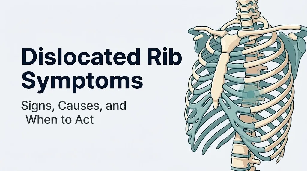
Dislocated rib symptoms typically include sharp or aching chest wall pain, pain in the upper or mid back, tenderness at a specific point along the rib, and discomfort that worsens with deep breathing, coughing, or twisting the torso. These symptoms arise from displacement or abnormal movement of a rib at one of its three possible articulations: the costovertebral joint at the spine, the costochondral junction between the bony rib and costal cartilage, or the sternocostal joint where cartilage meets the sternum.
According to the National Institute of Arthritis and Musculoskeletal and Skin Diseases (NIAMS), rib cage injuries are among the most commonly underdiagnosed sources of chest wall pain, in part because symptoms frequently mimic cardiac, pulmonary, or gastrointestinal conditions. The fact that the intercostal nerves running beneath each rib send pain signals both locally and into distant body regions means a rib problem can feel like abdominal pain, shoulder pain, or even referred flank pain.
This article covers every clinically recognized symptom pattern associated with rib displacement and slipping rib syndrome, explains the anatomical reason each symptom occurs, identifies which presentations are self-monitoring and which require urgent care, and clearly distinguishes rib dislocation from related conditions that share similar symptoms.
Dislocated Rib Symptoms: The Clinical Overview
Dislocated rib symptoms occur when a rib joint moves beyond its normal range of motion, irritating the joint capsule, periosteum, surrounding muscles, and the intercostal nerve running beneath the rib.
The rib cage is not a rigid static structure. Each of the 12 pairs of ribs moves with every breath, expanding outward and upward during inhalation and returning inward during exhalation. This constant motion means even a small disruption at any rib articulation becomes immediately apparent because the affected joint never gets rest. It’s a bit like having a pebble in your shoe: at rest it might be manageable, but every step amplifies the irritation.
Three distinct clinical entities share the label “dislocated rib” in common usage. First, a true rib dislocation involves complete displacement of the rib head from the costovertebral joint, typically caused by high-force trauma such as a motor vehicle collision or significant fall. Second, rib subluxation refers to partial displacement where the joint surfaces lose proper alignment but maintain contact, often caused by repetitive strain, athletic activity, or forceful coughing. Third, slipping rib syndrome (also called Cyriax syndrome) involves recurrent subluxation of the lower false ribs (typically ribs 8, 9, or 10) due to laxity of the fibrous interchondral ligaments.
According to a review published in the Clinical Journal of Pain, the symptom overlap between these three entities is substantial, which contributes to average diagnostic delays of years in patients with slipping rib syndrome. Understanding the specific clinical picture of each entity helps patients and clinicians recognize the pattern faster.
The most clinically relevant symptoms across all three entities include:
- Localized sharp or aching pain at the rib cage margin or spine
- Pain that worsens with deep inhalation, coughing, or sneezing
- A clicking, popping, or grinding sensation at the affected joint
- Tenderness to palpation at the specific rib level affected
- Referred pain into the upper abdomen, back, or shoulder that can mislead the clinical picture
- Restricted trunk rotation toward the affected side
Older adults should be aware that their symptom pattern may differ. Costal cartilage in adults over 65 frequently undergoes ossification, meaning it calcifies and loses flexibility. This structural change makes the cartilage more likely to fracture than to subluxate, so what presents clinically as “rib out of place” symptoms in this age group may actually involve a cartilage fracture rather than joint displacement.
What Does a Dislocated Rib Feel Like
A dislocated rib feels like a sharp, often sudden pain at a precise point on the chest wall or back, frequently accompanied by a sensation that something has shifted or snapped out of position.
The exact quality of the pain varies by the location and type of displacement. At the costovertebral joint near the spine, the pain typically feels deep, aching, and located in the mid-to-upper back approximately 2 to 4 centimeters lateral to the thoracic spinous process. At the costochondral junction along the lateral chest wall, the pain tends to be sharper and more localized, often described as a catch or a stabbing point that catches patients off guard with specific movements. At the sternocostal joint near the breastbone, the pain mimics cardiac chest pain closely enough that many patients present to emergency rooms before the correct musculoskeletal diagnosis is made.
The sensation of “something moving wrong” is a distinguishing feature. Many patients report they can actually feel or hear a clicking sound when they take a deep breath, lean forward, or rotate their torso. This audible or palpable click represents the rib slipping over the edge of an adjacent structure or a joint surface partially re-engaging. A 2021 review in Pain Medicine noted that this clicking sensation, combined with reproducible point tenderness along the costal margin, forms one of the most clinically reliable symptom combinations for identifying slipping rib syndrome specifically.
Pain typically increases in intensity during:
- Deep inhalation (the rib moves furthest during full inspiration)
- Coughing or sneezing (rapid high-pressure chest expansion)
- Bending or rotating the torso away from the affected side
- Lying on the affected side at night
- Activities that require arm reach overhead, which distract the rib cage and stress the costovertebral joint
People with hypermobile Ehlers-Danlos syndrome (hEDS) or generalized joint hypermobility often describe the sensation as more frequent and less dramatically acute than in the general population, because their joints routinely move into ranges that would be painful for most people. In this population, rib symptoms may be subtle and chronic rather than the sudden acute event typical in trauma-related cases.
Rib Out of Place Symptoms: Location and Character of Pain
Rib out of place symptoms cluster predictably by anatomical location, with the specific location of pain pointing toward which articulation has been disrupted.
Pain at the posterior rib head, felt as a deep ache between the shoulder blade and the spine, suggests costovertebral joint involvement. This pain often radiates laterally along the rib path following the course of the intercostal nerve, producing a band of discomfort that wraps from the spine toward the front of the chest. Patients frequently describe it as feeling like a tight band around the torso.
Pain at the lateral chest wall, felt as a sharp point of tenderness along the mid-axillary line approximately at the level of the affected rib, suggests disruption at the costochondral junction or at the junction between adjacent costal cartilages. This location is most common in slipping rib syndrome affecting ribs 8-10, where the costal margin is the anatomical landmark for the disrupted interchondral ligaments.
Pain near the sternum, felt as a sharp tenderness directly lateral to the breastbone and reproducible with pressure at that point, suggests sternocostal joint involvement. This presentation is the one most frequently confused with cardiac conditions, gastroesophageal reflux disease, or pleuritis.
According to Mayo Clinic clinical guidance on chest wall pain, the single most useful clinical feature distinguishing musculoskeletal rib pain from cardiac or visceral pain is reproducibility: rib pain can be precisely reproduced by pressing on a specific anatomical point, rotating the torso to a specific position, or taking a controlled deep breath, whereas cardiac pain is not reproducible by position or palpation in the same reliable way.
Pain character comparison by location:
| Pain Location | Most Likely Joint Involved | Pain Character | Worsened By |
|---|---|---|---|
| Mid-to-upper back, paravertebral | Costovertebral joint | Deep, aching, band-like | Rotation, side-bending |
| Lateral chest wall, costal margin | Costochondral / interchondral | Sharp, point-specific, catching | Deep breath, reaching |
| Near sternum, anterior chest | Sternocostal joint | Sharp or pressure-like | Bending forward, pressing on area |
| Lower chest/upper abdomen | False rib interchondral laxity | Intermittent sharp, clicking | Twisting, rising from chair |
Women may notice that rib out of place symptoms change across their menstrual cycle. Relaxin, a hormone elevated during the luteal phase and prominently during pregnancy, increases ligament laxity throughout the body, including the interchondral ligaments. This can make rib joint hypermobility symptoms more pronounced during certain hormonal phases.
Key Takeaway: The location of rib pain points directly to which of the three main rib articulations is involved, and reproducing the exact pain with specific pressure or movement is the single most useful clinical feature that distinguishes rib joint problems from cardiac or abdominal conditions.
Slipping Rib Syndrome Symptoms: The Underdiagnosed Pattern
Slipping rib syndrome symptoms are characterized by recurrent episodes of sharp lower chest or upper abdominal pain caused by hypermobile costal cartilage catching against the rib above it and irritating the adjacent intercostal nerve.
Slipping rib syndrome (Cyriax syndrome) specifically involves the false ribs: ribs 8, 9, and 10, which attach to the sternum not directly but through shared costal cartilage connected by fibrous interchondral ligaments. When these ligaments become lax or rupture, the anterior end of one of these ribs can move superiorly, hooking beneath the rib above it during movement. This mechanical catching motion compresses or stretches the intercostal nerve at that level, producing sudden sharp pain that is often described as being “caught by a knife” in the lower chest.
The symptoms follow a distinctive pattern that helps differentiate slipping rib syndrome from other rib conditions:
- Intermittent episodes of severe, sharp pain in the lower chest or upper abdomen
- A palpable or audible clicking or popping when the rib slips and returns to position
- Pain that reproduces precisely when the examiner hooks fingers under the costal margin and pulls anteriorly (the Cyriax hooking maneuver)
- Episodes triggered by specific movements: twisting, bending, rising from a seated position, or taking a deep breath at the end of range
- Relatively normal periods between episodes, unlike the constant pain of costochondritis or rib fracture
A 2019 retrospective study published in the Journal of Pediatric Surgery found that slipping rib syndrome is diagnosed most commonly in adolescents and young adults who are active in sports, and that women are represented at roughly twice the rate of men in clinical case series. The anatomical reason for this disparity is not definitively established, but researchers have proposed that differences in rib cage geometry, thoracic ligament elasticity, and hormonal effects on ligament laxity may all contribute.
The condition is notoriously underdiagnosed. According to research published in Pain Medicine, patients with slipping rib syndrome are seen by an average of five or more physicians before receiving a correct diagnosis, and some endure years of investigations for gastrointestinal, cardiac, or pulmonary conditions before the mechanical origin of their pain is identified.
Pregnant women experience a distinct risk period. As the uterus expands and the thoracic cage widens, the costal margin is mechanically stressed in ways that can precipitate slipping rib syndrome symptoms for the first time or worsen pre-existing instability. A board-certified obstetric care provider should be informed of any new rib-related chest or upper abdominal pain during pregnancy, as the differential diagnosis in this context requires excluding placental, hepatic, and pulmonary causes first.
Slipped Rib Symptoms: How They Develop Over Time
Slipped rib symptoms evolve in a predictable pattern: an acute onset phase with sharp pain, a subacute phase with persistent aching and hypersensitivity to movement, and a potential chronic phase if the underlying joint instability is not corrected.
In the acute phase, typically the first 24 to 72 hours after the displacement event, the primary symptoms are:
- Immediate sharp pain at the moment of displacement, sometimes accompanied by an audible pop or snap
- Severe pain with any movement of the rib cage, including normal breathing
- Local muscle spasm in the intercostal and paraspinal muscles surrounding the affected rib level as the body attempts to stabilize the joint
- Difficulty finding a comfortable resting position, with most patients finding partial relief by slightly hunching forward or bracing the affected side
In the subacute phase, between 3 days and 6 weeks, acute sharp pain typically transitions to:
- A persistent deep ache at the affected rib level, present even at rest but worse with activity
- Residual tenderness at the specific joint point, reproducible with direct palpation
- Pain with activities requiring trunk rotation or overhead arm movement
- Possible radiation of pain along the intercostal nerve path, producing a band of sensitivity following the rib course from back to front
The chronic phase, defined as symptoms persisting beyond 6 to 12 weeks, occurs when the underlying joint instability has not been adequately addressed. In this phase, patients often describe:
- Intermittent flares of sharp pain with seemingly minor provocations
- A background ache that fluctuates with activity level
- Psychological frustration and anxiety because the pain is often dismissed or misdiagnosed
- Compensatory movement patterns, such as avoiding trunk rotation, that create secondary pain in adjacent thoracic levels or the cervical spine
According to the American Academy of Orthopaedic Surgeons, untreated or mismanaged rib joint instability can lead to chronic intercostal neuralgia, a state of persistent nerve sensitization in which the intercostal nerve remains hypersensitive long after the initial tissue injury has resolved. This is mechanically analogous to the sensitization process seen in other peripheral nerve pain conditions.
Athletes in contact sports, rowing, gymnastics, and cheerleading face a specific risk of recurrent slipped rib symptoms because these activities repeatedly stress the costovertebral and costochondral articulations. Returning to sport without adequate rehabilitation of the surrounding intercostal and paraspinal musculature significantly increases the risk of re-injury at the same rib level.
Rib Subluxation Symptoms: Partial vs. Full Displacement
Rib subluxation symptoms reflect partial displacement of a rib joint, producing a clinical picture that is generally less severe than full dislocation but still clinically significant and often equally difficult to diagnose.
In a rib subluxation, the joint surfaces of the costovertebral or costochondral articulation lose their optimal alignment but maintain some contact. Think of it like a drawer that has been pulled out unevenly: it still sits in the track but no longer slides smoothly, creating friction and mechanical strain with every movement. The rib can still function, but every breath and every movement produces abnormal joint mechanics and continuous low-grade irritation of the local nociceptors and intercostal nerve.
The distinguishing symptom features of rib subluxation compared to full dislocation include:
| Feature | Rib Subluxation | Full Rib Dislocation |
|---|---|---|
| Pain onset | Gradual or with specific movement | Immediate, acute, severe |
| Pain severity | Moderate; worsens with activity | Severe even at rest |
| Audible pop at onset | Sometimes | Usually |
| Range of motion | Partially restricted | Severely restricted |
| Visible deformity | Rarely present | May be visible in thin patients |
| Muscle spasm | Mild to moderate | Severe, protective |
| Breathing limitation | Partial; shallow breathing adopted | Marked; shallow breathing required |
Rib subluxation frequently follows repetitive strain rather than a single acute event. Occupations requiring sustained forward bending (such as prolonged computer work with forward head posture), repetitive twisting motions, or heavy lifting with poor thoracic mechanics create cumulative stress on the costovertebral joints. Over time, this repetitive load degrades the joint capsule and the posterior costotransverse ligament, predisposing the joint to subluxation with progressively less provocation.
People with a history of prior rib fracture at the same level face a specific risk. Fracture healing deposits callus bone, which alters the geometry of the rib head and how it seats within the costovertebral joint. This altered anatomy can predispose the healed rib level to subluxation under mechanical loads that would not affect an anatomically normal rib.
Key Takeaway: Rib subluxation involves partial joint misalignment rather than complete displacement; it produces a milder but persistent pain pattern worsened by movement, and it frequently develops from repetitive strain rather than a single traumatic event.
Popped Rib Symptoms: What Happens at the Moment of Injury
Popped rib symptoms begin at the precise moment a rib joint shifts suddenly, producing an immediate and unmistakable sensation that something has physically moved in the chest wall.
The acute presentation is distinctive. Most people describe:
- A sudden sharp pain, sometimes preceded or accompanied by an audible or palpable pop, snap, or crunch
- An immediate sense that something in the chest wall is “wrong” or “out of position”
- Acute pain that can be severe enough in the first minutes to cause alarm about cardiac events
- Involuntary guarding, where the person curls forward or splints the side by pressing an arm against the rib cage
- Difficulty taking a normal breath immediately after the event, typically managed by taking shallow breaths until the initial acute pain begins to settle
The mechanism at the moment of the pop is specific. During a forceful cough, sneeze, or sudden torso rotation, the thoracic spine rotates and the rib must accommodate that rotation by gliding at both the costovertebral and costotransverse joints. If the capsular ligaments of one of these joints are already weakened or if the rotational force exceeds normal ligament tolerance, the rib head shifts suddenly out of its seated position. The pop sound and sensation come from rapid joint capsule distraction and the sudden shift of fluid pressure within the synovial joint space.
According to Cleveland Clinic clinical musculoskeletal content, a single forceful cough can generate enough intrathoracic pressure and rotational torso force to cause both rib fractures and rib joint displacement in individuals with normal rib structure, and the risk is substantially higher in people with osteoporosis, connective tissue hypermobility, or a prior chest wall injury at that level.
The immediate care steps after a perceived rib pop:
- Stop the activity and sit or lie down in the position of greatest comfort (typically slightly forward-flexed).
- Take controlled, moderate-depth breaths. Avoid forcibly expanding to maximum capacity in the first hour.
- Apply ice wrapped in a cloth to the affected area for 15 to 20 minutes to reduce local inflammation.
- Avoid pressing hard on the area to “test” the tenderness repeatedly, as this perpetuates local inflammatory mediator release.
- Seek same-day evaluation by a primary care physician or emergency medicine physician if breathing difficulty, severe pain, or any of the emergency symptoms listed later in this article are present.
- If pain is moderate and breathing is not significantly compromised, contact a primary care physician within 24 to 48 hours for clinical assessment and imaging referral if needed.
Adolescents in competitive sports, particularly gymnastics, cheerleading, and rowing, experience popped rib events at higher rates than the general population. The high-repetition twisting, bending, and impact forces of these sports generate cumulative stress on the costovertebral joints that can exceed their tolerance acutely.
Rib Out of Place in Back Symptoms: The Posterior Presentation
Rib out of place in back symptoms center on the costovertebral joint, the articulation at the back of the rib cage where the rib head meets the lateral surface of the thoracic vertebral body, and where the rib tubercle meets the transverse process at the costotransverse joint.
These two posterior articulations are the pivot points for every rib movement during breathing. When one shifts out of alignment, the resulting pain is felt in the mid or upper back, typically 2 to 5 centimeters lateral to the thoracic spinous processes at the affected level. Patients and clinicians frequently misidentify this as thoracic muscle strain, herniated thoracic disc, or referred pain from a cervical problem, because the location overlaps with these other posterior thoracic pain sources.
The distinguishing features of posterior rib displacement symptoms include:
- Deep, sometimes boring ache felt between the shoulder blade and the thoracic spine
- Pain that is sharply worse with rotation of the torso toward the affected side
- A sensation of stiffness or restriction in the mid-back that feels different from pure muscle spasm because it has a joint-quality “stuck” character
- Point tenderness directly over the costovertebral joint when pressed by an examiner’s thumb
- Pain that may radiate laterally along the rib and anteriorly following the intercostal nerve path
- Morning stiffness that eases with movement, followed by increased pain after prolonged activity (a pattern reflecting joint capsule stiffness that warms up but fatigues with load)
Thoracic disc herniation is the most commonly confused differential diagnosis for posterior rib symptoms. The clinical distinction matters because the management differs substantially. Thoracic disc problems produce pain that radiates in the distribution of the nerve root affected, often with neurological signs including altered sensation or weakness in the legs if severe. Rib joint problems produce pain that follows the intercostal nerve (wrapping around the torso), reproduce with direct joint palpation, and do not produce lower extremity neurological symptoms in uncomplicated cases.
According to the NIAMS, posterior chest wall pain that is reproducible with palpation of a specific spinal or paraspinal point and worsens with rib rotation exercises is a clinically reliable indicator of costovertebral joint pathology rather than discogenic or visceral pain.
People over 60 presenting with new posterior rib pain following even a minor twisting event should be evaluated by a primary care physician or orthopedic surgeon within 48 hours. At this age, the same mechanical event that causes a rib subluxation in a younger person may cause a thoracic compression fracture in someone with osteoporosis, and distinguishing between these two diagnoses requires imaging.
Displaced Rib Symptoms: Swelling, Bruising, and Visible Changes
Displaced rib symptoms in more severe cases include visible and palpable changes at the rib cage that go beyond pain alone, providing important clinical clues about the degree of injury.
A fully displaced rib, particularly one that has moved at the costovertebral joint, may produce:
- Visible or palpable asymmetry of the posterior rib cage, where one rib appears to protrude slightly more than the corresponding rib on the opposite side
- Local swelling over the affected joint due to acute synovial inflammation within the joint capsule and surrounding soft tissue edema
- Bruising (ecchymosis) in cases where significant soft tissue trauma has accompanied the displacement, typically appearing 24 to 48 hours after the injury as extravasated blood tracks through the tissue planes
- A visible or palpable step-off deformity in cases of complete dislocation, where the rib head sits in an abnormal position relative to the vertebral body
It is worth noting that bruising and significant swelling are more characteristic of traumatic rib fractures than of isolated rib dislocations or subluxations. When these features are prominent following chest trauma, the clinical priority is to rule out pneumothorax, hemothorax, or internal organ injury before focusing on the specific bony or joint diagnosis.
According to the Annals of Emergency Medicine, chest wall injury patients presenting to emergency departments with rib-level bruising following blunt trauma should be evaluated for associated pneumothorax even when respiratory symptoms appear mild at initial presentation, because tension pneumothorax can develop over hours following an initial injury that seemed straightforward.
The absence of visible bruising does not rule out a displaced rib. Posterior costovertebral joint displacements, in particular, occur in deep anatomical territory surrounded by substantial paraspinal musculature. The deep position of the joint means local swelling and bruising may be minimal or invisible despite significant joint disruption.
Swelling at the costochondral junction along the lateral chest wall should be distinguished from the swelling seen in Tietze syndrome, an inflammatory condition producing fusiform swelling at the second or third costochondral junction. Tietze syndrome produces a firm, sometimes warm swelling that is visually and palpably distinct from the localized soft-tissue edema seen with mechanical rib displacement.
Key Takeaway: Bruising and swelling after a rib injury are more consistent with rib fracture or significant soft-tissue trauma than with simple subluxation, and any chest wall bruising following blunt trauma warrants evaluation for pneumothorax regardless of how mild initial breathing symptoms appear.
Ribs Out of Alignment Symptoms: How Movement Changes the Pain
Ribs out of alignment symptoms are defined by a consistent relationship between specific movements and pain reproduction, a feature that is both diagnostically useful and practically important for understanding why everyday activities become so problematic.
The key insight about rib alignment and movement is mechanical. The rib cage expands and contracts approximately 12 to 20 times per minute during normal resting breathing. When one or more rib joints are misaligned, every single one of those cycles stresses the affected articulation. Unlike a knee injury, where the joint rests during non-weight-bearing periods, a misaligned rib joint never gets a true rest while the person is alive and breathing.
Specific movements that reliably worsen ribs-out-of-alignment symptoms include:
- Trunk rotation: The costovertebral joint depends on precise gliding mechanics during thoracic spine rotation. Misalignment disrupts this glide, producing sharp pain with any rotational movement, including turning to look over the shoulder or reaching across the body.
- Deep inhalation: Full inspiration requires the ribs to swing upward and outward (bucket-handle movement). A misaligned rib cannot complete this movement normally, producing a catch or sharp pain at the point of maximum respiratory expansion.
- Rising from a seated position: This movement combines thoracic extension and abdominal contraction, both of which stress the costal margin and lower rib joints, precisely the joints involved in slipping rib syndrome.
- Carrying weight on the affected side: Gravitational load through the arm transfers into the shoulder girdle and thoracic cage, increasing compressive forces at the ipsilateral costovertebral joints.
- Sustained forward bending: Prolonged thoracic flexion (as in sitting at a desk for hours) loads the posterior elements of the thoracic cage and gradually increases the stress on posterior rib joint structures.
Movements that typically provide relative relief include lying flat on the unaffected side, slight forward-flexed posture (which unloads the posterior rib joints), and gentle self-bracing by holding the affected side during coughing or sneezing.
Research published in the Clinical Journal of Pain documents that patients with rib joint dysfunction develop predictable compensatory movement patterns within days of injury, including reduced thoracic rotation range of motion on the affected side and increased reliance on diaphragmatic breathing to minimize rib cage wall excursion. A board-certified sports medicine physician or physical therapist trained in thoracic spine and rib assessment can identify these compensatory patterns through structured physical examination.
Intercostal Nerve Pain and Rib Dislocation: The Referred Pain Problem
Intercostal nerve pain from rib dislocation is one of the most clinically confusing aspects of this injury type, because it sends pain signals to locations in the body that have nothing to do with where the structural problem actually sits.
The intercostal nerves are a series of twelve paired nerves corresponding to the twelve thoracic spinal levels (T1 through T12). Each intercostal nerve runs in a groove along the inferior border of the corresponding rib, traveling in the intercostal space between the internal and innermost intercostal muscle layers. The nerve has two major cutaneous branches: the lateral cutaneous branch exits midway along the rib and supplies sensation to the lateral chest wall, and the anterior cutaneous branch exits near the sternum and supplies sensation to the front of the chest wall and upper abdomen.
When a rib is displaced, the mechanical distortion of the joint capsule and surrounding connective tissue directly irritates the intercostal nerve at the posterior aspect of the rib, near the costovertebral joint. But the pain the nerve signals does not stay localized to that point. The nerve’s terminal branches are in the anterior chest, lateral chest, or upper abdomen. So the brain receives pain signals from those distant terminal locations even though the source of irritation is at the back of the chest.
This is why rib dislocation or slipping rib syndrome so frequently presents as:
- Upper abdominal pain that is investigated as peptic ulcer disease, gallbladder disease, or kidney problems
- Left anterior chest pain that is worked up as a potential cardiac event
- Right anterior chest pain that is attributed to pleuritis or pleurisy
- Lateral chest or flank pain that mimics kidney stone or musculoskeletal injury
According to a case series published in Pain Medicine in 2020, over 40% of patients ultimately diagnosed with slipping rib syndrome had previously undergone gastrointestinal endoscopy, abdominal ultrasound, or cardiac evaluation for what turned out to be intercostal nerve referred pain from lower rib hypermobility.
The clinical test that specifically implicates the intercostal nerve as the pain source is the Cyriax hooking maneuver: the examiner places fingers beneath the costal margin and applies a gentle anterior and superior traction. Reproduction of the patient’s exact familiar pain with this maneuver strongly supports slipping rib syndrome as the diagnosis and localizes the intercostal nerve as the irritated structure.
Clinicians should be especially careful about referred intercostal pain in patients with diabetes. Diabetic neuropathy can alter the normal pain referral patterns from the intercostal nerves, making the location of perceived pain less reliable as a diagnostic marker. A board-certified pain management specialist or neurologist may be needed to accurately map the pain source in these cases.
Rib Out of Place Breathing Difficulty: Why Every Breath Hurts
Rib out of place breathing difficulty occurs because normal lung expansion requires the rib cage to move outward smoothly with each inhalation, and a displaced or unstable rib joint disrupts this movement at the same precise moment during every breath.
During inspiration, the diaphragm contracts and descends, and the external intercostal muscles contract to lift and flare the rib cage outward. This creates negative pressure inside the thoracic cavity, drawing air into the lungs. A displaced rib joint creates a mechanical catch or restriction at one point in this smooth outward expansion. Every time the chest wall reaches the angle where the displaced joint is stressed, the person encounters pain that reflexively limits inhalation.
The result is predictable and clinically important: patients adopt splinted breathing, a protective pattern where inhalation is consciously or unconsciously limited to the depth just before the point of pain. This splinted breathing pattern has direct consequences:
- Reduced tidal volume with each breath, requiring a faster respiratory rate to maintain oxygen delivery
- Failure to fully expand the lower lobes of the lungs, creating zones of reduced aeration
- Retained secretions in under-ventilated lung segments, which in patients with compromised immune function or underlying lung disease can increase the risk of respiratory infection
- Progressive diaphragm fatigue from compensatory increased respiratory rate
According to Mayo Clinic clinical guidance on chest wall injuries, splinted breathing following any rib injury should be taken seriously, and patients who are significantly limited in their ability to take a normal deep breath warrant clinical evaluation to rule out pneumothorax, hemothorax, and rib fracture before treating the presentation as simple musculoskeletal pain.
Deep breathing exercises are ultimately necessary to prevent the complications of splinted breathing, but they should be introduced only after adequate pain management and only under the guidance of a physical therapist or respiratory therapist for patients whose breathing is substantially compromised. In the acute phase, incentive spirometry (a simple device that measures inspired volume) is sometimes recommended by clinicians to monitor and encourage adequate respiratory depth.
Breathing difficulty that is severe, worsening, or accompanied by any of the emergency symptoms described in the emergency section below requires immediate evaluation. Do not attempt deep breathing exercises in the setting of possible pneumothorax.
Key Takeaway: Splinted breathing after a rib injury is a predictable protective reflex, but it compounds the injury by reducing lung ventilation; any breathing difficulty that is severe or worsening warrants urgent evaluation, not watchful waiting.
Rib Dislocation vs. Rib Fracture Symptoms: Key Differences
Rib dislocation symptoms and rib fracture symptoms overlap significantly, but several clinical features help distinguish them before imaging confirmation is available.
A rib fracture involves structural failure of the bone itself, while a rib dislocation or subluxation involves displacement at the joint while the bone remains structurally intact. This distinction matters because their management differs: fractures require different activity restrictions, take longer to heal, and carry a greater risk of complications including pneumothorax from sharp bone fragment displacement.
The clinical features that most reliably separate these two diagnoses include:
| Clinical Feature | Rib Dislocation/Subluxation | Rib Fracture |
|---|---|---|
| Point of maximum tenderness | Over the joint (back, front-sternal, or costochondral margin) | Directly over the rib shaft |
| Crepitus (grating sensation) | Clicking or popping quality | Bone-on-bone grinding quality |
| Visible deformity | Rare; possible asymmetry at back | Step deformity at fracture site if displaced |
| Pain pattern | Follows intercostal nerve path, wraps around | Localizes to fracture site; less radicular |
| Bruising location | May be absent or over joint | Often over rib shaft at fracture site |
| Response to position change | Significant variation; clicking with position | Less variation; constant ache plus movement pain |
| Risk of pneumothorax | Lower in simple subluxation | Higher, especially with displaced fracture |
| Imaging finding | Normal or subtle on plain X-ray; CT needed | Visible on X-ray in most cases |
One important practical point: plain chest X-ray misses a substantial proportion of rib fractures and virtually all rib subluxations in the acute phase. A 2022 analysis published in Annals of Emergency Medicine noted that chest CT scan detects rib fractures missed by plain X-ray in approximately 50% of cases following blunt chest trauma. For rib joint displacement specifically, dynamic ultrasound performed by a musculoskeletal radiologist or sports medicine specialist during provocative movement can visualize the abnormal rib motion directly.
This distinction is particularly relevant for athletes returning to contact sports. A rib subluxation that has been misidentified as a muscle strain may be cleared for return to sport prematurely, leading to repeated displacement events and, over time, progressive joint instability.
Slipping Rib Syndrome vs. Costochondritis: How to Tell Them Apart
Slipping rib syndrome and costochondritis are frequently confused with each other because both produce chest wall pain that worsens with movement and deep breathing, but their underlying anatomy, symptom quality, and clinical management are distinct.
Costochondritis is an inflammatory condition involving the costochondral junctions: the points where the bony ribs meet the costal cartilage. It most commonly affects ribs 2 through 5 at the anterior chest wall and produces pain that is located near the sternum, tender to direct pressure at the costochondral junction, and typically described as a persistent aching soreness that worsens with palpation, movement, or deep breathing. There is no clicking, no sense of movement or slipping, and the pain does not reproduce with the Cyriax hooking maneuver.
Slipping rib syndrome involves the lower false ribs (8-10) at the interchondral junctions near the costal margin, produces intermittent sharp pain with a distinctive clicking or slipping sensation, and is reproduced by the hooking maneuver. The pain episodes are sharp and sudden rather than the constant aching soreness of costochondritis.
Key distinguishing features:
| Feature | Slipping Rib Syndrome | Costochondritis |
|---|---|---|
| Ribs most often involved | 8, 9, 10 (false ribs) | 2, 3, 4, 5 (true ribs) |
| Location | Costal margin, lateral chest | Near sternum, anterior chest |
| Pain quality | Sharp, sudden episodes | Constant aching soreness |
| Clicking or slipping sensation | Yes, characteristic | No |
| Hooking maneuver result | Reproduces pain | Negative |
| Swelling at joint | Absent | Absent (Tietze syndrome has swelling) |
| Age and sex pattern | Young to middle-aged; women predominate | Any age; no strong sex predominance |
| Duration of episodes | Seconds to minutes per episode | Hours to days of constant pain |
According to the Cleveland Clinic, costochondritis often follows a respiratory illness with prolonged coughing, whereas slipping rib syndrome more commonly follows a physical strain, athletic activity, or repetitive movement pattern. Both can follow chest trauma, making history-taking critical for correct identification.
A primary care physician or board-certified sports medicine physician can perform the Cyriax hooking maneuver during physical examination to help distinguish these two conditions in clinic. When the diagnosis remains uncertain, referral to a musculoskeletal radiologist for dynamic ultrasound of the costal margin during provocative movement can provide definitive anatomical confirmation.
When to See a Doctor for a Rib Out of Place
Knowing when to see a doctor for a rib out of place is as important as recognizing the symptoms themselves, because some presentations require urgent evaluation while others can be scheduled within days.
See a doctor the same day or go to urgent care if:
- Pain is moderate to severe and not improving with rest and over-the-counter analgesics within 4 to 6 hours
- The injury followed a significant physical trauma (direct blow, fall, collision)
- You cannot take a comfortably deep breath due to pain
- You notice a sensation of clicking or slipping that is new and concerning
- The pain is located near the sternum and you cannot confidently rule out a cardiac cause
Schedule a primary care physician appointment within 48 to 72 hours if:
- Pain is manageable with over-the-counter analgesics but has not improved after 5 to 7 days
- You have a history of prior rib injuries or connective tissue disorder and this presentation follows that pattern
- You suspect a chronic or recurrent slipping rib syndrome based on prior similar episodes
- Pain is interfering with sleep, work, or normal daily activities
Request a specialist referral if:
- Your primary care physician evaluates you and the pain persists beyond 4 to 6 weeks without improvement
- The clinical picture matches slipping rib syndrome and a Cyriax hooking maneuver test is positive
- You are an athlete whose return-to-sport timeline depends on an accurate diagnosis
- You have been treated empirically for gastrointestinal or cardiac symptoms that have not responded to treatment and rib-source pain has not been investigated
A board-certified orthopedic surgeon or sports medicine physician is the appropriate specialist for structural rib joint assessment and management planning. If chronic intercostal neuralgia has developed, a pain management specialist may coordinate intercostal nerve block as part of the diagnostic and therapeutic approach. If imaging is needed, a referral to a musculoskeletal radiologist for dynamic ultrasound or CT of the thorax provides the most diagnostic detail.
What to bring to your appointment:
- A description of what you were doing when symptoms began
- A log of which specific movements reproduce the pain
- A description of the pain quality (sharp/aching/clicking), severity on a scale of 1-10, and duration
- Any prior chest wall imaging, even from years ago
- A list of all current medications, including blood thinners, which affect management options
Key Takeaway: Rib pain following significant trauma, rib pain with breathing difficulty, and rib pain that mimics cardiac symptoms should be evaluated the same day by a physician; chronic or recurrent rib pain matching the slipping rib syndrome pattern warrants referral to an orthopedic surgeon or sports medicine specialist.
Emergency Symptoms: When to Call 911 or Go to the ER
Certain symptoms associated with rib injury require immediate emergency evaluation. Do not wait to see if these resolve on their own.
Call 911 or go to the nearest emergency room immediately if you experience:
- Severe difficulty breathing that is getting worse rather than better, even at rest
- Sudden sharp chest pain with rapid heart rate, lightheadedness, or feeling faint
- Chest pain that radiates to the left arm, jaw, neck, or between the shoulder blades (these are cardiac warning signs that must be ruled out before attributing chest pain to a rib)
- Coughing up blood or blood-tinged sputum after a chest injury
- One side of the chest appearing visibly sunken or paradoxically moving inward when you breathe (a sign of flail chest or severe rib fracture)
- Rapid worsening breathing difficulty with decreased breath sounds on one side (signs of pneumothorax)
- Signs of shock following chest trauma: pale or bluish skin color, extreme rapid heart rate, confusion, loss of consciousness
- Severe pain in the chest following high-speed or high-force trauma regardless of whether you can breathe
These presentations can indicate pneumothorax, hemothorax, tension pneumothorax, flail chest, or acute myocardial infarction, and they require emergency medical assessment, not an urgent care appointment or a phone call.
Frequently Asked Questions About Dislocated Rib Symptoms
What does a dislocated rib feel like to the touch?
A dislocated rib typically feels sharply tender when you press directly on the affected joint, whether that is at the back near the thoracic spine, along the lateral costal margin, or near the sternum.
In some cases, you can feel a subtle asymmetry or protrusion at the site of the displaced joint, particularly with costovertebral joint displacement in slender individuals.
Pressing on the area typically reproduces the same pain that movement provokes, which is one of the most clinically useful features for distinguishing rib joint pain from visceral or cardiac pain.
Can a rib pop out of place without a direct injury?
A rib can pop out of place without direct trauma through mechanisms including forceful coughing, sneezing, repetitive twisting, or sustained poor posture that gradually fatigues the posterior rib joint ligaments.
Slipping rib syndrome, in particular, often develops without any identifiable single injury event and instead reflects cumulative ligament laxity at the lower costal cartilage level.
People with connective tissue hypermobility conditions, such as hypermobile Ehlers-Danlos syndrome, are especially susceptible to rib joint displacement without a clear traumatic trigger.
How do you know if your rib is out of place or just strained?
A muscle strain produces a broader area of aching tenderness across the intercostal muscle group, worsens diffusely with movement, and does not produce a clicking or slipping sensation.
A displaced rib produces sharp, point-specific tenderness at the exact joint location, may produce a clicking sensation with deep breathing or rotation, and often generates referred pain following the intercostal nerve path around the torso.
A physician can perform direct palpation of the rib joints and the Cyriax hooking maneuver to help clinically distinguish between these two presentations.
Can a dislocated rib fix itself on its own?
Minor rib subluxations sometimes self-reduce, particularly if the displacement was small and caused by a single strain event rather than chronic ligament laxity.
However, slipping rib syndrome involving true interchondral ligament laxity does not self-resolve without addressing the underlying joint instability, and untreated cases frequently progress to chronic intercostal neuralgia.
A primary care physician or sports medicine specialist should evaluate persistent rib pain beyond 5 to 7 days, even if the symptoms seem to be partially improving, to ensure the correct diagnosis and management approach.
What are the red flag symptoms with a rib out of place?
Red flag symptoms requiring same-day or emergency evaluation include severe difficulty breathing, coughing up blood, chest pain that radiates to the jaw or left arm, one-sided chest asymmetry with breathing, and any chest wall symptoms following high-force trauma.
Fever developing alongside rib pain should prompt evaluation for pneumonia or pleuritis, as splinted breathing after a rib injury creates conditions for lower lobe lung infection.
A new or sudden worsening of breathing difficulty after a rib injury, even hours after the initial event, should be treated as a potential pneumothorax and evaluated by an emergency medicine physician without delay.
How long does a dislocated or slipped rib take to heal?
A straightforward rib subluxation that receives appropriate clinical management, including manual therapy from a qualified practitioner and physical therapy for stabilizing muscle rehabilitation, typically improves substantially within 4 to 8 weeks.
Slipping rib syndrome involving true interchondral ligament laxity follows a more variable timeline; conservative management resolves symptoms in some patients over several months, while others with persistent instability require intercostal nerve block or surgical costal cartilage resection for lasting relief.
Full return to high-demand athletic activity following either presentation should be guided by a sports medicine physician and confirmed by restoration of pain-free range of motion and adequate intercostal muscle strength, not by elapsed time alone.
Closing
Rib pain that fits the patterns described here deserves a specific clinical assessment, not a generic “rest and ice” recommendation. Whether the symptoms point to a traumatic costovertebral joint displacement, a chronic slipping rib syndrome, or a partial subluxation from repetitive strain, the underlying anatomical mechanism is precise and the appropriate clinical pathway is well-defined.
The most important practical step for anyone experiencing persistent or recurrent rib pain with clicking, a band of pain wrapping from back to front, or abdominal or chest pain that has not responded to treatment for other suspected causes, is to request a hands-on physical examination from a primary care physician or sports medicine specialist who can perform direct joint palpation and the Cyriax hooking maneuver. Imaging alone is often insufficient; the clinical examination at the bedside remains the most sensitive diagnostic tool for rib joint pathology.
Your symptoms have a specific anatomical explanation. Getting the right assessment from the right clinician is the first step toward a management plan that addresses the structural source of the pain rather than its referred effects