Dog Pneumonia Symptoms: Warning Signs and When to Act
Dog pneumonia symptoms include a moist, deep cough, labored breathing at rest, fever, lethargy, reduced appetite, nasal discharge, and in severe cases, blue or pale gums. Recognizing these signs early, and understanding which ones require emergency veterinary care tonight versus a morning appointment, can determine whether your dog recovers fully or suffers preventable complications.
According to the American Veterinary Medical Association (AVMA), respiratory disease is among the most common reasons dogs require urgent veterinary care, and pneumonia carries a measurable mortality risk when treatment is delayed. Research published in the Journal of Veterinary Internal Medicine has documented that dogs with pneumonia presenting with oxygen saturations below 95% at the time of initial examination have substantially worse short-term outcomes than those evaluated before respiratory compromise becomes severe.
This article covers every recognized symptom of canine pneumonia, explains the specific veterinary mechanism behind each sign, distinguishes aspiration pneumonia from other subtypes, and gives you a clear framework for deciding whether your dog needs emergency care right now.
Dog Pneumonia Symptoms: What Every Owner Needs to Know
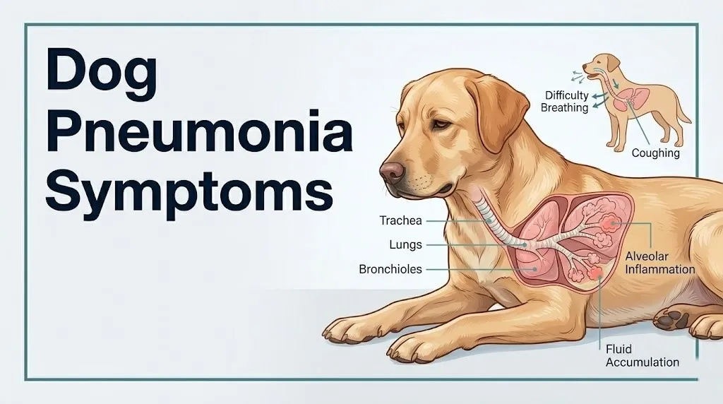
Dog pneumonia symptoms are the observable signs a dog produces when infection or aspiration has caused the lung’s air-filled alveoli to fill with inflammatory exudate, reducing the organ’s ability to supply the body with oxygen.
The lungs in a healthy dog function like millions of tiny balloons, each alveolus inflating with fresh air during each breath and transferring oxygen directly into the bloodstream across its thin wall. When pneumonia strikes, those tiny air sacs fill with a mixture of fluid, dead immune cells, and bacteria. They can no longer inflate or exchange gas. The dog’s body compensates by breathing faster and harder, and the symptoms you see are the downstream consequences of that failing compensation.
The full list of recognized symptoms, ranked from earliest-presenting to most severe:
- Moist, productive, deep cough (earlier and more reliable than fever in most dogs)
- Increased resting respiratory rate (above 30 breaths per minute)
- Lethargy and reluctance to exercise
- Reduced appetite or complete refusal of food
- Fever (rectal temperature above 103.5 degrees Fahrenheit)
- Mucopurulent nasal discharge (yellow, green, or blood-tinged)
- Labored, effortful breathing with visible chest wall movement
- Elbow abduction and neck extension (orthopneic posture)
- Reluctance to lie down flat
- Pale, gray, or blue gum color (cyanosis, a late and serious sign)
- Open-mouth breathing at rest (abnormal in dogs outside of panting context)
- Collapse or loss of consciousness (emergency)
Dogs do not complain of breathlessness the way humans do. Every one of these signs is your dog communicating physical distress through behavior and physical appearance rather than words.
What Is Pneumonia in Dogs?
Pneumonia in dogs is inflammation of the lung parenchyma (the functional gas exchange tissue of the lung, comprising the alveoli, bronchioles, and pulmonary interstitium) caused by infection, aspiration of foreign material, or less commonly by immune-mediated or toxic injury.
The canine lung is divided into lobes: four in the right lung (cranial, middle, caudal, and accessory) and two in the left lung (cranial and caudal). This lobe structure matters clinically because pneumonia rarely affects all lobes equally. Knowing which lobes are affected on a chest X-ray helps your veterinarian determine the likely cause and guides treatment decisions.
Think of a healthy lung like a fresh sponge full of air. Pneumonia turns parts of that sponge solid, replacing air with fluid and cellular debris. The more of the sponge that solidifies, the less gas exchange occurs, and the harder the dog’s body must work to extract enough oxygen to function.
The American College of Veterinary Internal Medicine (ACVIM) classifies canine pneumonia by cause: infectious (bacterial, viral, fungal, or parasitic), aspiration (caused by inhaling foreign material), and non-infectious (immune-mediated or lipoid). Each type produces a similar core set of respiratory symptoms, but the severity, progression rate, lobe distribution, and treatment approach differ meaningfully between them.
Quick Tip:
- Pneumonia is not a single disease. It is a pattern of lung inflammation caused by different underlying triggers.
- The type of pneumonia your dog has determines how quickly it will progress and what treatment is needed.
- Dogs with known predisposing conditions (megaesophagus, laryngeal paralysis) are at higher risk and should be evaluated at the first sign of respiratory symptoms.
Types of Pneumonia in Dogs
The four primary types of pneumonia in dogs are bacterial, viral, fungal, and aspiration, each with a distinct mechanism, typical patient profile, and clinical course.
Bacterial pneumonia is the most frequently diagnosed type in clinical veterinary practice, according to the Merck Veterinary Manual. The most common causative organisms include Bordetella bronchiseptica, Streptococcus equi subspecies zooepidemicus, and Mycoplasma cynos. These pathogens colonize the lower airways after overcoming or bypassing the mucociliary clearance system, the escalator-like mechanism by which ciliated respiratory epithelial cells sweep inhaled particles up and out of the airway.
Viral pneumonia in dogs is typically caused by canine influenza virus (strains H3N8 and H3N2) or canine distemper virus. Viral infection is particularly dangerous because it impairs alveolar macrophage function directly, creating a window of vulnerability during which secondary bacterial infection easily establishes. A dog that initially presents with influenza-like signs (runny nose, mild cough, low-grade fever) and then deteriorates rapidly within 48 to 72 hours is often experiencing viral-to-bacterial pneumonia progression.
Fungal pneumonia (also called mycotic pneumonia) is caused by environmental fungi including Aspergillus fumigatus, Blastomyces dermatitidis, Histoplasma capsulatum, and Cryptococcus neoformans. Geographic exposure matters: Blastomyces is endemic to the Mississippi and Ohio River valleys and Great Lakes regions. Histoplasma is also endemic to river valley soils across the central United States. Dogs in these regions who spend time outdoors, particularly digging in disturbed soil, face elevated exposure risk.
Aspiration pneumonia occurs when a dog inhales food, water, vomit, gastric acid, or a foreign liquid into the lower airway. It has a distinct mechanism and symptom profile covered in detail in a dedicated section below.
| Pneumonia Type | Most Common Cause | Typical Patient | Key Distinguishing Feature |
|---|---|---|---|
| Bacterial | Bordetella, Streptococcus, Mycoplasma | Any dog; kennel-exposed dogs | Productive cough, rapid fever onset |
| Viral | Canine influenza, distemper | Unvaccinated, young, shelter dogs | Rapid deterioration after mild onset |
| Fungal | Blastomyces, Histoplasma, Aspergillus | Outdoor dogs in endemic regions | Chronic, non-responsive to antibiotics |
| Aspiration | Inhaled material | Brachycephalic, megaesophagus, post-anesthetic dogs | Right-sided lobe distribution, sudden onset |
Key Takeaway: Bacterial pneumonia is the most common type in dogs, but viral and aspiration pneumonia can progress faster and require a different treatment approach. Type matters for treatment, and only your veterinarian can determine it.
Early Signs of Pneumonia in Dogs
The earliest signs of pneumonia in dogs appear before the classic labored breathing develops, and recognizing them at this stage offers the best chance of uncomplicated recovery.
A moist, productive-sounding cough is typically the first owner-observable symptom. It sounds wetter and deeper than the dry, honking cough characteristic of kennel cough (infectious tracheobronchitis). Some dogs bring up small amounts of mucus or phlegm during coughing episodes. Others swallow what they produce, which means you may not see mucus even when the cough is productive.
Behavioral changes often precede obvious respiratory signs. A dog in the early stages of pneumonia may seem quieter than usual, less interested in play or walks, slower to greet at the door, or more inclined to rest. These behavioral shifts reflect the systemic effect of interleukin-6 (IL-6) and tumor necrosis factor-alpha (TNF-alpha), pro-inflammatory cytokines released by alveolar macrophages during the initial immune response. These mediators cross the blood-brain barrier and suppress normal activity and appetite centrally.
Reduced food intake in the first 24 to 48 hours of illness is a common early sign, particularly in dogs that are normally enthusiastic eaters. Food refusal combined with a new or worsening cough should prompt a call to your veterinarian, not a wait-and-see approach.
Quick Tip:
- A new, wet-sounding cough in a dog that was healthy the day before warrants a veterinary call within 24 hours, not a full week of monitoring.
- Early-stage pneumonia is significantly easier and less expensive to treat than pneumonia that has progressed to respiratory distress.
- Puppies under 6 months may skip the early behavioral signs and deteriorate directly to moderate or severe respiratory distress within 24 to 48 hours due to their immature immune systems.
Coughing in Dogs With Pneumonia
Coughing in dogs with pneumonia is typically moist, productive, and deep-chested, originating from the lower airways rather than the trachea, and it persists throughout the day rather than occurring in isolated episodes.
The cough reflex in dogs is triggered by irritant receptors in the bronchial mucosa and lower trachea. In pneumonia, those receptors are continuously stimulated by the presence of exudate (inflammatory fluid and cellular debris) in the airways. The dog coughs in an attempt to clear that material upward and out. The productive quality of the cough, its wet sound and the occasional visible mucus, reflects the volume of exudate the lower airways are generating.
Some dogs with pneumonia cough so forcefully and frequently that they retch or vomit after coughing bouts. This is sometimes mistaken for a gastrointestinal problem rather than a respiratory one, delaying veterinary evaluation. A cough that is consistently followed by retching or gagging, particularly with any new respiratory symptoms, warrants prompt veterinary assessment.
Cough frequency and character change as pneumonia progresses. According to research reviewed in Veterinary Clinics of North America: Small Animal Practice, dogs with early pneumonia may cough intermittently and remain comfortable between episodes. As consolidation increases and airway secretions thicken, coughing becomes more frequent, more effortful, and more distressing to the dog.
Dogs on immunosuppressive medications (corticosteroids such as prednisone, or chemotherapy agents) may have a blunted cough despite active pneumonia. The neutrophil response that generates the exudate is suppressed, so there is less material to trigger the cough reflex. These dogs may appear less obviously ill while their lung disease progresses silently.
Fever and Lethargy: Systemic Signs of Dog Pneumonia
Fever and lethargy in dogs with pneumonia result from the same cytokine cascade that drives alveolar inflammation, and they are important early warning signs that the body is mounting a systemic immune response to infection.
Normal canine rectal temperature ranges from 101.0 to 102.5 degrees Fahrenheit. A temperature above 103.5 degrees Fahrenheit is considered a fever in dogs. The fever in pneumonia is driven by prostaglandin E2 (PGE2), produced from arachidonic acid by cyclooxygenase enzymes in response to circulating IL-1 and IL-6. PGE2 acts directly on the hypothalamic preoptic area to raise the thermal set-point, triggering heat-conservation mechanisms (vasoconstriction, shivering) until the body temperature reaches the new, elevated set-point.
To check your dog’s temperature at home accurately:
- Use a digital rectal thermometer designated for pet use.
- Lubricate the tip with a small amount of petroleum jelly or water-soluble lubricant.
- With a second person gently restraining the dog in a standing position, insert the thermometer tip approximately one inch into the rectum.
- Wait for the thermometer to beep or signal completion (typically 30 to 60 seconds for a digital model).
- Any reading consistently above 103.5 degrees Fahrenheit in a resting, non-overheated dog warrants a call to your veterinarian the same day.
Lethargy in pneumonia reflects both the direct effect of inflammatory cytokines on the central nervous system and the metabolic cost of the immune response. A dog with moderate lethargy will still respond to you, eat if offered especially palatable food, and move to preferred resting spots, but will do so slowly and with less enthusiasm. A dog with severe lethargy may refuse to stand, show no interest in food or interaction, and may not respond normally to its name. Severe lethargy with respiratory symptoms is an urgent finding.
According to the Merck Veterinary Manual, fever combined with a productive cough and any degree of respiratory rate increase constitutes a clinical presentation consistent with pneumonia and warrants same-day veterinary evaluation rather than home monitoring.
Key Takeaway: Fever above 103.5 degrees Fahrenheit combined with a new, wet-sounding cough in your dog is a same-day veterinary call, not a wait-and-see situation.
Breathing Problems: Respiratory Signs of Pneumonia in Dogs
Breathing problems in dogs with pneumonia range from a mildly elevated resting respiratory rate in early stages to open-mouth breathing, elbows-out posture, and cyanotic gums in severe cases, and tracking these signs is the most reliable way to gauge how quickly your dog is deteriorating.
A healthy dog at rest breathes 15 to 30 times per minute. Counting your dog’s resting respiratory rate (RRR) is the single most actionable thing you can do at home to assess respiratory status. To count it: observe the rise and fall of the chest wall or belly for 30 seconds while the dog is resting quietly and not panting, then multiply by two. A consistently elevated RRR above 30 breaths per minute in a resting dog is abnormal and warrants veterinary evaluation.
As pneumonia progresses and more alveoli fill with exudate, the compensatory mechanisms escalate. The dog begins actively recruiting its accessory respiratory muscles: the intercostal muscles between the ribs become visibly active, producing a pronounced chest wall heaving. The diaphragm flattens fully with each breath. The nostrils may flare. These are signs that the dog is working hard to breathe rather than breathing comfortably.
The orthopneic posture is a specific and clinically important sign in dogs with respiratory compromise. The dog stands (or sits) with its front legs spread wider than normal (elbows abducted), its neck extended forward, and its head lowered slightly. This posture mechanically opens the thoracic inlet and allows the chest wall to expand to its maximum capacity. A dog that adopts this posture and resists lying down is not being difficult. It is physically unable to breathe adequately in a recumbent position. This sign warrants emergency veterinary evaluation.
Research published in the Journal of Veterinary Internal Medicine has found that dogs presenting with pneumonia in orthopneic posture have a higher rate of requiring oxygen supplementation within the first hour of hospital evaluation than dogs presenting without this posture. Do not wait to see if it resolves.
Nasal Discharge and Other Visible Signs in Dogs With Pneumonia
Nasal discharge in dogs with pneumonia is typically mucopurulent (containing mucus and pus), appearing yellow, green, or occasionally blood-tinged, and it differs from the clear, watery discharge typical of viral upper respiratory infection.
The mucopurulent quality reflects the neutrophil-rich exudate being produced in the lower airways. Some of that material migrates upward through the mucociliary escalator and exits via the nasal passages. The color and thickness of the discharge provides clinical information: thin and clear suggests viral irritation of the upper tract; thick, colored, and opaque suggests active bacterial infection with substantial neutrophil infiltration.
Beyond nasal discharge, several other visible signs help assess the severity of pneumonia:
- Mucous membrane color: The gums (gingival mucosa) should be a healthy salmon-pink in dogs. Press on the gums with your fingertip; color should return within 1 to 2 seconds (normal capillary refill time). Pale pink, white, or gray gums suggest reduced perfusion or anemia. Blue or purple gums (cyanosis) indicate life-threatening oxygen deprivation and require immediate emergency veterinary care. Do not drive to call ahead; go directly to the nearest emergency veterinary hospital.
- Eye appearance: Dogs with systemic illness from pneumonia often have a sunken, dull, or glazed eye appearance. The third eyelid (nictitating membrane) may be partially visible at the inner corner of the eye, which can indicate pain, illness, or significant systemic stress.
- Body condition: Rapid weight loss over several days, visible ribbing, or coat changes may appear in dogs with prolonged or untreated pneumonia as the metabolic cost of fighting infection exceeds caloric intake.
Nasal discharge in brachycephalic breeds (such as Pugs, French Bulldogs, or English Bulldogs) may be less visible because their short nasal passages and restricted nasal anatomy reduce nasal airflow, causing these dogs to swallow discharge rather than expel it. In these breeds, the first visible sign of respiratory illness may be labored breathing rather than obvious nasal discharge.
Aspiration Pneumonia Symptoms in Dogs
Aspiration pneumonia in dogs occurs when foreign material (food, water, vomit, gastric acid, or liquid medication) is inhaled past the epiglottis and into the lower airway, triggering chemical inflammation and secondary bacterial infection in the lung parenchyma.
The mechanism is specific to canine anatomy. The right caudal bronchus in dogs angles more steeply and has a larger caliber relative to the tracheal diameter than in humans, meaning that aspirated material traveling down the trachea disproportionately enters the right middle and right caudal lung lobes. This is why thoracic radiographs in dogs with aspiration pneumonia typically show consolidation patterns in the right-sided dependent lobes rather than the bilateral or diffuse patterns more common in bacterial pneumonia.
The initial chemical injury from aspirated gastric acid causes an intense local inflammatory response within minutes to hours, producing the early symptoms: sudden-onset coughing, retching, gagging, and distress immediately or within a few hours of a known aspiration event. Secondary bacterial contamination from the aspirated material then establishes over the following 12 to 72 hours, adding fever and progressive respiratory compromise.
Aspiration pneumonia symptoms that may distinguish it from primary bacterial pneumonia include:
- Sudden onset following a known aspiration event (vomiting, forced medication administration, regurgitation in dogs with megaesophagus, or immediately post-anesthesia recovery)
- Initial acute coughing and gagging that may partly settle before returning with fever 24 to 48 hours later
- Right-sided or right-predominant abnormal lung sounds on veterinary auscultation
- History of a predisposing condition: megaesophagus (the esophagus is dilated and lacks coordinated peristalsis, causing food to pool rather than pass to the stomach, with chronic regurgitation and constant aspiration risk), laryngeal paralysis (failure of the arytenoid cartilages to abduct during inspiration, leaving the glottis inadequately protected), or brachycephalic obstructive airway syndrome (BOAS) (anatomically narrowed upper airway increasing aspiration risk during eating, drinking, and vomiting)
According to a clinical review published in Veterinary Clinics of North America: Small Animal Practice, aspiration pneumonia carries a higher short-term mortality rate in dogs with megaesophagus than in dogs with aspiration pneumonia from other causes, due to the difficulty of preventing repeated aspiration events in megaesophagus patients.
If you suspect your dog has aspirated a toxic substance (petroleum product, cleaning solution, plant extract, or medication), call the ASPCA Animal Poison Control Center at 888-426-4435 immediately in addition to seeking emergency veterinary care.
Key Takeaway: If your dog has megaesophagus, laryngeal paralysis, or is a brachycephalic breed, any new cough or breathing change should be evaluated by a licensed veterinarian the same day, not monitored at home.
How Dogs With Pneumonia Behave Differently
Dogs with pneumonia behave differently from healthy dogs in ways that go beyond obvious respiratory signs, and these behavioral changes are often the first cues that something is seriously wrong.
A dog with early pneumonia typically shows a reduction in voluntary activity that owners often describe as the dog “acting off” or “just not being herself.” This reflects the central nervous system effects of circulating cytokines. The dog may be slower to wake from sleep, less interested in walks it normally enjoys, and less engaged with toys or play. These behavioral changes can appear 12 to 24 hours before fever or audible respiratory signs develop.
Eating and drinking patterns change in ways that carry diagnostic significance. Complete food refusal in a dog that normally eats readily is more clinically concerning than partial reduced intake. Dogs with painful, frequent coughing may drink less because swallowing triggers additional coughing bouts. Reduced water intake combined with fever creates a risk of dehydration that worsens the dog’s ability to clear airway secretions, because well-hydrated dogs maintain thinner mucus that is easier to cough up.
Sleep posture changes are often overlooked by owners but are meaningful. A dog with respiratory compromise will avoid lying on its side (lateral recumbency), which compresses the dependent lung and further reduces functional gas exchange. Instead, the dog may sleep in a sternal position (on its chest, not its side), prop itself against a wall or piece of furniture, or repeatedly shift positions rather than settling into comfortable sleep. Observing sleep posture is one of the most practical things an owner can do to assess nighttime respiratory status without waking the dog to count breaths.
Senior dogs over 8 years of age may show blunted fever responses due to age-related changes in immune signaling, meaning their rectal temperature may remain in or near the normal range even when pneumonia is active. These dogs may appear primarily lethargic and anorexic without the obvious fever that would prompt a younger dog’s owner to seek care more quickly.
Puppy and Senior Dog Pneumonia Symptoms
Puppies and senior dogs present with dog pneumonia symptoms differently from healthy adult dogs, and both groups deteriorate faster, making early veterinary evaluation especially time-sensitive.
Puppies under 4 to 6 months of age have an immature adaptive immune system. Their alveolar macrophage function is reduced, their antibody production capacity is still developing, and the passive maternal antibody protection they received at birth declines sharply between 6 and 16 weeks of age. This creates a window of vulnerability during which respiratory pathogens encounter reduced resistance at the alveolar level.
Puppies with pneumonia may present with:
- Rapid deterioration over 12 to 24 hours from mild coughing to respiratory distress
- Open-mouth breathing appearing earlier in the course than in adult dogs
- Hypothermia (low body temperature below 100 degrees Fahrenheit) rather than fever in very young or severely ill puppies, because their thermoregulatory system is also immature
- Failure to nurse or eat, appearing as weakness or crying rather than the behavioral withdrawal seen in adult dogs
- Audible crackling or gurgling sounds from the chest heard without a stethoscope in severe cases
Neonatal pneumonia (in puppies under 2 to 3 weeks of age) is a veterinary emergency with a narrow survival window. Any neonate showing breathing irregularities, crying persistently, or failing to nurse requires immediate veterinary evaluation.
Senior dogs over 8 to 10 years have decreased immune surveillance at the alveolar level and may carry multiple chronic diseases that alter how pneumonia presents:
- Reduced fever response means temperature may be only slightly elevated or even low-normal despite active infection
- Chronic kidney disease or cardiac disease in senior dogs may contribute to fluid accumulation in the lungs (pulmonary edema) that is difficult to distinguish from consolidation on clinical examination alone, requiring chest X-ray for differentiation
- Senior dogs on long-term nonsteroidal anti-inflammatory drugs (NSAIDs) may have blunted systemic inflammatory markers, masking the CBC changes (neutrophilia, left shift) that help confirm bacterial pneumonia
According to the Merck Veterinary Manual, senior dogs with concurrent systemic disease have longer hospital stays and slower response to treatment than otherwise healthy adult dogs with the same pathogen. A senior dog showing any combination of new cough, reduced activity, and altered eating pattern warrants same-day veterinary evaluation without waiting for fever or obvious breathing changes to develop.
Breeds and Conditions at Highest Risk for Dog Pneumonia
Certain dog breeds and dogs with specific underlying health conditions face substantially elevated risk of developing pneumonia or deteriorating more rapidly once pneumonia is established.
Brachycephalic breeds carry the highest structural risk. The anatomical abnormalities of brachycephalic obstructive airway syndrome (BOAS), including stenotic nares, an elongated soft palate, a narrowed trachea, and everted laryngeal saccules, impair normal mucociliary clearance and increase the mechanical difficulty of breathing under any respiratory load. Breeds most affected include the English Bulldog, French Bulldog, Pug, Boston Terrier, Shih Tzu, Cavalier King Charles Spaniel, and Boxer.
Dogs with the following conditions are at elevated risk for aspiration pneumonia specifically:
- Megaesophagus: The esophagus loses coordinated muscular contraction, becomes dilated, and pools ingested food and water above the lower esophageal sphincter. Regurgitation (passive return of material from the esophagus without the active muscular effort of vomiting) occurs frequently, and the regurgitated material is easily inhaled. Breeds with higher megaesophagus prevalence include the German Shepherd, Great Dane, Irish Setter, and Labrador Retriever.
- Laryngeal paralysis: The arytenoid cartilages that normally open the glottis widely during inspiration fail to abduct, narrowing the airway and creating turbulent, high-velocity airflow that increases aspiration risk during eating and drinking. Older large and giant breed dogs (Labrador Retrievers, Golden Retrievers, Saint Bernards, Irish Setters) are disproportionately affected, typically presenting between 9 and 13 years of age.
- Dogs receiving long-term corticosteroid therapy (for immune-mediated disease, allergic conditions, or inflammatory bowel disease): Corticosteroids impair alveolar macrophage function and neutrophil migration into alveolar spaces, reducing the lung’s first-line defense against inhaled pathogens.
| Risk Category | Breeds or Conditions | Specific Risk Mechanism |
|---|---|---|
| BOAS-related | English Bulldog, French Bulldog, Pug, Boston Terrier | Impaired mucociliary clearance, aspiration risk |
| Megaesophagus | German Shepherd, Great Dane, Labrador | Chronic regurgitation and aspiration |
| Laryngeal paralysis | Labrador, Golden Retriever, Saint Bernard (older) | Glottis protection failure during swallowing |
| Immunosuppressed | Dogs on corticosteroids or chemotherapy | Reduced alveolar macrophage function |
| Age-related | Puppies under 6 months, seniors over 8 years | Immature or reduced immune surveillance |
Key Takeaway: If your dog is a brachycephalic breed or has megaesophagus or laryngeal paralysis, any new respiratory symptom should go to a licensed veterinarian the same day, because these dogs have less reserve and deteriorate faster than other dogs.
How Veterinarians Diagnose Pneumonia in Dogs
A veterinarian diagnoses pneumonia in dogs through a combination of physical examination findings, thoracic radiography, blood work, and in some cases lower airway sampling, with thoracic radiography being the most diagnostically definitive single test.
The diagnostic process typically unfolds as follows:
- Physical examination: The veterinarian auscultates (listens to) the lungs with a stethoscope. Normal lung sounds in dogs are soft and barely audible in the lower lobes. Pneumonia produces abnormal sounds: crackles (a crackling or rattling sound from fluid movement in airways), wheezes (a high-pitched sound from narrowed or inflamed airways), or, in severely consolidated regions, complete absence of breath sounds (because air no longer flows through the affected tissue).
- Thoracic radiography (chest X-ray): Two views (right lateral and ventrodorsal or dorsoventral) provide the most information. On radiographs, consolidated lung tissue appears as an opacity (a white area where normal air-filled lung should appear dark). The distribution of the opacity helps identify the pneumonia type: right middle and caudal lobe opacity suggests aspiration pneumonia; diffuse bilateral opacity with a specific pattern suggests fungal disease or severe bacterial pneumonia.
- Complete blood count (CBC): Bacterial pneumonia typically produces neutrophilia (elevated neutrophil count) with a left shift (the presence of immature band neutrophils in the circulation), indicating that the bone marrow is releasing neutrophils faster than they can mature. Viral pneumonia may initially show lymphopenia (reduced lymphocyte count). A normal or low neutrophil count in a dog that appears clinically ill with respiratory signs may indicate immunosuppression or a very early-stage process.
- Pulse oximetry: A non-invasive clip sensor placed on the tongue, ear, or paw web measures arterial oxygen saturation (SpO2). Normal SpO2 in dogs is 95 to 100%. A reading below 95% indicates hypoxemia. Below 90%, supplemental oxygen therapy is required.
- Tracheal wash or bronchoalveolar lavage (BAL): For dogs that do not respond to initial broad-spectrum antibiotic therapy, or when fungal pneumonia or an unusual pathogen is suspected, sampling of the lower airway fluid allows cytology (microscopic cell examination) and culture with antibiotic sensitivity testing. This guides targeted antibiotic selection.
According to the American College of Veterinary Internal Medicine (ACVIM), thoracic radiography combined with CBC and physical examination correctly identifies pneumonia in the majority of dogs presenting with compatible clinical signs. Culture and sensitivity testing of BAL or tracheal wash fluid is recommended for all dogs that fail to improve within 48 to 72 hours of initial antibiotic treatment.
Dog Pneumonia vs Kennel Cough: Key Differences
Dog pneumonia and kennel cough (infectious tracheobronchitis, caused most commonly by Bordetella bronchiseptica and canine parainfluenza virus) are frequently confused, but they differ in the location of inflammation, the severity of illness, and the urgency of veterinary care required.
Kennel cough is primarily an upper airway and tracheal disease. The hallmark is a harsh, dry, honking cough that sounds like a goose, often triggered by excitement or pressure on the trachea (collar contact). Dogs with kennel cough typically remain alert, active, eating, and afebrile (without fever) despite their spectacular-sounding cough. The infection is largely self-limiting in otherwise healthy adult dogs, resolving over 1 to 3 weeks.
Pneumonia, by contrast, involves the lung parenchyma rather than the trachea. The cough is wet and productive rather than dry and honking. The dog appears systemically unwell: lethargic, febrile, anorexic. Breathing rate is elevated at rest. The dog does not bounce back between coughing bouts; it remains subdued.
| Feature | Kennel Cough | Pneumonia |
|---|---|---|
| Cough character | Dry, harsh, honking | Moist, productive, deep |
| Activity level | Often normal between coughs | Lethargic, reduced |
| Appetite | Usually maintained | Reduced or absent |
| Fever | Absent or low-grade | Present (above 103.5°F) |
| Resting respiratory rate | Normal | Elevated above 30 breaths/minute |
| Breathing effort | Normal at rest | Labored, effortful |
| Gum color | Normal pink | May be pale, gray, or blue in severe cases |
| Urgency | Vet within a few days if persists | Same-day to emergency |
Kennel cough can progress to bacterial pneumonia in puppies, senior dogs, immunocompromised dogs, or dogs with pre-existing respiratory conditions. A dog initially diagnosed with kennel cough that becomes lethargic, stops eating, develops a fever, or shows elevated resting respiratory rate has progressed beyond simple tracheobronchitis and requires immediate reassessment by a licensed veterinarian.
Emergency Signs: When to Take Your Dog to the Vet Immediately
Certain dog pneumonia symptoms represent a life-threatening emergency and require immediate veterinary care. Do not wait until morning, do not monitor at home, and do not attempt home treatment.
Take your dog to a veterinary emergency hospital immediately if you observe any of the following:
- Blue, purple, gray, or white gum color: This is cyanosis or severe pallor, indicating the dog’s blood oxygen level has fallen to a dangerous level. Normal gum color is salmon-pink. Any deviation toward blue or gray is a respiratory emergency.
- Resting respiratory rate consistently above 40 breaths per minute: At this rate, the dog is in respiratory distress. Count the rate when the dog is resting and not panting; if it remains above 40, go to the emergency hospital without delay.
- Open-mouth breathing in a resting, cool dog: Dogs breathe through their noses at rest. Open-mouth breathing outside of panting after exercise or heat is abnormal and indicates the dog cannot move enough air through its nasal passages to meet its oxygen demand.
- Orthopneic posture that does not resolve: A dog standing with elbows spread wide, neck extended, and refusing to lie down is physically unable to breathe adequately when recumbent. This is a medical emergency, not a behavioral quirk.
- Collapse or inability to stand: Hypoxemia severe enough to prevent the dog from supporting its own weight requires immediate resuscitative veterinary care.
- Loss of consciousness or unresponsiveness: This level of compromise indicates near-complete respiratory failure. Take your dog to the nearest emergency veterinary hospital immediately. Do not call ahead if it would delay departure.
- Sudden marked deterioration after apparent stabilization: A dog that seemed to be improving and then suddenly worsens may have developed a complication such as sepsis, pleural effusion (fluid around the lung), or pneumothorax (air outside the lung in the chest cavity).
If aspiration of a toxic substance is suspected in addition to the respiratory emergency, call the ASPCA Animal Poison Control Center at 888-426-4435 on the way to the emergency hospital. Simultaneous call and transport is appropriate for this level of urgency.
These signs can indicate life-threatening hypoxemia or septic shock, and they require emergency evaluation at a veterinary hospital equipped with oxygen supplementation, not a scheduled clinic appointment.
What to Tell Your Veterinarian About Your Dog’s Symptoms
Giving your veterinarian a complete, organized clinical history when you arrive is one of the most useful things you can do for your dog, because it shapes the diagnostic approach from the first minute of evaluation.
To prepare the most useful symptom report:
- Write down when the symptoms started: Include the exact day the cough began, when lethargy appeared, when appetite changed, and the order in which signs developed. Sequence matters: a dog whose cough appeared before lethargy followed a different course than one whose lethargy came first.
- Record the resting respiratory rate at home: The rate you measured at home before the dog was stressed by the car ride or clinic environment is more representative of baseline status than the in-clinic rate, which is typically elevated by anxiety.
- Describe the cough specifically: Wet or dry? Productive (you saw mucus)? How many times per hour? Does it occur at rest, during activity, or after eating?
- Note any known aspiration events: Did the dog vomit and then inhale? Were you administering liquid medication and the dog coughed during it? Did the dog regurgitate food without warning?
- List all current medications and supplements: Include corticosteroids, NSAIDs, antibiotics (if started by a prior vet), antiparasitic medications, and any supplements. Some of these affect the immune picture on blood work.
- Bring vaccination records: The veterinarian needs to know whether the dog has been vaccinated against Bordetella, canine influenza, and distemper, because an up-to-date vaccine status changes the differential diagnosis priorities.
- Mention recent exposures: Dog park visits, boarding, grooming salon, contact with sick dogs, travel to another region (relevant for fungal pneumonia from endemic areas).
Quick Tip:
- Take a short video of your dog’s resting breathing and coughing episodes before leaving for the vet. What looks dramatic at home sometimes resolves in the car from the excitement of travel, and the video gives your vet objective evidence of what you observed.
- For brachycephalic or megaesophagus dogs, bring feeding records: how much, what consistency, any regurgitation noted, and timing relative to symptom onset.
Frequently Asked Questions About Dog Pneumonia Symptoms
How do I know if my dog has pneumonia or just a bad cough?
A wet, productive, deep-chested cough accompanied by lethargy, reduced appetite, or fever above 103.5 degrees Fahrenheit in a resting dog suggests pneumonia rather than simple tracheobronchitis.
Dogs with kennel cough typically remain alert and active between coughing bouts and do not develop elevated resting respiratory rates.
Any dog with a new cough and either systemic signs (fever, lethargy, food refusal) or elevated resting respiratory rate (above 30 breaths per minute) should be evaluated by a licensed veterinarian the same day.
What does a dog with pneumonia sound like when breathing?
A dog with pneumonia may produce audible crackles or a wet rattling sound during breathing, which reflects fluid and inflammatory exudate moving in the airways.
Breathing may be faster and more effortful than normal, with visible chest wall heaving and, in severe cases, open-mouth breathing or gasping.
A veterinarian will hear crackles or decreased breath sounds on stethoscope auscultation over the affected lung lobes, which is one of the core physical examination findings used to identify pneumonia.
Can my dog recover from pneumonia at home without going to the vet?
Dogs cannot recover from bacterial or aspiration pneumonia with home care alone because they require antibiotic therapy, and in most cases oxygen supplementation, intravenous fluids, or airway management that is not possible outside a veterinary facility.
Home monitoring before a veterinary appointment is appropriate only if the dog is alert, still eating some food, has a normal or mildly elevated resting respiratory rate, and shows normal pink gum color.
Any dog with pale or blue gums, a resting respiratory rate above 40 breaths per minute, or open-mouth breathing requires emergency veterinary care, not home observation.
How fast does pneumonia progress in dogs?
Bacterial pneumonia in dogs can progress from mild coughing to respiratory distress within 24 to 72 hours, particularly in puppies, senior dogs, brachycephalic breeds, and immunocompromised dogs.
Aspiration pneumonia may produce severe symptoms within hours of the aspiration event, because the chemical injury from gastric acid is immediate and the secondary bacterial infection follows within 12 to 24 hours.
Fungal pneumonia typically has a slower course, presenting as a gradual weeks-long decline rather than a sudden deterioration, which is one reason it is often initially mistaken for other conditions.
Is pneumonia in dogs contagious to other dogs or to people?
Bacterial pneumonia caused by Bordetella bronchiseptica is contagious to other dogs and should be treated as an isolation situation until your veterinarian confirms the pathogen and advises on precautions.
Canine influenza virus (H3N8 and H3N2) is highly contagious between dogs but does not transmit to humans; canine distemper virus is not transmissible to humans.
Fungal and aspiration pneumonia are not contagious to other dogs or people, as they arise from environmental exposure or mechanical aspiration rather than direct transmission.
What is the normal breathing rate for a dog, and when is it too fast?
A healthy resting dog breathes 15 to 30 times per minute; count by watching the chest wall rise and fall for 30 seconds and multiplying by two.
A resting respiratory rate above 30 breaths per minute measured when the dog is calm, cool, and not recently exercised is abnormal and warrants a same-day veterinary call.
A resting rate consistently above 40 breaths per minute, or any rate accompanied by blue or gray gums, open-mouth breathing, or orthopneic posture, requires immediate emergency veterinary evaluation.
Recognizing the Signs That Matter Most
Dog pneumonia symptoms exist on a spectrum from subtle behavioral changes to life-threatening respiratory failure, and the difference between a dog that walks out of the veterinary clinic in 24 hours versus one that requires oxygen therapy for days often comes down to how early that first veterinary call was made.
Your job as an owner is not to diagnose pneumonia. Your job is to recognize when something is wrong, assess the resting respiratory rate, check the gum color, note the behavioral changes, and get that information to a licensed veterinarian without delay. A wet, productive cough with fever and lethargy is a same-day appointment. Blue gums, open-mouth breathing, or elbows-out orthopneic posture at 11 pm means driving to the emergency veterinary hospital tonight.
The information in this article gives you the framework to make that judgment call with confidence. Your dog cannot tell you how it feels. You are its best and only advocate in the room.