Symptoms of Gallbladder: What They Mean and When to Act
The symptoms of gallbladder disease range from brief episodic right upper abdominal pain after fatty meals to fever, jaundice, and sepsis requiring emergency surgery, and the difference between those two presentations is not just severity but the specific physiological stage of the disease. Recognizing which pattern you’re in determines whether you need a scheduled ultrasound or a trip to the emergency room tonight.
According to the National Institute of Diabetes and Digestive and Kidney Diseases (NIDDK), gallstones affect an estimated 10 to 15% of adults in the United States, and approximately 1 million new gallstone diagnoses are made each year. Women are approximately twice as likely as men to develop gallstones during their reproductive years, driven by hormonal mechanisms discussed in detail below.
This article covers every clinically recognized symptom of gallbladder disease with the physiological mechanism behind each one, explains how symptoms differ between the sexes, identifies which presentations are self-limiting and which require emergency evaluation, and tells you exactly what to bring to your appointment when you’re ready to get answers.
Symptoms of Gallbladder Problems: What the Research Says
The symptoms of gallbladder problems are caused by three primary mechanisms: obstruction of the cystic duct by gallstones, inflammation of the gallbladder wall (cholecystitis), and obstruction of the common bile duct with resultant cholestasis, each producing a distinct and clinically recognizable symptom pattern.
The NIDDK reports that approximately 80% of people with gallstones remain asymptomatic for years, and an estimated 1 to 4% of those with asymptomatic gallstones develop symptoms each year. The point of first symptoms is called the transition to symptomatic cholelithiasis, and once symptoms begin, the natural history tends toward recurrence rather than spontaneous resolution.
The clinical spectrum of gallbladder symptoms is wider than most people expect:
| Gallbladder Condition | Primary Symptoms | Clinical Urgency |
|---|---|---|
| Asymptomatic cholelithiasis | None | Monitoring; no immediate intervention needed |
| Biliary colic | Episodic RUQ pain, nausea, resolves within 6 hours | Scheduled evaluation within 1 to 2 weeks |
| Acute cholecystitis | Persistent RUQ pain over 6 hours, fever, Murphy’s sign | Same-day or emergency evaluation |
| Choledocholithiasis | RUQ pain, jaundice, dark urine, pale stools | Urgent evaluation, same-day or ER |
| Ascending cholangitis | Fever, jaundice, RUQ pain (Charcot’s triad), possible sepsis | Emergency: 911 or ER immediately |
| Gallstone pancreatitis | Severe epigastric pain radiating to back, vomiting | Emergency: 911 or ER immediately |
Individual variation matters here from the start. People with diabetes mellitus are disproportionately represented in the group who progress from asymptomatic to complicated gallbladder disease without the typical warning of escalating pain, because diabetic peripheral neuropathy blunts visceral afferent signaling. Their first presentation may be septic cholecystitis rather than a prodrome of biliary colic.
How the Gallbladder Works and Why It Causes Symptoms
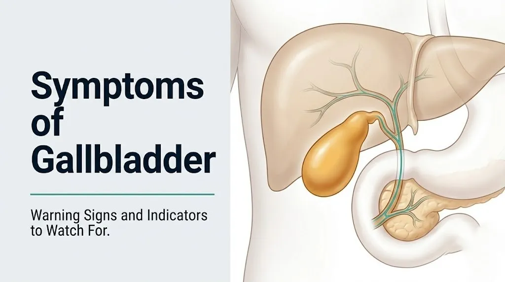
The gallbladder is a pear-shaped muscular sac approximately 7 to 10 cm long, located in the gallbladder fossa on the inferior surface of the right lobe of the liver, whose function is to store and concentrate bile between meals and then forcefully contract to deliver concentrated bile into the duodenum when fat is ingested.
Think of the gallbladder like a pressurized storage tank with a single drainage valve. During fasting, bile produced by hepatocytes flows down the left and right hepatic ducts, through the common hepatic duct, and backs up through the cystic duct into the gallbladder for storage. The gallbladder concentrates this bile 5 to 10 fold by absorbing water. When a fatty meal arrives in the duodenum, duodenal I-cells detect the fat and release cholecystokinin (CCK) into the bloodstream. CCK binds to CCK-1 receptors on gallbladder smooth muscle, triggering a coordinated contraction that propels concentrated bile through the cystic duct, through the common bile duct (choledochus), past the sphincter of Oddi at the ampulla of Vater, and into the duodenum where it emulsifies dietary fat.
When a gallstone sits in the cystic duct and that drainage valve is blocked, the force of gallbladder contraction against the obstruction creates pressure inside the gallbladder. That pressure is transmitted to the gallbladder wall. The wall’s visceral afferent nerve fibers, traveling with the T5-T9 greater splanchnic nerves, carry the pain signal to the spinal cord, where it’s perceived as pain in the right upper quadrant and epigastrium.
The specific bile composition that predisposes to stone formation involves an imbalance between cholesterol, bile salts, and phospholipids in the stored bile. Normally, bile salts and phospholipids keep cholesterol in solution. When cholesterol concentration exceeds the solubilizing capacity, crystals precipitate and aggregate into stones. This supersaturation can be caused by excess hepatic cholesterol secretion, reduced bile salt synthesis, or bile stasis (incomplete gallbladder emptying).
Older adults over 70 have lower bile salt synthesis rates than younger adults, which partially explains why gallstone prevalence increases with age in both sexes, reaching approximately 30 to 40% in adults over age 70, according to NIDDK surveillance data.
Gallbladder Pain: Location, Character, and What It Feels Like
Gallbladder pain is located in the right upper quadrant of the abdomen, directly beneath the right rib cage, and may radiate to the epigastrium (the central upper abdomen), the right flank, and the right shoulder, producing a characteristic pattern that helps clinicians distinguish it from other causes of abdominal pain.
The pain of gallbladder disease has specific characteristics that differentiate it from, say, peptic ulcer pain or cardiac pain. Patients typically describe it as:
- A deep, cramping, or aching pressure rather than a sharp stabbing sensation initially
- Building in intensity over 15 to 60 minutes to a plateau, then either resolving or escalating further
- Located in the right upper quadrant or epigastrium, not in the lower abdomen or pelvis
- Radiating around the right side to the right flank or to the right shoulder blade area
- Accompanied by nausea and sometimes vomiting, though vomiting does not relieve the pain
- Unrelieved by antacids or change of body position (distinguishing it from musculoskeletal or reflux pain)
- Often beginning 30 minutes to 2 hours after a fatty or large meal
The radiation to the right shoulder blade and shoulder tip is a specific clinical feature that deserves its own mechanistic explanation (covered in the referred pain section below). What’s worth noting here is that the location of gallbladder pain is not always exactly where people expect it. Some patients feel the worst of it in the epigastrium (central upper abdomen) rather than the right upper quadrant, which is why gallbladder disease is one of the most frequently misattributed causes of “stomach pain” or “indigestion” in clinical practice.
According to the American College of Gastroenterology, right upper quadrant or epigastric pain lasting more than 15 minutes and less than 6 hours that resolves spontaneously is the defining clinical feature of biliary colic, the most common symptomatic gallbladder presentation. Pain that exceeds 6 hours indicates progression to acute cholecystitis until proven otherwise.
Biliary Colic Symptoms: The Intermittent Gallbladder Attack
Biliary colic is the most common symptomatic presentation of gallstone disease, occurring when a gallstone temporarily lodges in the cystic duct during gallbladder contraction and produces a self-limited episode of right upper quadrant pain lasting typically 30 minutes to 6 hours before the stone dislodges and pain resolves.
The name “colic” is actually a misnomer that has persisted in clinical usage. True colic implies fluctuating wavelike pain (as in renal colic or intestinal colic), but biliary colic is more accurately a sustained crescendo pain that builds to a plateau and holds there rather than coming in waves. This distinction matters clinically because patients expecting waves of pain sometimes dismiss a sustained aching pressure as “not bad enough to be my gallbladder.”
The triggering mechanism is consistent: CCK released after fat ingestion contracts the gallbladder against an obstructed cystic duct. The pain begins 30 minutes to 2 hours after the fatty or large meal. It builds over 15 to 60 minutes. It plateaus. It then resolves over 30 to 90 minutes as the stone shifts back out of the duct opening and gallbladder pressure drops.
Key features that identify a biliary colic episode:
- Pain duration: 30 minutes to 6 hours; spontaneous resolution
- Triggers: fatty meals, large meals, occasionally lying flat or bending forward
- Nausea present in the majority of patients; vomiting occurs but does not relieve pain
- No fever (fever indicates progression to cholecystitis)
- Pain does not respond to antacids or movement changes
- May be accompanied by diaphoresis (sweating) during the pain peak
Biliary colic recurs. According to data cited in the Annals of Surgery, approximately 70% of patients who have one biliary colic episode will have another within 2 years if the gallstones are not removed. Each recurrence carries a small cumulative risk of progressing to acute cholecystitis, so repeated episodes are not something to indefinitely monitor at home.
Pregnant women in the second or third trimester who experience biliary colic episodes should be evaluated by a board-certified obstetrician-gynecologist in collaboration with a gastroenterologist or general surgeon, because the management approach and any procedural intervention timing requires specialist coordination during pregnancy.
Key Takeaway: Biliary colic produces a self-limited pain episode of 30 minutes to 6 hours, triggered by fatty meals and resolving spontaneously; any episode lasting beyond 6 hours, or any episode with fever, indicates progression to acute cholecystitis requiring same-day medical evaluation.
Acute Cholecystitis Symptoms: When the Gallbladder Becomes Inflamed
Acute cholecystitis occurs when a gallstone becomes persistently impacted in the cystic duct, causing gallbladder wall ischemia, transmural inflammation, and bacterial superinfection, producing a clinical picture of persistent right upper quadrant pain lasting more than 6 hours, fever, and a positive Murphy’s sign on physical examination.
Acute cholecystitis is not just a longer or more severe biliary colic episode. It is a mechanistically different condition. In biliary colic, the stone temporarily obstructs and dislodges; the gallbladder wall remains intact; inflammation is minimal; fever is absent. In acute cholecystitis, the stone stays impacted long enough that the gallbladder wall becomes progressively ischemic (because the distended gallbladder compresses its own blood supply), the ischemic tissue becomes colonized by bacteria migrating from the intestine via the biliary system, and a full transmural inflammatory process develops.
This transmural inflammation is why acute cholecystitis hurts differently than biliary colic. When the gallbladder wall inflammation extends to the adjacent parietal peritoneum, the somatic pain fibers of the lower intercostal nerves are now involved. The pain becomes constant rather than plateauing and potentially resolving; it worsens with movement, deep breathing, and palpation; voluntary guarding and rigidity of the right upper quadrant muscles develop.
Murphy’s sign is a clinically important physical examination finding: the examiner presses beneath the right costal margin while the patient takes a deep breath. The descending diaphragm pushes the inflamed gallbladder against the examiner’s fingers, producing a sharp pain that causes the patient to involuntarily stop inhaling. A positive Murphy’s sign has approximately 65% sensitivity and 87% specificity for acute cholecystitis, according to data in the Gastroenterology journal.
Observable symptoms of acute cholecystitis include:
- Persistent right upper quadrant pain lasting more than 6 hours without resolution
- Fever typically above 38°C (100.4°F), often higher
- Nausea and vomiting, typically more pronounced than in biliary colic
- Right upper quadrant tenderness and voluntary guarding on gentle palpation
- Loss of appetite and inability to tolerate food
- Positive Murphy’s sign
- Elevated white blood cell count (neutrophilia) and elevated C-reactive protein on blood tests
Older adults over 65 with acute cholecystitis may have lower fever responses than younger patients, and their white blood cell count elevation may be less pronounced, making clinical recognition harder. The Mayo Clinic notes that elderly patients with acute cholecystitis present atypically often enough that a high index of clinical suspicion is warranted even without classic fever or leukocytosis.
Referred Shoulder Pain as a Gallbladder Symptom
Right shoulder pain and right shoulder blade pain are genuine gallbladder symptoms produced by a specific anatomical convergence between the biliary visceral afferent pathways and the right phrenic nerve (arising from cervical nerve roots C3, C4, and C5), which shares central nervous system processing pathways with the skin and muscles of the right shoulder region.
This is one of the most confusing and underexplained features of gallbladder disease. People feel pain in their right shoulder or right shoulder blade, assume it is a muscle or rotator cuff issue, take ibuprofen, apply heat, and never consider that the origin is in the upper right abdomen. The mechanism deserves a clear explanation.
When the gallbladder becomes distended or inflamed, the inflammatory process can irritate the adjacent underside of the right diaphragm. The right diaphragm is innervated by the right phrenic nerve, which carries sensory signals from C3, C4, and C5 nerve roots. Those same spinal cord levels (C3-C5) also receive sensory input from the right shoulder and right upper arm skin and muscles. When diaphragmatic irritation sends signals into the C3-C5 dorsal horn, the brain cannot reliably distinguish whether the signal originated from the diaphragm or the shoulder. The pain is perceived as coming from the shoulder and shoulder blade area, even though the actual site of pathology is the gallbladder and subdiaphragmatic space.
This is called referred pain, specifically phrenic nerve-mediated referred pain. It occurs in other conditions affecting the right hemidiaphragm and subphrenic space as well, including right-sided pleural effusion, hepatic abscess, and right lower lobe pneumonia.
Clinically important notes about shoulder pain as a gallbladder symptom:
- Right shoulder or shoulder blade pain occurring alongside right upper quadrant or epigastric abdominal discomfort after meals is the pattern that points toward a biliary source
- Shoulder pain without any abdominal symptoms is less likely to be gallbladder related, though isolated referred pain without prominent abdominal pain is documented in the literature
- Right shoulder pain with fever and jaundice is a combination that requires emergency evaluation, because this triad can indicate ascending cholangitis
Women are more likely than men to present with shoulder pain as a prominent gallbladder symptom relative to abdominal pain, according to clinical observation data published in the Annals of Surgery. This sex-specific presentation pattern contributes to delayed diagnosis in women, who may be evaluated for rotator cuff pathology or cervical spine disease before gallbladder disease is considered.
Nausea, Vomiting, and Digestive Symptoms of Gallbladder Disease
Nausea is present in the majority of patients with symptomatic gallbladder disease, occurring because biliary tract distension and inflammation activate the same visceral afferent pathways that trigger the brainstem’s vomiting center (the area postrema in the medulla oblongata) through the vagus nerve (cranial nerve X), producing nausea and vomiting that do not relieve the underlying pain.
This last point is clinically useful. In gastroenteritis or food poisoning, vomiting typically produces some temporary relief of nausea. In biliary colic and acute cholecystitis, vomiting provides no pain relief because the pain source is the obstructed bile duct and the distended gallbladder, not gastric irritation. A patient who vomits repeatedly during an abdominal pain episode and notes that vomiting doesn’t help at all should flag this for their physician, because it points away from gastric causes and toward biliary or pancreatic pathology.
Additional digestive symptoms associated with gallbladder disease include:
- Bloating and abdominal fullness, particularly after meals, from delayed gastric emptying related to biliary dysfunction
- Intolerance to fatty, greasy, or fried foods, producing upper abdominal discomfort predictably after such meals
- Heartburn and belching that are sometimes attributed to gastroesophageal reflux disease (GERD) but originate from the biliary system
- Diarrhea in some patients with bile acid malabsorption resulting from altered biliary function
- Pale or clay-colored stools if bile duct obstruction is present (absent bile pigment in stool)
- Dark amber or tea-colored urine if conjugated bilirubin is being excreted through the kidneys (bilirubinuria)
According to the Cleveland Clinic, nausea without prominent pain is one of the presentations that leads to delayed gallbladder diagnosis, particularly in women who may present with nausea as the dominant symptom and are initially evaluated for peptic ulcer disease, gastroparesis, or cyclic vomiting syndrome. If nausea is consistently worse after fatty meals and is accompanied by right upper quadrant discomfort, a gallbladder ultrasound is the appropriate first diagnostic step even if pain is not the most prominent complaint.
Key Takeaway: Vomiting that fails to relieve nausea during an episode of upper abdominal pain is a key clinical clue pointing toward a biliary or pancreatic source rather than a gastric cause, because gastric vomiting typically provides at least transient relief.
Gallbladder Symptoms After Eating: The Fat-Pain Connection
Gallbladder symptoms occurring specifically after eating, particularly after fatty, greasy, or large meals, are the hallmark pattern of gallstone disease because dietary fat triggers cholecystokinin (CCK) release from duodenal I-cells, which causes the gallbladder to contract forcefully; when a stone is obstructing the cystic duct, this contraction produces pain.
The timing is specific and diagnostically useful. Biliary colic pain typically begins 30 minutes to 2 hours after a fatty or large meal, corresponding to the time required for dietary fat to reach the duodenum, trigger CCK release, produce gallbladder contraction, and generate enough pressure to produce symptoms. Pain that begins immediately with the first bite, during eating rather than after eating, is less likely to be biliary and more likely to be esophageal or gastric.
High-fat meal triggers that consistently provoke gallbladder symptoms include fried foods, fatty meats, cheese-heavy dishes, cream sauces, and rich desserts. However, a large meal of any composition can trigger symptoms because gastric distension alone can stimulate some degree of CCK-mediated gallbladder contraction, even without high fat content.
Nighttime gallbladder attacks deserve specific mention. Some patients experience biliary colic predominantly at night, particularly during the 12 to 3 a.m. window. This reflects the physiology of delayed gastric emptying and bile concentration: a late heavy dinner triggers gallbladder contraction hours later, when the patient is supine, and the horizontal position may also slightly increase intra-gallbladder pressure by altering bile distribution.
Quick Tip:
- Keep a symptom diary for 2 to 4 weeks before your appointment, noting the time of each pain episode, the time of the preceding meal, what you ate, and how long the pain lasted; this information significantly improves the diagnostic efficiency of your first appointment with a primary care physician or gastroenterologist
- Fatty meal avoidance can reduce the frequency of biliary colic episodes but does not eliminate gallstones or prevent eventual progression to acute cholecystitis
- Patients with diabetes mellitus should be aware that their typical meal-to-symptom pattern may be blunted or absent due to autonomic neuropathy, meaning their first symptomatic presentation may be more severe than the usual biliary colic prodrome
Gallbladder Symptoms in Women: Hormonal Mechanisms and Differences
Gallbladder symptoms in women are more likely to include nausea, back pain, and epigastric discomfort as prominent features rather than the textbook right upper quadrant pain, and women are approximately twice as likely as men to develop gallstones because of estrogen-driven increases in hepatic cholesterol secretion and progesterone-mediated reductions in gallbladder motility.
The hormonal mechanisms behind this sex difference operate on two distinct pathways. Estrogen binds to estrogen receptor alpha (ERalpha) in hepatocytes and upregulates the expression of HMG-CoA reductase (the rate-limiting enzyme in cholesterol synthesis) while simultaneously increasing expression of the LDL receptor pathway, delivering more cholesterol substrate to the liver for secretion into bile. The result is bile with higher cholesterol saturation, which is more likely to precipitate cholesterol crystals and form gallstones. Progesterone acts on gallbladder smooth muscle, inhibiting CCK-mediated contraction and slowing the emptying rate. Bile sits in the gallbladder longer between meals, concentrating further and providing more time for crystal nucleation.
During pregnancy, both estrogen and progesterone surge dramatically. According to a study published in Gut, bile lithogenicity (stone-forming tendency) is measurably highest in the third trimester, and biliary sludge (a precursor to gallstones) develops in approximately 30% of pregnant women by the third trimester. New gallstone formation during pregnancy occurs in approximately 2 to 4% of pregnant women per trimester.
Gallbladder symptoms in women that differ from typical male presentations:
- Nausea as the primary or most prominent complaint, with abdominal pain secondary
- Pain described as pressure or fullness in the upper abdomen rather than sharp cramping
- Right-sided back pain or right shoulder blade pain without clear abdominal pain
- Symptoms attributed to irritable bowel syndrome, GERD, or dyspepsia for months before gallbladder disease is considered
- Higher rates of atypical presentation leading to longer diagnostic delays
Women using oral contraceptives containing estrogen face a higher risk of gallstone formation than women not using hormonal contraception, due to the same hepatic cholesterol pathway described above. Postmenopausal women using estrogen-containing hormone replacement therapy face a similar, though somewhat attenuated, elevated risk compared to untreated postmenopausal women.
Gallbladder Symptoms in Men: Why Male Presentations Are Underrecognized
Gallbladder symptoms in men are less frequently recognized as biliary in origin because men present at lower rates than women, are diagnosed later on average, and tend to present with more classic right upper quadrant pain with less of the nausea-dominant or back-pain-dominant atypical pattern seen in women, but their lower recognition often stems from lower clinical index of suspicion rather than genuinely milder disease.
The male-specific clinical picture matters for a specific reason. Men who present with right upper quadrant pain are sometimes evaluated for musculoskeletal causes, hepatitis, or peptic ulcer disease before gallbladder disease reaches the differential. The delayed diagnosis is not because men have fewer symptoms but because the overall lower prevalence (approximately 6 to 8% lifetime in American men versus 12 to 16% in American women, per NIDDK data) means clinicians statistically encounter the diagnosis less often in male patients and may not reach for it as quickly.
Men with gallbladder disease who are at above-average risk include:
- Men over 60 (gallstone prevalence rises substantially with age in both sexes)
- Men with obesity (BMI over 30) who have higher hepatic cholesterol output
- Men with cirrhosis or chronic hemolytic conditions (higher pigment stone formation)
- Men who have experienced rapid weight loss through very low calorie diets or bariatric surgery
- Men of Native American heritage, who have among the highest gallstone prevalence of any demographic group in the United States regardless of sex
According to NIDDK population data, the gallstone prevalence sex gap narrows considerably after age 60 as female hormonal advantage from reproductive estrogen declines with menopause, making older men an important group for whom gallbladder disease should be actively considered when upper abdominal symptoms are present.
Men with diabetes mellitus face the same blunted visceral pain risk as diabetic women, with the additional complication that diabetic men may attribute upper abdominal discomfort to gastroparesis (which is a genuine complication of diabetic autonomic neuropathy) and delay evaluation for a biliary source. If a diabetic man is experiencing episodic upper abdominal discomfort with or without nausea, a gallbladder ultrasound should be considered alongside evaluation for gastroparesis, not instead of it.
Key Takeaway: Women develop gallstones at approximately twice the rate of men due to estrogen-driven bile cholesterol supersaturation and progesterone-driven gallbladder dysmotility, and their symptoms are more likely to present atypically as nausea or back pain rather than classic right upper quadrant pain.
Atypical Gallbladder Symptoms: Older Adults, Diabetic Patients, and Silent Disease
Atypical gallbladder symptoms occur when the normal visceral pain signaling from the gallbladder is blunted or absent, as happens in patients with diabetic peripheral neuropathy or age-related autonomic dysfunction, producing presentations that range from mild vague abdominal discomfort to no symptoms at all until a serious complication like septic cholecystitis or gallbladder perforation develops.
This is perhaps the most clinically important gap in all published gallbladder symptom content, because the patients most likely to have silent or atypical disease are also the ones most likely to have serious complications when their disease finally declares itself.
Older adults over 65 have reduced visceral afferent sensitivity from age-related autonomic nervous system changes, reduced fever response from blunted inflammatory signaling, and reduced white blood cell reactivity, all of which conspire to produce atypical acute cholecystitis presentations. An elderly patient with septic acute cholecystitis may present primarily with confusion, reduced appetite, and mild discomfort rather than severe pain and high fever. According to the Annals of Surgery, elderly patients with acute cholecystitis have higher rates of gangrenous cholecystitis and gallbladder perforation at presentation precisely because pain-blunted earlier stages are not recognized.
Patients with type 2 diabetes have peripheral neuropathy affecting the small autonomic fibers that carry visceral pain from the gallbladder via the splanchnic nerves. These patients are also at elevated risk of acalculous cholecystitis (cholecystitis without gallstones), particularly during critical illness, sepsis, or prolonged fasting, because gallbladder wall ischemia can occur independent of stone obstruction in hemodynamically compromised states.
Patients on chronic opioid therapy experience opioid-induced sphincter of Oddi dysfunction: opioid receptor activation in the sphincter of Oddi smooth muscle produces sustained contraction, elevating biliary pressure even without mechanical stone obstruction and producing biliary colic-like pain that does not respond to conventional gallbladder management. Any clinician evaluating upper abdominal pain in a patient on chronic opioids should consider this in the differential before attributing pain to gallstones.
Chronic Cholecystitis Symptoms: The Long-Term Gallbladder Pattern
Chronic cholecystitis is a long-standing, low-grade inflammation of the gallbladder wall resulting from repeated episodes of biliary colic or subclinical gallbladder irritation, producing a symptom pattern of persistent but less severe right upper quadrant discomfort, postprandial bloating, fat intolerance, and intermittent nausea that is often confused with functional dyspepsia or irritable bowel syndrome.
Chronic cholecystitis differs from acute cholecystitis in severity and mechanism. Whereas acute cholecystitis is a sudden, intense, infection-driven inflammation, chronic cholecystitis represents the cumulative scarring and fibrosis of a gallbladder wall subjected to repeated low-level injury. The gallbladder in chronic cholecystitis is typically thickened, fibrotic, and poorly contractile, with impaired emptying even when the cystic duct is not acutely obstructed.
The symptom timeline matters diagnostically. Patients with chronic cholecystitis often describe months to years of:
- Recurring right upper quadrant or epigastric discomfort that never quite resolves
- Persistent intolerance to fatty foods producing bloating and upper abdominal fullness
- Vague nausea that worsens after meals without a single dramatic attack
- Frequent belching and bloating attributed to “gas” or “indigestion” for years
- Fatigue and reduced appetite persisting between episodes
This diffuse, chronic symptom picture is why chronic cholecystitis is systematically under-diagnosed relative to acute cholecystitis. A HIDA scan (hepatobiliary iminodiacetic acid scan) that measures gallbladder ejection fraction is the most useful diagnostic tool for chronic cholecystitis when an ultrasound shows gallstones or wall thickening but the clinical picture seems less dramatic than acute cholecystitis. A gallbladder ejection fraction below 35% at 30 minutes post-CCK stimulation is generally considered abnormal and consistent with gallbladder dysmotility or chronic dysfunction.
Patients with fibromyalgia or functional gastrointestinal disorders may have overlapping symptom pictures with chronic cholecystitis, which can make clinical differentiation challenging. A board-certified gastroenterologist should evaluate patients with chronic upper abdominal symptoms that have not responded to standard treatments for functional dyspepsia.
Choledocholithiasis Symptoms: When Stones Reach the Common Bile Duct
Choledocholithiasis is the presence of gallstones in the common bile duct (choledochus), producing a clinical picture of right upper quadrant pain combined with jaundice, dark urine, and pale stools that indicates the stone is blocking bile drainage from the liver into the duodenum, causing conjugated bilirubin to accumulate in the systemic circulation.
The distinction from gallbladder-confined gallstone disease matters enormously because the consequences of common bile duct obstruction extend well beyond gallbladder pain. When the common bile duct is obstructed, bile backs up through the intrahepatic ductal system into the liver. Conjugated (direct) bilirubin, which has been processed by hepatocytes and made water-soluble, regurgitates into the bloodstream, producing the yellow skin and scleral coloration of jaundice. Because bile pigment is absent from the intestine, stool becomes pale or clay-colored (acholic stools). Because conjugated bilirubin is water-soluble and filtered by the kidneys, urine becomes dark amber or tea-colored (bilirubinuria).
Distinguishing choledocholithiasis from gallbladder-confined disease:
| Feature | Biliary Colic (Cystic Duct) | Choledocholithiasis (Common Bile Duct) |
|---|---|---|
| Pain location | Right upper quadrant, epigastrium | Right upper quadrant, epigastrium |
| Jaundice | Absent | Present (yellowing of skin and sclera) |
| Urine color | Normal | Dark amber or tea-colored |
| Stool color | Normal | Pale or clay-colored |
| Liver enzymes | Typically normal | ALP, GGT, bilirubin elevated |
| Fever | Absent in uncomplicated cases | May be present if cholangitis develops |
| Urgency | Scheduled evaluation | Same-day evaluation or ER |
The most dangerous complication of choledocholithiasis is ascending cholangitis, which occurs when bacteria ascend from the duodenum into the obstructed bile duct system and cause systemic sepsis. Ascending cholangitis is characterized by Charcot’s triad: right upper quadrant pain, fever with rigors (shaking chills), and jaundice. This triad constitutes a medical emergency.
According to the American College of Gastroenterology guidelines, any patient presenting with Charcot’s triad should receive emergency evaluation and treatment, as ascending cholangitis untreated has a mortality rate that can exceed 50% in severe cases with septic shock.
Gallbladder Symptoms That Are Medical Emergencies
Certain symptoms associated with gallbladder disease require immediate emergency evaluation. Do not wait to see if these resolve on their own.
Call 911 or go to the nearest emergency room immediately if you experience:
- Fever with right upper quadrant pain lasting more than 6 hours (indicating acute cholecystitis with possible bacterial superinfection requiring IV antibiotics and urgent surgical evaluation)
- Fever, jaundice, and right upper quadrant pain occurring together (Charcot’s triad) (indicating ascending cholangitis, a bacterial infection of the bile duct system that can progress to septic shock within hours)
- Severe epigastric pain radiating to the back, accompanied by vomiting that does not relieve the pain (indicating gallstone pancreatitis, where a stone impacted at the ampulla of Vater is obstructing both the bile duct and the pancreatic duct, triggering pancreatic autodigestion)
- Sudden severe worsening of right upper quadrant pain followed by a brief false calm (indicating possible gallbladder perforation, a surgical emergency where the devitalized gallbladder wall ruptures; the brief improvement can reflect pain nerve decompression before peritonitis develops)
- Fever, confusion, low blood pressure, and rapid heart rate combined with abdominal pain (indicating septic shock from ascending cholangitis or gangrenous cholecystitis, a life-threatening systemic emergency)
- Jaundice developing over hours to days with any abdominal pain (indicating common bile duct obstruction requiring urgent evaluation to rule out ascending cholangitis and rule in the need for emergency ERCP or surgery)
These presentations can indicate gangrenous acute cholecystitis, ascending cholangitis, gallstone pancreatitis, or gallbladder perforation with bile peritonitis, all of which require emergency medical assessment and cannot be safely managed with an urgent care visit or a next-available appointment.
How Gallbladder Disease Is Diagnosed: Tests and Clinical Examination
Gallbladder disease is diagnosed through a combination of clinical history, physical examination findings (particularly Murphy’s sign), and imaging studies, with abdominal ultrasound as the first-line and most accurate non-invasive test for identifying gallstones, gallbladder wall thickening, and pericholecystic fluid.
Understanding the diagnostic process helps you prepare for your appointment and know what to expect after it.
How the diagnostic process typically unfolds from first visit to confirmed diagnosis:
- Your primary care physician takes a detailed history, asking about pain location, timing relative to meals, duration, associated symptoms (fever, jaundice, vomiting), and any previous similar episodes
- Physical examination includes palpation of the right upper quadrant for tenderness and assessment for Murphy’s sign (inspiratory arrest with deep palpation below the right costal margin)
- Blood tests are ordered: a complete blood count (CBC) to assess white blood cell count (elevated in acute cholecystitis), liver function tests (ALT, AST, ALP, GGT, total and direct bilirubin) to assess for cholestasis or hepatic involvement, and C-reactive protein (CRP) as an inflammation marker
- An abdominal ultrasound is ordered; this test has approximately 95% sensitivity for detecting gallstones 3 mm or larger and can identify gallbladder wall thickening (over 3 mm suggests cholecystitis), pericholecystic fluid, and a sonographic Murphy’s sign
- If ultrasound is inconclusive but gallbladder dysfunction is suspected, a HIDA scan is ordered to measure gallbladder ejection fraction under CCK stimulation
- If choledocholithiasis (common bile duct stones) is suspected, magnetic resonance cholangiopancreatography (MRCP) provides detailed imaging of the bile duct system without radiation or invasive procedures
- If common bile duct stones are confirmed, endoscopic retrograde cholangiopancreatography (ERCP) allows both diagnosis and therapeutic stone removal in a single procedure
A board-certified gastroenterologist evaluates patients with confirmed gallstone disease, functional gallbladder disorders (low ejection fraction on HIDA without stones), and complex presentations. A board-certified general surgeon evaluates patients for cholecystectomy (gallbladder removal), which is the definitive treatment for symptomatic gallstone disease.
Key Takeaway: Abdominal ultrasound is the correct first test to request when gallbladder disease is suspected; a normal ultrasound does not rule out gallbladder dysfunction if symptoms persist, and a HIDA scan measuring gallbladder ejection fraction is the next appropriate step.
When to See a Doctor for Symptoms of Gallbladder Problems
You should contact a primary care physician within 1 to 2 weeks if you have experienced even one episode of right upper quadrant pain lasting more than 30 minutes, particularly if it was triggered by a fatty meal, accompanied by nausea, and resolved on its own, because this pattern is consistent with biliary colic and warrants imaging even if the episode has passed.
The clinical threshold for seeking care varies by the severity and associated features of your symptoms. Here’s how to triage your own situation:
| Your Symptom Pattern | Urgency | Action |
|---|---|---|
| Mild RUQ discomfort after meals, no fever, resolves within 30 minutes | Low urgency | Call primary care physician for appointment within 1 to 2 weeks |
| Pain lasting 30 min to 6 hours, nausea, no fever, resolves | Moderate urgency | Call primary care physician within 1 to 3 days; bring symptom diary |
| Pain lasting more than 6 hours, without fever or jaundice | High urgency | Call primary care physician same day or go to urgent care |
| Pain with low-grade fever, nausea, vomiting, 4 to 6 hours | High urgency | Go to urgent care or emergency department the same day |
| Pain with fever above 38.5°C (101.3°F), or with jaundice | Emergency | Go to emergency department immediately |
| Charcot’s triad: pain, fever with chills, jaundice | Emergency | Call 911 or go to ER immediately |
| Severe epigastric pain radiating to the back | Emergency | Call 911 or go to ER immediately |
When you see your primary care physician for gallbladder symptom evaluation, bring the following information to make your visit as productive as possible:
- A list of all episodes: date, time, what you ate, when pain started relative to eating, pain severity, duration, and how it resolved
- Any changes in urine color (dark or tea-colored)
- Any changes in stool color (pale or clay-colored)
- Any yellowing of skin or eyes you’ve noticed
- Your complete medication list, including oral contraceptives, fibrates, somatostatin analogues, or opioid medications
- Any family history of gallbladder disease, as there is a heritable component to gallstone risk
A board-certified gastroenterologist should be involved in evaluation if your primary care physician’s initial workup is inconclusive, if your symptoms recur despite low-fat dietary modification, or if a HIDA scan reveals a gallbladder ejection fraction below 35%. The decision about cholecystectomy involves a board-certified general surgeon and should be based on symptom burden, imaging findings, ejection fraction results, and your overall surgical risk profile.
Frequently Asked Questions About Symptoms of Gallbladder
What does a gallbladder attack feel like?
A gallbladder attack (biliary colic) feels like a deep, sustained cramping or aching pressure in the right upper abdomen or central upper abdomen, building over 15 to 60 minutes to an intense plateau, then either resolving or intensifying.
The pain often radiates to the right shoulder blade or right shoulder, is accompanied by nausea and sometimes vomiting, and begins 30 minutes to 2 hours after a fatty or large meal.
A gallbladder attack that lasts more than 6 hours without resolving, or that comes with fever, indicates progression to acute cholecystitis and warrants same-day evaluation at an emergency department or urgent care.
Can gallbladder symptoms come and go for years?
Gallbladder symptoms can come and go for months to years in people with symptomatic gallstones whose stones intermittently obstruct and dislodge from the cystic duct during gallbladder contraction.
This intermittent pattern is called biliary colic, and while individual episodes are self-limiting, approximately 70% of patients who have one episode will have another within 2 years according to data published in the Annals of Surgery.
Each recurrence carries a cumulative risk of progression to acute cholecystitis, making ongoing monitoring or surgical evaluation appropriate rather than indefinitely waiting out the pattern.
What are the symptoms of gallbladder problems in women specifically?
Women with gallbladder disease are more likely to present with nausea as the dominant symptom, right-sided back pain, and vague epigastric discomfort rather than the classic right upper quadrant pain that is typically described in clinical teaching.
This atypical presentation in women is partly because women tend to describe visceral pain differently from men and partly because estrogen and progesterone-related changes in gallbladder function produce a symptom picture that overlaps with GERD, IBS, and musculoskeletal back pain.
A woman who has persistent nausea after fatty meals, recurring right-sided back pain, and a history of pregnancy or oral contraceptive use should ask her primary care physician specifically about gallbladder ultrasound evaluation.
Can you have gallbladder problems without gallstones?
Yes, gallbladder disease can occur without gallstones in conditions including biliary dyskinesia (impaired gallbladder motility with a reduced ejection fraction on HIDA scan), acalculous cholecystitis (acute gallbladder inflammation without stones, most common in critically ill or hospitalized patients), and sphincter of Oddi dysfunction.
Symptoms in these conditions may be identical to stone-related biliary colic but a gallbladder ultrasound will appear normal, which is why a HIDA scan to measure gallbladder ejection fraction is ordered when ultrasound is negative but symptoms persist.
A board-certified gastroenterologist evaluates these presentations and can guide the decision about whether cholecystectomy is appropriate when there are no stones but there is documented gallbladder dysfunction.
Where exactly is gallbladder pain located on the body?
Gallbladder pain is located in the right upper quadrant (RUQ) of the abdomen, directly beneath the right rib cage, though in many patients it is felt equally or more prominently in the epigastrium (the central upper abdomen, just below the sternum).
The pain commonly radiates around the right side to the right flank or upward to the right shoulder blade and right shoulder tip through the referred pain pathway of the right phrenic nerve (C3-C5 nerve roots).
Pain that is in the right lower abdomen, the left side, or the pelvis is unlikely to be gallbladder in origin and warrants evaluation for other causes including appendicitis, ovarian pathology, or colonic disease.
What are the warning signs that your gallbladder needs to be removed?
The clearest warning signs that gallbladder removal (cholecystectomy) is likely warranted include recurrent biliary colic episodes that disrupt daily function, a first episode of acute cholecystitis, a HIDA scan ejection fraction below 35% with symptoms consistent with chronic cholecystitis, or any episode of choledocholithiasis or gallstone pancreatitis.
According to Society of American Gastrointestinal and Endoscopic Surgeons guidelines, symptomatic gallstone disease is an indication for laparoscopic cholecystectomy, and the decision involves weighing the frequency and severity of symptoms against the patient’s surgical risk profile.
This decision should be made in consultation with a board-certified general surgeon who reviews your specific imaging, ejection fraction, symptom history, and any previous complications.
Closing
The symptoms of gallbladder disease follow a pattern that, once you know it, is recognizable: right upper quadrant or epigastric pain triggered by fatty meals, accompanied by nausea, radiating to the right shoulder, and either resolving within 6 hours or escalating to fever and persistent pain. That distinction between self-limiting biliary colic and acute cholecystitis is the most practically useful clinical fact in this entire article.
If you’ve had even one episode matching the biliary colic description, the right step is a phone call to your primary care physician requesting an abdominal ultrasound. Don’t wait for a second attack to confirm the pattern. Bring a symptom diary to that appointment: what you ate, when the pain started, how long it lasted, whether it has happened before.
If your pain is accompanied by fever, jaundice, or dark urine right now, don’t schedule an appointment. Go to the emergency department today.