What Causes Itchy Legs Below the Knee: 2026 Guide
The most common causes of itchy legs below the knee are dry skin, chronic venous insufficiency, atopic dermatitis, contact dermatitis, diabetic peripheral neuropathy, and stasis dermatitis, though in some cases the itch signals a systemic condition affecting the liver, kidneys, or peripheral nervous system that needs prompt clinical investigation. Understanding which cause is driving your itch matters because the physiological mechanism is different for each one, the pattern of the itch looks and feels distinctly different depending on the cause, and several causes require medical treatment rather than home management.
The American Academy of Dermatology (AAD) reports that chronic pruritus, defined as itch lasting more than six weeks, affects approximately 13% of the global population at any given time, with the lower extremities being among the most frequently reported locations. The below-the-knee distribution is not random. This region has specific anatomical vulnerabilities in its venous drainage, skin barrier, and nerve supply that make it disproportionately susceptible to multiple distinct itch-producing conditions.
This article covers the full clinical picture: the neurophysiology of how itch forms, the 12 most clinically relevant causes ranked from most to least common, the situational triggers that produce episodic itching, the population-specific patterns in older adults and pregnant women, the medication-related causes most readers don’t consider, and the specific symptoms that mean this is more than a skin issue.
What Causes Itchy Legs Below the Knee
The most frequent causes of itchy legs below the knee, ranked from most to least common in the general adult population, are dry skin, chronic venous insufficiency with stasis dermatitis, atopic dermatitis, contact dermatitis, and diabetic peripheral neuropathy.
Less common but clinically important causes include psoriasis, lichen simplex chronicus, folliculitis, medication-induced pruritus, exercise-induced urticaria, cholestatic pruritus, and uremic pruritus from chronic kidney disease. Rarely, an infestation (scabies) or fungal infection can produce persistent localized itch below the knee without an immediately obvious rash.
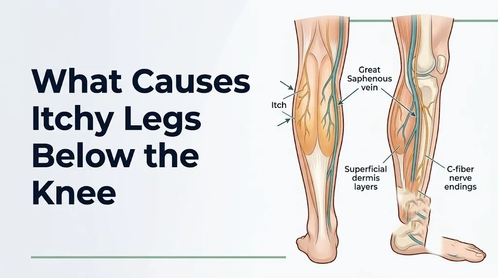
The below-the-knee distribution is not incidental. The skin of the lower leg is thinner than the thigh, has fewer sebaceous glands per square centimeter (meaning less natural oil production), is subject to greater hydrostatic venous pressure due to gravity, and receives sensory nerve supply from the sural nerve, saphenous nerve, and superficial peroneal nerve. All three of these nerve supply features make this region uniquely prone to both barrier-related and neuropathic itch.
| Cause Category | Mechanism | Most Affected Group | Self-Resolving? |
|---|---|---|---|
| Dry skin (xerosis) | Impaired stratum corneum barrier, increased TEWL | Older adults, winter months | Yes, with emollients |
| Venous insufficiency/stasis dermatitis | Venous hypertension, capillary leakage, mast cell activation | Adults over 50, people with obesity | No; requires compression and medical care |
| Atopic dermatitis | Filaggrin mutation, Th2 immune activation, IL-31 | Children and young adults; can persist | No; requires dermatological management |
| Contact dermatitis | Allergen or irritant activating epidermal immune response | Anyone exposed to trigger | Yes, if trigger removed |
| Diabetic peripheral neuropathy | Small C-fiber axonal degeneration, ectopic nerve firing | Adults with type 2 diabetes, especially over 50 | No; requires diabetes management |
| Peripheral neuropathy (non-diabetic) | Nerve fiber damage from various systemic causes | Varies by cause | Depends on underlying cause |
| Cholestatic/uremic pruritus | Systemic pruritogens (bile salts, uremic toxins) acting on peripheral C-fibers | People with liver or kidney disease | No; requires systemic treatment |
How the Itch Signal Travels From the Lower Leg to the Brain
The itch sensation in the lower leg is generated when pruritogens (itch-producing chemical mediators) bind to specialized C-fiber pruritoceptors in the dermis and epidermis of the skin.
These C-fibers are unmyelinated, slow-conducting sensory nerve fibers distinct from the C-fibers that carry pain signals. Think of them as a dedicated biological alarm wire running from the skin’s surface directly to the spinal cord. When this wire detects a pruritogen, it fires. The signal travels from the skin of the lower leg through the dorsal root ganglia at the lumbar and sacral spinal levels (primarily L4 through S2), enters the dorsal horn of the spinal cord, and crosses to the contralateral side, ascending through the spinothalamic tract to the thalamus. From there, the signal reaches the somatosensory cortex and anterior cingulate cortex, where it becomes the conscious perception of itch and the almost irresistible urge to scratch.
The key pruritogens driving this process include histamine (released by dermal mast cells in allergic and inflammatory conditions), interleukin-31 (IL-31) (a cytokine released by activated T-helper 2 lymphocytes that is strongly implicated in atopic dermatitis), thymic stromal lymphopoietin (TSLP) (released by stressed keratinocytes in the epidermis), and substance P (released from primary afferent neurons in neuropathic conditions).
According to research published in JAMA Dermatology, the discovery of histamine-independent itch pathways, particularly the IL-31 and TSLP pathways, has fundamentally changed the understanding of chronic itch. Many people with persistent leg itch do not respond to antihistamines precisely because their itch is driven by IL-31 or TSLP rather than histamine, and those pathways require different clinical approaches.
In neuropathic itch, the mechanism is different entirely. Damaged nerve fibers fire spontaneously without any external pruritogen, generating an itch signal from within the nervous system itself. This is why neuropathic itch often has no visible skin change, no rash, and no obvious trigger, and why antihistamines have essentially no effect on it.
Older adults experience more intense itch responses for a given pruritogen load because age-related thinning of the stratum corneum and reduced skin lipid content lower the threshold at which C-fibers are activated by external triggers.
Dry Skin as a Cause of Itchy Legs Below the Knee
Dry skin, clinically termed xerosis cutis, is the single most common cause of itchy legs below the knee in the general adult population, and it produces itch specifically because a compromised skin barrier allows pruritogens to access C-fiber nerve endings that are normally protected by intact epidermal layers.
The lower leg below the knee has significantly fewer sebaceous glands than the thigh or upper body. Sebaceous glands secrete sebum, a lipid-rich fluid that coats the skin surface and forms a critical component of the stratum corneum barrier. With fewer glands and less sebum output, the lower leg’s skin barrier is structurally thinner and more susceptible to transepidermal water loss (TEWL), the evaporative loss of water from the skin’s surface that occurs when the barrier is compromised.
The key molecular event is disruption of the filaggrin protein network within the stratum corneum. Filaggrin breaks down into natural moisturizing factors (amino acids and their derivatives) that hold water within the skin. When cold air, low humidity, excessive washing, or aging reduces filaggrin production or function, TEWL increases, the barrier opens, and external pruritogens gain access to the nerve endings beneath.
According to the AAD, xerosis affects an estimated 29% to 85% of adults over age 60, with the lower extremities being the most commonly affected body site. Postmenopausal women are at particularly elevated risk because the decline in estrogen after menopause reduces skin lipid synthesis and sebaceous gland activity, accelerating barrier thinning.
Quick Tip:
- Dry skin itch from xerosis typically worsens in winter due to low ambient humidity and indoor heating, both of which accelerate TEWL.
- The lower shin (anterior lower leg) tends to be the driest area because it receives the least mechanical contact and sebum distribution from normal movement.
- People with hypothyroidism experience xerosis more severely due to reduced sweating and sebum production driven by low thyroid hormone levels; a primary care physician should be contacted if leg itching co-occurs with fatigue, weight gain, and cold intolerance.
Key Takeaway: Dry skin causes itchy legs below the knee by disrupting the filaggrin-based stratum corneum barrier, allowing pruritogens to reach C-fiber nerve endings that are normally shielded by intact skin, and this is worst in older adults, postmenopausal women, and anyone in low-humidity environments.
Atopic Dermatitis and Contact Dermatitis Causing Itchy Lower Legs
Atopic dermatitis (eczema) and contact dermatitis both cause intense itching in the lower leg but through distinct immune mechanisms, and telling them apart is the first step toward appropriate management.
Atopic dermatitis is driven by a filaggrin gene mutation that impairs the skin barrier, combined with an overactive T-helper 2 (Th2) immune response. In people with this condition, the compromised barrier allows environmental allergens to penetrate the epidermis and trigger Th2 lymphocyte activation, which releases interleukin-4, interleukin-13, and IL-31 in large quantities. IL-31 binds directly to receptors on C-fiber pruritoceptors in the dermis, generating an intense, often relentless itch without requiring histamine as an intermediary. This is why antihistamines typically provide little relief in atopic dermatitis.
Contact dermatitis operates differently. Allergic contact dermatitis occurs when a specific allergen (nickel in metal clasps, fragrances in skin lotions, rubber chemicals in sock elastic, chromate in leather shoes) triggers a type IV delayed hypersensitivity reaction mediated by sensitized CD4+ T lymphocytes. The reaction takes 24 to 96 hours to develop after allergen exposure, producing a red, itchy, sometimes blistering rash in the exact distribution of contact. Irritant contact dermatitis produces a similar-looking rash through direct epidermal damage rather than immune activation, and it occurs immediately without prior sensitization.
In the lower leg specifically, the most frequent contact allergen culprits are:
- Rubber components in sock elastic bands (producing a band-like itch and rash at the mid-calf where elastic contacts skin)
- Nickel in ankle bracelet hardware or shoe buckles
- Fragrance and preservative chemicals in moisturizing creams applied to the lower leg
- Topical antibiotic components (neomycin, bacitracin) applied to wounds or ulcers
The AAD states that contact dermatitis of the lower extremity is frequently misidentified as atopic dermatitis or stasis dermatitis without patch testing, leading to ineffective treatment. A board-certified dermatologist should perform standardized patch testing when contact allergen exposure is suspected.
People with pre-existing atopic dermatitis have a higher baseline rate of contact allergen sensitization due to their impaired skin barrier, meaning they can develop both conditions simultaneously in the same area of skin.
Psoriasis and Lichen Simplex Chronicus on the Lower Leg
Psoriasis and lichen simplex chronicus (LSC) are two distinct dermatological conditions that produce chronic itching in the lower leg, often in the same patient population, and both are frequently underdiagnosed when they occur below the knee.
Psoriasis in the lower leg produces well-defined, raised, silvery-scaled plaques on a red base, most commonly on the anterior shin (the pre-tibial surface). The itch in plaque psoriasis results from a T-cell-mediated inflammatory cascade driven by tumor necrosis factor-alpha (TNF-alpha), interleukin-17, and interleukin-23, which accelerate keratinocyte proliferation and create the thickened scaly plaques. The itch is typically persistent, often described as burning, and worsens with dryness. Pre-tibial psoriasis is a specific subtype that can be particularly resistant to topical treatment due to the thickness of the scales on the lower leg skin.
Lichen simplex chronicus develops through a different mechanism, one that is self-perpetuating. LSC begins with an initial itch (from any cause) that triggers scratching. Repeated mechanical scratching causes the skin to thicken through a process called lichenification, where the epidermis responds to chronic trauma by increasing the production of keratinocytes and structural proteins. The thickened skin then generates its own itch signal through sensitized C-fibers embedded in the lichenified tissue, creating a scratch-itch cycle that becomes independent of the original trigger.
According to the British Journal of Dermatology, LSC most commonly affects the lower leg and nape of the neck, with the anterior shin being the most frequently reported location in the lower extremity. Women are diagnosed with LSC approximately twice as frequently as men, though this may partly reflect healthcare-seeking behavior differences rather than true biological sex differences in incidence.
To tell psoriasis from LSC at a glance:
- Psoriasis: Well-defined silver-scaled plaques with clear edges; may have nail involvement or joint symptoms
- LSC: Single or few leather-textured, hyperpigmented, thickened patches with excoriation marks; edges less defined
- Shared feature: Both produce intense itch and are worsened by scratching
- Key distinction: Psoriasis is symmetrical and follows a predictable anatomical distribution; LSC often follows where the dominant hand reaches most easily
Stasis Dermatitis and Venous Insufficiency Causing Itchy Legs Below the Knee
Stasis dermatitis is one of the most clinically important causes of itchy legs below the knee in adults over 50, and it is produced by a specific vascular mechanism involving venous valve failure, venous hypertension, and chronic inflammatory changes in the lower leg skin.
The great saphenous vein and small saphenous vein drain blood from the lower leg back toward the heart. These veins contain one-way valves that prevent blood from flowing backward under the influence of gravity. When these valves fail (a condition called chronic venous insufficiency or CVI), blood pools in the superficial venous system of the lower leg, raising the hydrostatic pressure in the small capillary beds of the dermis and subcutaneous tissue.
Think of it like a backed-up drain. When the outflow is obstructed and pressure builds, fluid and cellular components that normally stay inside the capillaries begin leaking into the surrounding tissue. Red blood cells that escape deposit hemosiderin (an iron-containing pigment from red blood cell breakdown) into the dermis, producing the characteristic brownish discoloration seen in stasis dermatitis. The leaked fluid triggers activation of local mast cells, which release histamine and other pruritogens directly into the affected dermal tissue.
The result is itch that is localized specifically to the medial lower leg (inner ankle and lower inner calf), the area drained by the great saphenous vein’s tributaries and perforating veins, combined with skin that appears brown, thickened, and sometimes weeping or crusty.
According to the American Venous Forum, chronic venous insufficiency affects approximately 25% of women and 15% of men in the United States, making stasis dermatitis one of the most prevalent causes of lower leg skin disease in the adult population. People with obesity, a history of DVT, prolonged standing occupations, or multiple pregnancies are at substantially elevated risk due to increased venous pressure in the lower extremity.
People with stasis dermatitis should not be managed with emollients alone. Compression therapy addressing the underlying venous hypertension is the mechanistically necessary intervention, and this requires evaluation by a primary care physician or board-certified vascular medicine specialist to determine the appropriate compression class and rule out coexisting peripheral arterial disease before compression is applied.
Key Takeaway: Stasis dermatitis causes intense, medially distributed lower leg itch through venous valve failure and venous hypertension that drives hemosiderin deposition and mast cell histamine release into the dermis, and it requires compression therapy, not just moisturizers, to address the underlying mechanism.
Varicose Veins and Itchy Legs Below the Knee
Varicose veins cause itchy legs below the knee through a mechanism closely related to stasis dermatitis: elevated venous pressure in the superficial venous system produces local inflammatory changes and mast cell activation in the overlying skin.
Varicose veins are the visibly dilated, tortuous branches of the superficial venous system that appear when the great saphenous vein or small saphenous vein valves are incompetent. Unlike the deep internal changes of CVI, varicose veins are visible through the skin, typically appearing as blue-green, rope-like or cord-like elevations on the inner calf, the back of the knee, or the medial ankle area. The skin directly overlying a varicose vein cluster is particularly susceptible to itching because the capillary beds immediately beneath that skin are under the highest local venous pressure.
The itch from varicose veins has a characteristic distribution: it typically follows the anatomical course of the varicose vein itself rather than covering the entire lower leg. Patients often describe a burning or crawling itch along the visible varicose cord, which worsens in the evening after prolonged standing or sitting with the legs dependent (hanging downward).
The American Venous Forum reports that symptomatic varicose veins affect approximately 23% of adults in Western populations, and pruritus is one of the most common patient-reported symptoms alongside aching and heaviness. Importantly, the itch from varicose veins often appears without any visible skin change (no rash, no redness), which leads many people to search for a dermatological explanation when the cause is vascular.
People with varicose veins who are pregnant face a compound risk: the growing uterus increases pressure in the inferior vena cava, reducing venous return from the lower extremities, worsening venous hypertension in the superficial veins, and intensifying both the varicose vein dilation and the associated itch. A board-certified obstetrician-gynecologist should be informed of worsening lower leg varicose vein symptoms during pregnancy.
Scratching varicose vein-related itch is clinically hazardous. The skin overlying varicose veins is thin, chronically inflamed, and poorly vascularized, and scratching can produce a venous ulcer that heals poorly and is prone to infection.
Diabetic Neuropathy and Itchy Legs Below the Knee
Diabetic peripheral neuropathy causes itchy legs below the knee through the progressive axonal degeneration of small C-fiber and A-delta nerve fibers in the dermis and epidermis, producing spontaneous itch signals from the nerve fibers themselves rather than from any external pruritogen applied to the skin surface.
The pathophysiology begins with chronic hyperglycemia (elevated blood glucose). Sustained high glucose levels drive excess glucose into the polyol pathway within nerve cells, where it is converted to sorbitol by aldose reductase. Sorbitol accumulation depletes myo-inositol and impairs Na+/K+-ATPase pump function in the nerve axon, disrupting the electrochemical gradient essential for normal nerve firing. This metabolic injury, combined with the accumulation of advanced glycation end-products (AGEs) in the nerve’s surrounding tissue and the development of endoneurial hypoxia from microvascular disease in the vasa nervorum (the small blood vessels supplying the peripheral nerves), leads to progressive axonal degeneration of the smallest-diameter fibers first.
The clinical result of this selective small-fiber degeneration is a spectrum of sensory abnormalities in the feet and lower legs: burning, tingling, numbness, and in some patients, a persistent diffuse itch that has no corresponding visible skin change. The itch tends to be bilateral, affecting both lower legs, and is often described as deep, burning, or insect-crawling in quality rather than the surface-level itch of a skin condition.
According to the National Institutes of Health (NIH), approximately 60% to 70% of people with diabetes develop some form of diabetic neuropathy over the course of their disease, with peripheral neuropathy of the lower extremities being the most common presentation.
People with poorly controlled type 2 diabetes who develop new lower leg itching without an obvious dermatological cause should discuss this symptom specifically with their primary care physician, asking for a full diabetic neuropathy evaluation that includes a 10-gram monofilament test and vibration threshold testing at the lower extremity.
Peripheral Neuropathy and Systemic Causes of Itchy Lower Legs
Beyond diabetes, several systemic conditions produce itchy legs below the knee through either peripheral nerve damage or circulating chemical mediators that activate C-fiber pruritoceptors throughout the body, with the lower legs often affected first or most prominently.
Uremic pruritus occurs in people with chronic kidney disease (CKD), particularly those at stage 3b or higher or those on dialysis. Impaired kidney function allows uremic toxins (including secondary metabolites of protein catabolism that the kidneys normally excrete) to accumulate in the bloodstream. These toxins act on peripheral C-fiber pruritoceptors and may also cause mast cell activation in the skin, generating a chronic, often severe itch that affects the entire body but is frequently most intense in the lower extremities. Research published in the Journal of the American Academy of Dermatology has found that uremic pruritus affects approximately 40% of people with end-stage renal disease on dialysis.
Cholestatic pruritus occurs when the liver fails to excrete bile acids normally, either from primary liver disease (primary biliary cholangitis, primary sclerosing cholangitis) or from drug-induced cholestasis. Retained bile acids enter the systemic circulation and activate TGR5 bile acid receptors on cutaneous C-fiber pruritoceptors, producing a generalized itch that is classically worst in the palms, soles, and lower extremities. The lower leg is a common initial complaint.
Other systemic causes of lower leg itch without primary skin pathology include:
- Polycythemia vera (excess red blood cell production causing aquagenic pruritus, particularly after contact with water)
- Iron deficiency anemia (iron depletion may reduce itch-threshold regulation in dermal nerve fibers)
- Thyroid disease (both hypothyroidism via xerosis and hyperthyroidism via increased skin blood flow and mast cell activation)
- Lymphoma (Hodgkin’s lymphoma classically causes pruritus, sometimes localized to the lower extremities, as an early presenting symptom)
The Mayo Clinic states that persistent unexplained itch lasting more than 6 weeks, particularly without visible skin changes, warrants a systemic workup including a complete blood count, comprehensive metabolic panel, liver function tests, thyroid function tests, and serum creatinine.
Key Takeaway: Itchy legs below the knee without any visible rash or skin change is a specific clinical pattern that points toward neuropathic itch, uremic pruritus, or cholestatic pruritus rather than a dermatological cause, and it warrants blood work and systemic evaluation rather than topical treatment.
Itchy Legs Below the Knee After Exercise
Itchy legs below the knee after exercise are caused by two physiologically distinct mechanisms depending on whether the itch occurs during the first few minutes of activity or in the minutes to hours following intense exercise.
The first mechanism is exercise-induced urticaria, a condition in which the rise in core body temperature and the increase in sweating during physical activity causes the release of acetylcholine from sympathetic nerve fibers in the skin. This acetylcholine binds to mast cell surface receptors and triggers the sudden release of histamine into the dermal tissue, producing a characteristic pattern of small, intensely itchy wheals (hives) or diffuse itching across the lower extremities. Clinicians refer to this specific variant as cholinergic urticaria, and it is most pronounced in the lower legs during walking or running because these muscles generate significant heat and require high-volume sweat gland activation.
The second mechanism relates to the rapid vasodilation of capillary beds in the lower leg during aerobic exercise. Blood flow to the exercising muscle increases dramatically, and the sudden dilation of previously underused capillaries in the dermis is registered by adjacent C-fiber nerve endings as an itch signal. This is particularly noticeable in deconditioned individuals returning to exercise after a period of inactivity; as cardiovascular fitness improves and capillary networks adapt to regular exercise demands, this type of itch typically reduces in intensity over several weeks.
Research published in the Journal of Allergy and Clinical Immunology has documented exercise-induced urticaria in approximately 0.5% of the general population, with peak occurrence in the second and third decades of life. Women report it slightly more frequently than men, though the biological reason for this sex difference is not fully established.
People who experience lower leg itching so severe during exercise that it forces them to stop, particularly if accompanied by throat tightness, difficulty breathing, or facial swelling, should stop exercise immediately and seek emergency evaluation. This presentation suggests exercise-induced anaphylaxis, a potentially life-threatening reaction that is clinically distinct from cholinergic urticaria.
Itchy Legs Below the Knee at Night
Itchy legs below the knee that are consistently worse at night, specifically after getting into bed, involve two separate clinical patterns that are mechanistically distinct and clinically important to separate.
The first pattern is circadian variation in skin physiology. Skin temperature rises slightly in the evening as part of the normal circadian thermoregulatory cycle, and this small temperature increase enhances mast cell degranulation and C-fiber pruritoceptor sensitivity. At the same time, the skin loses slightly more water (higher TEWL) in the early evening than at other times of day, meaning existing dry skin or barrier-compromised skin produces more itch at night. Bedding materials (wool, synthetic fabrics) that contact the lower leg add contact irritation. The absence of daytime distractions allows the itch perception to dominate conscious awareness more fully.
The second pattern is restless legs syndrome (RLS), which produces an uncomfortable sensation in the lower legs, described by many patients as a crawling, burning, or itching feeling deep within the leg, most intense when sitting or lying down and partially relieved by moving the legs. The sensation originates in the peripheral nervous system and is closely linked to dopaminergic pathway dysregulation in the brain’s basal ganglia. This is not skin-surface itch, it is a neurological sensation that patients frequently describe using itch-adjacent language.
According to the National Institute of Neurological Disorders and Stroke (NINDS), RLS affects approximately 7% to 10% of the U.S. population, with a higher prevalence in women and in adults over 45. A substantial proportion of people with RLS first seek care believing they have a skin condition, because the sensation is most naturally described as itching or crawling.
The clinical distinction matters: nocturnal lower leg itch that is relieved by scratching the skin surface points toward a dermatological or vascular cause. Nocturnal lower leg discomfort that is only partially or temporarily relieved by moving the legs and is not specifically surface-level itch points toward RLS and warrants evaluation by a primary care physician or neurologist.
Itchy Legs Below the Knee During Pregnancy
Itchy legs below the knee during pregnancy can result from three distinct physiological mechanisms that must be evaluated separately, because one of them, intrahepatic cholestasis of pregnancy (ICP), carries real risks to the fetus.
The most common cause of lower leg itching in pregnancy is venous pressure changes. As the uterus enlarges, it compresses the inferior vena cava, reducing venous return from the lower extremities. This raises venous pressure in the leg veins, worsening any pre-existing venous insufficiency and creating new superficial venous dilation and the associated stasis-related itch at the medial lower leg.
The second cause is polymorphic eruption of pregnancy (PEP), formerly called pruritic urticarial papules and plaques of pregnancy (PUPPP). PEP produces an intensely itchy, raised rash that typically begins in stretch marks on the abdomen and can spread to the thighs and lower legs. It is driven by maternal immune activation in response to fetal cell microchimerism in the abdominal skin and is not associated with fetal harm.
The third and most clinically serious cause is intrahepatic cholestasis of pregnancy (ICP). ICP occurs when elevated estrogen and progesterone during pregnancy impair the hepatic transport of bile acids, causing them to accumulate in the maternal bloodstream. These bile acids activate cutaneous C-fiber pruritoceptors, producing intense, generalized itch that is typically worst in the palms and soles, often worst at night, and notably lacks a primary skin rash. ICP is associated with an increased risk of preterm birth, fetal distress, and in severe cases, intrauterine fetal death.
The American College of Obstetricians and Gynecologists (ACOG) recommends that any pregnant woman with persistent generalized itch, particularly in the third trimester, be evaluated with serum bile acid testing to exclude ICP. A pregnant woman experiencing intense lower leg and plantar itch without a visible rash should contact her obstetrician-gynecologist the same day for bile acid level measurement.
Key Takeaway: Itchy lower legs during pregnancy require differentiation between benign venous pressure changes, PEP (harmless), and intrahepatic cholestasis of pregnancy, because ICP involves elevated bile acids that increase fetal risk and must be evaluated by an obstetrician-gynecologist with same-day serum bile acid testing.
Medications That Cause Itchy Legs Below the Knee
Several classes of medications cause itchy legs below the knee as a direct pharmacological effect or as an immune-mediated drug reaction, and this cause is frequently missed because both patients and clinicians focus on skin or vascular explanations before reviewing the medication list.
The lower leg may be preferentially affected in drug-induced itch because it has reduced skin barrier function, higher venous pressure promoting drug accumulation in local tissues, and higher C-fiber density in certain distributions that may be selectively sensitized by some drug classes.
Medications most commonly associated with lower extremity pruritus:
- Opioid analgesics (morphine, hydrocodone, oxycodone): stimulate mu-opioid receptors on mast cells and spinal cord neurons, triggering histamine release and direct neurological itch pathway activation without a skin rash
- ACE inhibitors (lisinopril, enalapril, ramipril): cause accumulation of bradykinin by inhibiting its breakdown; bradykinin directly activates C-fiber pruritoceptors
- Statins (atorvastatin, rosuvastatin): can produce myopathy and associated pruritus through mechanisms involving muscle enzyme disruption and inflammatory cytokine release
- Diuretics (hydrochlorothiazide, furosemide): can cause drug-induced xerosis through systemic dehydration and electrolyte shifts that reduce skin hydration
- Calcium channel blockers (amlodipine, nifedipine): produce peripheral edema in the lower legs, and the resulting tissue congestion can trigger local inflammatory itch through the same mechanism as venous hypertension
- Nonsteroidal anti-inflammatory drugs (NSAIDs): inhibit cyclooxygenase enzymes, which diverts arachidonic acid into leukotriene pathways that can trigger mast cell activation and pruritus, particularly in people with aspirin-exacerbated respiratory disease
Anyone who develops new lower leg itching within 4 to 8 weeks of starting a new medication should bring the complete list of all current medications (prescription, over-the-counter, and supplements) to their primary care physician appointment and ask specifically whether any of the new additions are associated with pruritus as a recognized adverse effect.
Itchy Legs Below the Knee With Swelling or Redness
Itchy legs below the knee with swelling or redness represent a clinical pattern that must be urgently differentiated because some causes in this group are medical emergencies while others are manageable conditions that still require prompt evaluation.
The combination of itch, swelling, warmth, and redness in one lower leg (unilateral presentation) is the classic clinical presentation of deep vein thrombosis (DVT). A DVT occurs when a blood clot forms in the deep venous system of the lower leg, typically within the tibial veins, peroneal veins, or popliteal vein. The clot triggers an inflammatory response in the vessel wall and surrounding tissue, producing unilateral swelling (edema), skin warmth, and sometimes surface-level itch or tenderness along the inner calf.
The reason this is an emergency: a DVT that propagates proximally into the femoral or iliac veins can break off and travel as a pulmonary embolism (PE), blocking the pulmonary arterial circulation. PE can be fatal. Any unilateral lower leg presentation of itch, swelling, warmth, and redness must be evaluated as a potential DVT until imaging rules it out.
Bilateral leg swelling with itch is a different clinical picture. It points toward:
| Cause | Key Features | Provider to Contact |
|---|---|---|
| Chronic venous insufficiency | Bilateral, worse in evening, medial lower leg, brown skin changes | Primary care physician or vascular medicine specialist |
| Cardiac edema (heart failure) | Bilateral, pitting edema, shortness of breath, worse when lying flat | Primary care physician or cardiologist urgently |
| Medication-related edema | Bilateral, began after new medication, no skin color changes | Primary care physician |
| Hypoalbuminemia (liver or kidney disease) | Bilateral, generalized swelling, possible jaundice or urinary changes | Primary care physician urgently |
| Lymphedema | Unilateral or bilateral, non-pitting, skin thickening over time | Vascular medicine specialist or lymphedema-certified physical therapist |
| Cellulitis (skin infection) | Unilateral, red, hot, tender, rapidly spreading, possible fever | Emergency medicine physician or primary care physician same day |
Cellulitis of the lower leg, a bacterial skin infection usually caused by Streptococcus pyogenes or Staphylococcus aureus, produces unilateral warmth, redness, swelling, and itch or tenderness that spreads rapidly. Unlike DVT, the redness in cellulitis is in the skin itself (not deep), the area is hot to touch, and the person often develops fever and chills. Cellulitis requires antibiotic treatment and should be evaluated the same day it is suspected.
Emergency Symptoms: When to Call 911 or Go to the ER
Certain symptoms associated with lower leg itching require immediate emergency evaluation. Do not wait to see if these resolve on their own.
Call 911 or go to the nearest emergency room immediately if you experience:
- Unilateral lower leg itching, swelling, warmth, and redness accompanied by calf pain or tenderness, particularly if you have been on bed rest, taken a long flight, or are pregnant (possible deep vein thrombosis with pulmonary embolism risk)
- Sudden onset of severe shortness of breath, chest pain, rapid heart rate, or coughing up blood in the context of leg swelling and lower leg symptoms (possible pulmonary embolism)
- Lower leg itching during exercise accompanied by throat tightness, difficulty breathing, swelling of the face or lips, or feeling of faintness (possible exercise-induced anaphylaxis)
- Rapidly spreading red, warm, painful area on one lower leg with fever above 38.3°C (101°F) and chills (possible necrotizing fasciitis or severe cellulitis requiring emergency evaluation)
- Lower leg itching in a pregnant woman accompanied by dark urine, yellowing of the skin or eyes, or pale stools (possible intrahepatic cholestasis of pregnancy with acute hepatic decompensation)
These presentations can indicate deep vein thrombosis, pulmonary embolism, anaphylaxis, necrotizing fasciitis, or acute hepatic cholestasis, and require emergency medical assessment, not an urgent care appointment or a phone call.
When Itchy Legs Below the Knee Signal Something Serious
Certain patterns of lower leg itch are clinically significant and require prompt evaluation by a physician even when no emergency features are present.
The most telling pattern is chronic itch without a visible skin change. When the skin of the lower leg looks entirely normal but the itch is persistent, this strongly points toward either neuropathic itch (from diabetic or other peripheral neuropathy), systemic itch (from CKD, cholestasis, lymphoma, or polycythemia vera), or a vascular cause (venous insufficiency before visible skin changes develop). All of these require clinical evaluation rather than topical treatment.
Red flags that warrant a primary care physician appointment within one to two weeks:
- Itch lasting more than 6 weeks without a clear dermatological explanation
- Itch accompanied by unexplained weight loss (possible lymphoma or internal malignancy)
- Itch accompanied by persistent fatigue, decreased urination, or urinary foam (possible CKD)
- Itch accompanied by yellowing of the eyes or skin, dark urine, or pale stools (possible cholestasis or liver disease)
- Itch accompanied by night sweats, enlarged lymph nodes, or fever (possible lymphoma)
- New lower leg itch in a person with diagnosed type 2 diabetes who has not had a neuropathy evaluation in the past year
- Lower leg itch that is progressively worsening over weeks to months in a person over 60 with no previous skin conditions
According to the U.S. National Library of Medicine (MedlinePlus), pruritus that is generalized or that persists without a dermatological cause warrants a systematic clinical workup. A primary care physician should be the first point of contact, and they will determine whether referral to a board-certified dermatologist, board-certified nephrologist, board-certified hepatologist, or board-certified neurologist is appropriate based on the full clinical picture.
How Doctors Diagnose What Causes Itchy Legs Below the Knee
Doctors diagnose the cause of itchy legs below the knee through a structured process that begins with the clinical history, proceeds to physical examination, and uses targeted testing to identify the specific underlying mechanism.
The diagnostic process typically unfolds in this order:
- Detailed symptom history: The clinician asks about the duration of the itch, its exact distribution (inner ankle, shin, outer calf, entire lower leg), whether a visible rash is present, the timing pattern (constant, nocturnal, exercise-triggered, episodic), and any recent medication changes, new skin product use, or environmental exposures.
- Medical history review: Past or current diabetes, kidney disease, liver disease, thyroid disease, lymphoma, prior DVT, and varicose veins are all directly relevant. A complete medication list (including supplements and over-the-counter products) is reviewed specifically for known pruritic agents.
- Physical examination of the skin: The clinician examines the skin for specific patterns: lichenification suggests LSC; silvery plaques suggest psoriasis; brown discoloration and eczematous changes at the medial ankle suggest stasis dermatitis; visible varicose veins with localized itch suggest vascular origin; normal-appearing skin with itch suggests neuropathic or systemic cause.
- Vascular examination: Pulses, capillary refill, and presence of varicose veins or leg edema are assessed. If venous insufficiency is suspected, a venous duplex Doppler ultrasound is ordered.
- Neurological examination: Vibration threshold, monofilament testing, and deep tendon reflexes at the ankle are assessed when neuropathy is suspected.
- Blood tests: Depending on the clinical picture, these may include a complete blood count (CBC), comprehensive metabolic panel (CMP), liver function tests (LFTs), serum bile acids, thyroid function tests (TSH, free T4), serum creatinine, estimated GFR, and a complete blood count looking for polycythemia vera.
- Patch testing: When allergic contact dermatitis is suspected, a board-certified dermatologist applies a standardized panel of common allergens to the skin under occlusion for 48 hours and reads the reaction at 48 and 96 hours.
- Skin punch biopsy: When the clinical picture does not match a clear diagnosis, a 3 to 4mm punch biopsy of the affected lower leg skin, examined by a dermatopathologist, can distinguish psoriasis, lichen simplex chronicus, stasis dermatitis, and other histologically distinct conditions.
Key Takeaway: The diagnostic process for lower leg itch starts with the presence or absence of a visible skin change: visible rash points toward dermatological causes (patch testing, biopsy), absent rash in chronic itch points toward neuropathic or systemic causes (blood work, nerve conduction studies, venous Doppler ultrasound).
Frequently Asked Questions About Itchy Legs Below the Knee
Why do my legs itch below the knee but there’s no rash?
Itchy legs below the knee without a visible rash most commonly indicate neuropathic itch, venous insufficiency in its early stage, or a systemic cause such as early chronic kidney disease, cholestasis, or a medication side effect.
In neuropathic itch, damaged C-fiber nerve fibers fire spontaneously without an external skin trigger, producing an itch sensation in otherwise normal-appearing skin.
Any persistent itch lasting more than 6 weeks without a visible rash warrants evaluation by a primary care physician who should order blood work including liver function tests, kidney function tests, thyroid function, and a complete blood count.
Can varicose veins cause itching below the knee?
Varicose veins cause itching below the knee by elevating venous pressure in the superficial venous system of the lower leg, which triggers mast cell activation and histamine release into the surrounding dermal tissue.
The itch typically follows the anatomical course of the varicose vein and worsens in the evening after prolonged standing or sitting with the legs in a dependent position.
A board-certified vascular medicine specialist can confirm this with a venous duplex Doppler ultrasound and discuss whether compression therapy or procedural treatment is appropriate.
Is itchy lower legs a sign of diabetes?
Itchy lower legs can be a sign of diabetic peripheral neuropathy, which affects approximately 60% to 70% of people with diabetes over the course of their disease, according to the National Institutes of Health.
The neuropathic itch in diabetes results from progressive degeneration of small C-fiber nerve endings in the lower leg dermis and epidermis, producing spontaneous itch signals without an external skin trigger.
A person with diabetes who develops new lower leg itching without an obvious dermatological cause should ask their primary care physician for a formal peripheral neuropathy evaluation including monofilament testing and vibration threshold testing.
Why do my legs itch so badly at night below the knee?
Nocturnal lower leg itch intensifies because skin temperature and transepidermal water loss both increase slightly in the evening, enhancing mast cell degranulation and C-fiber sensitivity, while bedding materials add contact irritation and the absence of daytime distraction allows itch perception to dominate.
If the nighttime sensation is specifically a crawling, burning, or uncomfortable feeling deep within the leg that is partially relieved by moving the leg rather than scratching the surface, restless legs syndrome is a strong clinical consideration.
Restless legs syndrome, which affects approximately 7% to 10% of the U.S. population, is driven by dopaminergic pathway dysregulation and requires evaluation by a primary care physician or neurologist rather than dermatological treatment.
When should I see a doctor for itchy legs below the knee?
A primary care physician should be contacted for lower leg itch that has lasted more than 6 weeks, occurs without a visible skin change, is accompanied by leg swelling or skin color changes, or appears alongside systemic symptoms such as fatigue, urinary changes, or jaundice.
Immediate emergency evaluation is required if one leg is itchy, swollen, warm, and red simultaneously, as this combination can indicate deep vein thrombosis with pulmonary embolism risk.
A board-certified dermatologist is the appropriate specialist referral when a visible rash persists despite standard care, when contact allergen testing (patch testing) is needed, or when psoriasis, lichen simplex chronicus, or atopic dermatitis is suspected.
Can itchy legs below the knee be a sign of DVT?
Itchy legs below the knee can be an associated symptom of deep vein thrombosis, particularly when the itch is accompanied by unilateral swelling, warmth, and redness in the same leg.
The itch in DVT results from the inflammatory response triggered by the thrombus within the deep venous system and the resulting increase in local tissue fluid pressure.
Call 911 or go to the nearest emergency room immediately if unilateral lower leg itching occurs together with swelling, warmth, calf pain or tenderness, and especially if breathing difficulty or chest pain develops, as these signs indicate possible DVT with pulmonary embolism.
The Pattern of the Itch Is the Diagnosis
The physiological cause behind itchy legs below the knee is almost always identifiable when the right questions are asked about the itch’s distribution, timing, relationship to visible skin changes, and associated symptoms. Bilateral itch with brown skin discoloration at the medial ankle points to venous disease. Normal-appearing skin with bilateral itch in a person with diabetes points to neuropathy. Itch that follows the course of a visible varicose vein points to venous pressure. Itch without any skin change and without venous or neurological risk factors warrants blood work for systemic causes.
The step that matters most: note whether the itch is accompanied by swelling, redness, or warmth in one leg. If it is, treat it as a DVT until a venous ultrasound proves otherwise.
For persistent itch without those vascular features, start with a primary care physician visit armed with a clear description of the itch pattern, timing, and any associated symptoms. That single appointment, with the right history and targeted blood work, will identify the mechanism behind most cases and point toward the correct specialist and treatment path.