Thumb Pain Causes: Why Your Thumb Hurts in 2026
Thumb pain causes range from tendon inflammation and joint cartilage breakdown to ligament tears and systemic diseases like rheumatoid arthritis, and identifying the correct cause determines whether you need rest and a splint or an urgent visit to a hand surgeon. The thumb is not a simple digit. It accounts for roughly 40 percent of total hand function, according to the American Society for Surgery of the Hand (ASSH), and its complex anatomy makes it vulnerable to a wider range of conditions than any other finger.
According to the Centers for Disease Control and Prevention (CDC), arthritis conditions alone affect more than 58 million adults in the United States as of the most recent reporting period, and thumb joint involvement is one of the earliest and most functionally limiting presentations. Thumb pain is not just inconvenient. When the underlying cause goes unidentified, it can progress to permanent joint damage, tendon rupture, or chronic instability that significantly limits grip strength and daily function.
This article covers 16 specific thumb pain causes, ranked by frequency and clinical importance. For each cause, you’ll find the exact anatomical structure involved, the physiological mechanism producing your pain, who is most at risk, and when that specific presentation requires professional evaluation.
Thumb Pain Causes Overview
Thumb pain causes span at least a dozen distinct clinical diagnoses, affecting different anatomical structures, producing different pain patterns, and requiring different treatments. Understanding which structure is actually hurting — the joint surface, the tendon sheath, a ligament, a nerve, or the bone itself — is the first step toward an accurate diagnosis.
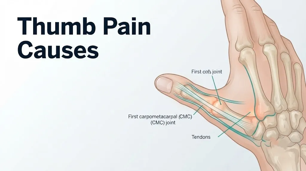
The thumb has three bones: the proximal phalanx, the distal phalanx, and the first metacarpal. It moves through four major joints: the interphalangeal (IP) joint at the tip, the metacarpophalangeal (MCP) joint at the knuckle, the first carpometacarpal (CMC) joint at the base where the thumb meets the wrist, and the carpometacarpal articulation with the trapezium bone. Each of these joints can develop arthritis, sustain injury, or be affected by systemic disease.
The tendons running along the thumb include the abductor pollicis longus (APL), the extensor pollicis brevis (EPB), the extensor pollicis longus (EPL), and the flexor pollicis longus (FPL). Any of these can become inflamed, compressed, or torn. Two major nerve branches supply sensation to the thumb: the median nerve, which covers the palmar surface and tip, and the superficial branch of the radial nerve, which runs along the back and radial side of the thumb.
| Anatomical Structure | Conditions Associated | Pain Location |
|---|---|---|
| First CMC joint (basal joint) | Basal joint osteoarthritis, trauma | Base of thumb near wrist |
| MCP joint | Gamekeeper’s thumb, rheumatoid arthritis, gout | Thumb knuckle |
| APL and EPB tendons | De Quervain’s tenosynovitis | Radial (outer) side of wrist extending to thumb |
| Flexor pollicis longus tendon | Trigger thumb | Palm side of thumb base |
| Ulnar collateral ligament | Skier’s thumb, gamekeeper’s thumb | Inner (ulnar) side of MCP joint |
| Median nerve | Carpal tunnel syndrome | Thumb tip, index and middle fingers |
| Proximal phalanx or first metacarpal | Fracture | Bone-deep, anywhere along thumb shaft |
People over 40 are disproportionately affected by degenerative causes. Those under 40 are more likely experiencing traumatic or inflammatory causes. This distinction matters when building a differential diagnosis.
What Causes Pain at the Base of the Thumb
Pain at the base of the thumb almost always originates at the first carpometacarpal joint, the saddle-shaped joint where the first metacarpal meets the trapezium bone of the wrist, though De Quervain’s tenosynovitis running along the same region can mimic it closely.
The first CMC joint is unique in the human skeleton. Its saddle shape allows the wide rotational range of motion needed for thumb opposition, pinching, and gripping. That same mobility, however, makes it the most mechanically stressed small joint in the hand. Research published in the Journal of Hand Surgery has documented that forces across the first CMC joint during pinching can reach up to 12 times the applied pinch force, due to the lever arm mechanics of the thumb.
When cartilage at this joint degrades, the subchondral bone beneath it becomes exposed and reactive, forming osteophytes (bone spurs) and narrowing the joint space. The result is pain precisely at the base of the thumb, a deep aching quality that worsens when gripping a jar, turning a key, or picking up a heavy object.
Distinguishing first CMC joint pain from De Quervain’s tenosynovitis requires a clinical examination. CMC joint pain is reproduced by the grind test: the examiner axially loads and rotates the thumb metacarpal against the trapezium. De Quervain’s pain is reproduced with the Finkelstein test: tucking the thumb inside a fisted hand and deviating the wrist toward the small finger side.
Women experience basal joint arthritis at a rate approximately four times that of men, a difference partly explained by the broader angle of the first CMC joint in female anatomy and the effects of estrogen decline on articular cartilage after menopause.
De Quervain’s Tenosynovitis as a Cause of Thumb Pain
De Quervain’s tenosynovitis is caused by progressive narrowing and inflammation of the first dorsal extensor compartment, the fibrous tunnel formed by the extensor retinaculum that houses the abductor pollicis longus (APL) and extensor pollicis brevis (EPB) tendons as they cross the wrist toward the thumb.
Think of the first dorsal compartment as a narrow pipe through which two tendons must constantly slide. When the pipe’s inner lining becomes inflamed and thickened from repetitive friction or overuse, the tendons no longer glide smoothly. Every movement that stretches or slides these tendons — lifting a baby, gripping a golf club, swiping a phone screen — creates friction against the narrowed compartment wall, generating pain on the radial (outer) side of the wrist that radiates into the thumb.
The pain from De Quervain’s is characteristically reproduced with the Finkelstein test, which has a positive predictive value of approximately 85 to 90 percent for De Quervain’s diagnosis when performed correctly, according to clinical hand surgery literature. The pain typically starts at the radial styloid process (the bony prominence on the outer side of the wrist) and travels distally into the thumb.
According to the National Institute of Arthritis and Musculoskeletal and Skin Diseases (NIAMS), De Quervain’s tenosynovitis is most common in adults between 30 and 50 years of age. Women are affected at a rate approximately six times higher than men, with the highest incidence occurring during pregnancy and the first year postpartum.
- Pain on the outer (radial/thumb) side of the wrist extending into the thumb
- Swelling over the radial styloid that may be visible without pressing
- Weakness when pinching or gripping
- A catching or snapping sensation with thumb movement in some cases
- Pain that worsens with lifting, opening jars, or carrying bags
For people with diabetes, the inflammatory response in the tendon sheath can be more persistent and resistant to corticosteroid injections, according to clinical observations published in the Journal of Hand Surgery.
Basal Joint Arthritis and First CMC Joint Osteoarthritis
Basal joint arthritis, the clinical term for osteoarthritis of the first carpometacarpal joint, is the most common form of arthritis affecting the hand and one of the most common thumb pain causes in adults over 40. It involves the gradual breakdown of the articular cartilage lining the saddle-shaped articulation between the first metacarpal and the trapezium.
Healthy articular cartilage at this joint acts as a frictionless shock absorber, distributing the extreme compressive forces generated during pinch and grip. As the cartilage degrades, the joint loses its smooth gliding surface. Subchondral bone becomes exposed, inflammatory mediators including prostaglandins and interleukin-1 beta accumulate in the joint space, and the surrounding synovium becomes reactive. Bone spurs form at the joint margins as the body attempts to stabilize a mechanically compromised joint.
The Arthritis Foundation estimates that roughly 40 percent of women and 25 percent of men will have X-ray evidence of first CMC joint osteoarthritis by age 75, though the presence of radiographic changes does not always correlate directly with pain severity. Some people with marked joint space narrowing report only mild discomfort, while others with early changes have functionally limiting pain.
| OA Stage | X-Ray Finding | Symptoms | Clinical Management |
|---|---|---|---|
| Stage 1 | Normal joint space, slight synovitis | Mild aching with heavy use | Splinting, activity modification |
| Stage 2 | Mild joint space narrowing, small osteophytes | Moderate pain with pinching/gripping | Splinting, injection, PT |
| Stage 3 | Significant narrowing, osteophyte formation, subluxation | Constant pain, weakness, deformity | Injection, possible surgery |
| Stage 4 | Bone on bone, scaphotrapezial OA | Severe pain at rest and with use | Surgical reconstruction |
People with prior thumb trauma, hypermobile joints, or a family history of hand arthritis develop this condition earlier and more aggressively. A board-certified orthopedic surgeon specializing in hand and upper extremity conditions should evaluate patients who fail conservative management or who present with Stage 3 or 4 disease on imaging.
Key Takeaway: Pain at the base of the thumb that worsens with pinching or gripping in adults over 40 most often points to basal joint osteoarthritis at the first CMC joint, but De Quervain’s tenosynovitis and these two conditions can coexist in the same thumb.
Rheumatoid Arthritis Causing Thumb Pain
Rheumatoid arthritis (RA) causes thumb pain through a fundamentally different mechanism than osteoarthritis: it is an autoimmune condition in which the immune system attacks the synovial lining of the joints, not wear-based cartilage degradation.
In RA, immune cells driven by elevated tumor necrosis factor alpha (TNF-alpha) and interleukin-6 (IL-6) infiltrate the synovial membrane of the thumb joints, triggering synovitis. This inflamed synovium, called pannus, secretes enzymes including matrix metalloproteinases that directly erode articular cartilage and the periarticular bone. At the thumb specifically, the MCP joint and IP joint are frequently the first sites of involvement, producing swelling, warmth, and morning stiffness lasting longer than 60 minutes.
According to the Arthritis Foundation, RA affects approximately 1.5 million people in the United States, with women developing it at three times the rate of men. The peak onset in women is between ages 30 and 60, often during hormonally active periods. In the hand, RA commonly causes a characteristic deformity called boutonniere deformity of the thumb (flexion at the IP joint with MCP hyperextension) in advanced, untreated disease.
The critical difference from osteoarthritis: RA pain is typically worst in the morning and improves with movement. OA pain worsens with use and improves with rest. Both can affect the same person simultaneously.
Diagnosis relies on blood testing for rheumatoid factor (RF) and anti-citrullinated protein antibodies (anti-CCP), which are positive in approximately 70 and 80 percent of RA patients respectively, according to the American College of Rheumatology’s published diagnostic criteria. People experiencing symmetrical joint pain, morning stiffness, fatigue, and swelling in multiple small joints should be referred to a board-certified rheumatologist promptly. Early treatment with disease-modifying antirheumatic drugs (DMARDs) prevents the joint destruction that becomes permanent once pannus erosion is established.
Trigger Thumb Causes and Mechanism
Trigger thumb occurs when the A1 pulley, a ring of connective tissue at the base of the thumb that holds the flexor pollicis longus (FPL) tendon close to the metacarpal bone, becomes thickened and narrowed. The FPL tendon must pass through this ring with every flexion and extension of the thumb. When the ring narrows, the tendon no longer slides freely.
The mechanical picture is similar to a frayed rope being forced through a ring that’s too small for it. Initially the tendon catches, creating a clicking or snapping sensation. As the A1 pulley thickens further, the thumb may lock in a flexed position and require passive straightening with the opposite hand. In severe cases, the thumb locks completely.
The Mayo Clinic notes that trigger thumb is more common in women than men and most frequently affects adults between 40 and 60. People with type 2 diabetes are approximately three to four times more likely to develop trigger thumb than the general population, a relationship attributed to the effects of chronic hyperglycemia on collagen cross-linking and tendon sheath tissue composition.
Other risk factors include:
- Rheumatoid arthritis (synovitis can thicken the A1 pulley)
- Hypothyroidism
- Gout (urate crystal deposition in the tendon sheath)
- Occupations involving prolonged gripping of tools
- Carpal tunnel syndrome (the two conditions coexist in approximately 8 percent of trigger finger cases, according to hand surgery literature)
Children can also develop trigger thumb, though through a different mechanism: a congenital nodule on the FPL tendon rather than acquired A1 pulley stenosis. Pediatric trigger thumb often resolves spontaneously before age four, according to the American Academy of Orthopaedic Surgeons (AAOS). Adults with persistent triggering that does not respond to corticosteroid injection should be evaluated by a hand surgeon for surgical A1 pulley release.
Carpal Tunnel Syndrome and Thumb Pain
Carpal tunnel syndrome causes thumb pain through compression of the median nerve as it passes through the carpal tunnel, the narrow fibro-osseous passage at the base of the palm bounded by the carpal bones on the bottom and the transverse carpal ligament on top.
The median nerve carries both sensory and motor signals to the thumb. Specifically, the nerve’s recurrent motor branch innervates the thenar muscles (abductor pollicis brevis, opponens pollicis, and superficial head of flexor pollicis brevis) that control thumb opposition and abduction. Its sensory branches supply the palmar surface of the thumb, index finger, middle finger, and the radial half of the ring finger.
When elevated pressure within the carpal tunnel compresses the median nerve, the first symptoms are typically tingling or numbness in the thumb tip and index finger, often waking the person at night. This nocturnal pattern occurs because the wrist naturally flexes during sleep, which increases carpal tunnel pressure and reduces nerve blood flow.
According to the National Institutes of Health (NIH), carpal tunnel syndrome affects an estimated 3 to 6 percent of adults in the general population, making it the most common peripheral nerve compression disorder. Women are affected at a rate 3:1 over men, partly because of a naturally narrower carpal tunnel anatomically and partly due to fluid retention effects during pregnancy and menopause on the transverse carpal ligament.
The thumb pain in carpal tunnel syndrome has a distinctive character: it is predominantly described as burning, tingling, or “electric” rather than the deep aching of joint pain or the sharp pain of a tendon problem. Weakness of the thenar musculature — difficulty holding a coffee cup or buttoning a shirt — signals advanced median nerve compression and warrants urgent evaluation by a board-certified neurologist or hand surgeon, as prolonged compression can cause permanent thenar muscle atrophy.
Key Takeaway: Carpal tunnel syndrome causes thumb tingling and numbness that wakes you at night, while De Quervain’s causes radial-sided wrist pain with gripping. These two conditions produce thumb symptoms through completely different anatomical mechanisms and should not be confused.
Gamekeeper’s Thumb and Ulnar Collateral Ligament Injury
Gamekeeper’s thumb describes a complete or partial tear of the ulnar collateral ligament (UCL) at the metacarpophalangeal (MCP) joint of the thumb, producing instability of the pinch mechanism that can become permanent without proper treatment.
The UCL is the primary stabilizer against radial-directed stress at the thumb MCP joint. It runs from the base of the proximal phalanx to the first metacarpal head on the ulnar (inner) side of the joint. When the thumb is forcefully pushed away from the palm (radially deviated), the UCL is placed under extreme tensile load and can rupture.
The acute version, called skier’s thumb, occurs when a skier falls on an outstretched hand while still holding a ski pole, forcing the thumb into radial abduction. Historically, gamekeepers developed the chronic version through repeated stress of the UCL when dispatching game. Both produce pain and swelling at the thumb knuckle on the inner (ulnar) side and, critically, a feeling of weakness or instability when pinching.
A key clinical complication is the Stener lesion: when the UCL tears completely, the aponeurosis of the adductor pollicis muscle can interpose between the torn ligament ends, physically preventing the ligament from healing in anatomical position without surgical repair. Research published in clinical hand surgery literature suggests Stener lesions occur in approximately 64 to 87 percent of complete UCL ruptures.
Testing UCL integrity at home: pinching paper between the thumb and index finger and assessing whether the thumb buckles outward suggests instability. However, only a clinical examination by a board-certified orthopedic surgeon with stress radiography or MRI can determine whether the tear is complete, whether a Stener lesion is present, and whether surgical repair is needed. Partial tears may be treated with thumb spica casting for 4 to 6 weeks. Complete tears with a Stener lesion typically require surgical UCL reconstruction.
Thumb Fractures and Acute Bone Injury
Thumb fractures cause acute, immediate pain that is bone-deep, worsens with any attempted movement, and is typically associated with a clear injury event. The most common thumb bones fractured are the proximal phalanx, the distal phalanx (often at the tip from a crushing injury), and the base of the first metacarpal.
Two specific fracture patterns at the base of the first metacarpal deserve special attention because they behave differently from simple breaks:
The Bennett’s fracture is an intra-articular fracture-dislocation at the base of the first metacarpal, where a fragment of bone remains attached to the anterior oblique ligament while the metacarpal shaft is displaced proximally by the pull of the abductor pollicis longus. Without anatomical reduction, the deformity disrupts the first CMC joint mechanics long-term.
The Rolando fracture is a comminuted (multi-fragment) intra-articular fracture at the same location, generally associated with high-energy trauma and a more difficult surgical reconstruction.
A scaphoid fracture, technically a wrist fracture but frequently presenting as radial-sided thumb and wrist pain after a fall on an outstretched hand, is one of the most commonly missed fractures in emergency medicine. The scaphoid’s blood supply enters distally, meaning proximal pole fractures carry a risk of avascular necrosis if not immobilized promptly. Initial X-rays miss scaphoid fractures in up to 20 percent of cases, according to data published in clinical emergency medicine literature. An MRI or CT scan within 5 to 7 days is recommended when clinical suspicion is high and initial X-rays are negative.
Any thumb injury with visible deformity, marked swelling, bony tenderness, or inability to move the thumb should be evaluated in an emergency room or urgent care setting with same-day imaging, not monitored at home.
Key Takeaway: A scaphoid fracture after a fall on an outstretched hand can look like a simple wrist sprain initially, but negative X-rays do not rule it out. If the radial side of your wrist and thumb base are tender after a fall, push for MRI or CT imaging within a week.
Gout and Other Systemic Causes of Thumb Joint Pain
Gout causes thumb joint pain when monosodium urate (MSU) crystals deposit within the joint space or periarticular soft tissues, triggering an acute, intensely painful inflammatory response mediated by the NLRP3 inflammasome and subsequent release of interleukin-1 beta (IL-1 beta) and IL-6 from activated macrophages.
Gout most commonly affects the first metatarsophalangeal joint of the foot, but hand and wrist involvement, including the thumb MCP joint, occurs in approximately 30 to 40 percent of people with recurring gout, according to data published in the Annals of the Rheumatic Diseases. An acute gout flare produces extreme pain, redness, warmth, and swelling that typically peaks within 12 to 24 hours of onset. The affected joint is often so painful that even the weight of a bed sheet is intolerable.
Three additional systemic causes of thumb pain are less common but clinically important:
Psoriatic arthritis affects approximately 30 percent of people with psoriasis and can involve the thumb joints with an inflammatory pattern that includes dactylitis (sausage-like swelling of the entire digit), nail changes, and enthesitis (inflammation at tendon and ligament insertion points). Unlike RA, psoriatic arthritis is often seronegative (RF and anti-CCP are typically negative).
Raynaud’s phenomenon causes episodes of thumb and finger pain tied to vasospasm of the digital arteries in response to cold temperatures or emotional stress. The characteristic color change (white to blue to red as the digit goes from ischemic to deoxygenated to reperfused) differentiates it from arthritic causes. Raynaud’s affects approximately 5 to 10 percent of women in temperate climates, according to the NIH National Institute of Arthritis and Musculoskeletal and Skin Diseases (NIAMS).
Tendinopathy from chronic overuse, distinct from acute tenosynovitis, involves degenerative changes within the tendon substance itself (the APL or EPL most commonly) rather than inflammation of the surrounding sheath, and requires targeted physical therapy and load management rather than anti-inflammatory treatment.
Repetitive Use, Occupational Risk, and Technology-Related Thumb Pain
Repetitive use is a thumb pain cause that operates through two distinct mechanisms: acute inflammatory tenosynovitis from rapid, high-frequency tendon loading, and chronic tendinopathic change from accumulated microtrauma without adequate tissue recovery time.
Occupations carrying the highest documented thumb injury and pain risk include dental hygienists, assembly line workers, musicians (particularly guitarists and cellists), construction workers, and healthcare workers who frequently perform manual tasks. A 2022 analysis of occupational musculoskeletal injury data found that workers performing repetitive pinch or grip tasks for more than 4 hours per day had significantly higher rates of De Quervain’s and basal joint pathology than controls.
Smartphone and tablet use has generated a newer presentation pattern. A 2023 observational study examining thumb pain in adults who averaged more than 6 hours of daily smartphone use found a higher prevalence of De Quervain’s-like tendon pain and first CMC joint discomfort compared to lower-use controls. The primary mechanical culprits are the sustained APL and EPB loading during screen swiping, the sustained grip needed for holding a large phone one-handed, and the repetitive short-amplitude thumb movements during texting.
Quick Tip:
- When texting extensively, alternate thumb use and hold the phone at a height that reduces neck and wrist flexion
- Set a device-down break every 30 to 45 minutes of continuous phone use if thumb base pain is developing
- Musicians and manual workers experiencing early thumb tendon symptoms should consult a certified hand therapist before symptoms progress to functional limitation
- Ergonomic modification at a workstation is more durable than symptom treatment without addressing the mechanical cause
Younger adults (18 to 35) who present with thumb pain and no history of trauma or systemic disease should specifically be asked about daily screen time, gaming hours, and instrument practice intensity, as these are the primary occupational and behavioral causes in that age group.
Thumb Pain Causes in Women, During Pregnancy, and Postpartum
Women experience several thumb pain causes at higher rates than men, and the physiological reasons are specific rather than coincidental. The most important to understand in 2026 are De Quervain’s in the postpartum period, basal joint OA in the perimenopausal years, and the amplifying role of hormonal changes on tendon and joint tissue.
During pregnancy, the hormone relaxin is produced by the corpus luteum and placenta to prepare the pelvic ligaments and soft tissues for childbirth. Relaxin acts on collagen-producing cells throughout the body, altering connective tissue laxity systemically. In the hand, this can lead to tendon sheath swelling and reduced clearance within the first dorsal compartment, directly predisposing to De Quervain’s tenosynovitis. Fluid retention during pregnancy also increases pressure within the carpal tunnel, which is why carpal tunnel syndrome in pregnancy is reported in approximately 31 to 62 percent of pregnant women, according to a review published in the Journal of Orthopaedics and Traumatology.
Postpartum De Quervain’s peaks in the first 3 to 6 months after delivery. Repetitive wrist radial deviation during breastfeeding, lifting a newborn with radially deviated wrists, and sustained APL and EPB loading during infant care creates the mechanical overload on already-sensitized tendon sheaths. New mothers who present with pain on the radial side of the wrist should specifically be asked about breastfeeding mechanics and infant lifting technique.
After menopause, declining estrogen accelerates articular cartilage degradation at the first CMC joint and reduces synovial fluid viscosity, which partly explains the four-fold higher rate of basal joint OA in postmenopausal women compared to age-matched men. Women in the perimenopausal years (typically 45 to 55) who develop new basal joint pain should have baseline X-rays taken to stage the joint if conservative management fails within 6 to 8 weeks.
Key Takeaway: New mothers with wrist and thumb pain on the radial side should be assessed for postpartum De Quervain’s tenosynovitis before assuming carpal tunnel syndrome, since the tendon mechanism and the correct treatment differ substantially between the two.
Thumb Pain Causes in Older Adults
In adults over 60, the most common thumb pain causes shift decisively toward degenerative conditions, with basal joint osteoarthritis, trigger thumb, and multi-joint rheumatoid disease accounting for the majority of presentations. The clinical picture is also more frequently complicated by comorbidities and medications that alter both the pain experience and the treatment options.
Basal joint OA in older adults is often bilateral, though one side typically becomes symptomatic before the other. By age 70, X-ray evidence of first CMC joint OA is present in approximately 25 percent of men and 40 percent of women, according to epidemiological data cited by the AAOS. Pain may be constant rather than activity-related in advanced stages, and nighttime pain indicates periarticular inflammation or Stage 3 to 4 disease.
Trigger thumb in older adults carries a higher rate of incomplete response to corticosteroid injection compared to younger patients, and a greater likelihood of requiring surgical A1 pulley release. Older adults with diabetes, hypothyroidism, or RA face a compounded risk.
An additional cause specific to older adults is gout with tophaceous deposits: chronic urate crystal accumulation in soft tissues can produce hard, chalky nodules (tophi) around the thumb joints that are palpable, occasionally drain white chalky material, and cause chronic low-grade pain punctuated by acute flares. This presentation is more common in older men on diuretic therapy for hypertension, as thiazide and loop diuretics reduce renal urate excretion, raising serum uric acid levels.
Older adults with thumb pain should be specifically asked about:
- Current diuretic or aspirin use (both can elevate uric acid)
- History of gout or kidney stones (uric acid stones)
- Morning stiffness duration (a marker of inflammatory vs. degenerative joint disease)
- Hand dominance and occupational history
- Falls or recent trauma (fracture risk increases with age-related bone density loss)
A board-certified rheumatologist or hand surgeon should evaluate older adults whose thumb pain has not responded to 6 to 8 weeks of conservative management or who have developed functional impairment in daily activities.
How Doctors Diagnose the Cause of Thumb Pain
Accurate diagnosis of thumb pain causes requires a structured clinical history, a focused physical examination, and targeted imaging or laboratory testing, because multiple conditions can produce overlapping symptoms in the same location.
The diagnostic process typically follows this sequence:
- History taking: Duration, onset (sudden vs. gradual), pain character (aching, burning, sharp, throbbing), exact location (base, knuckle, tip, radial side, ulnar side), aggravating activities, hand dominance, occupation, recent trauma, and any systemic symptoms such as morning stiffness, fatigue, or rash.
- Physical examination: The examiner palpates specific anatomical landmarks, checks active and passive range of motion, tests grip and pinch strength with a dynamometer, and performs provocative tests: Finkelstein test for De Quervain’s, grind test for basal joint OA, Tinel’s sign and Phalen’s test for carpal tunnel syndrome, and stress testing of the UCL for gamekeeper’s thumb.
- Plain X-ray (radiograph): First-line imaging for joint space narrowing, osteophyte formation, fractures, and dislocation. Standard views include AP, lateral, and stress views of the thumb.
- Ultrasound: Preferred for evaluating tendon integrity, tendon sheath fluid, and the A1 pulley. It is dynamic (can assess tendon movement in real time) and radiation-free.
- MRI: Ordered when the clinical picture suggests soft tissue injury not visible on X-ray, including UCL tears (to detect a Stener lesion), scaphoid fractures (X-ray-negative), or early inflammatory arthritis with synovitis.
- Laboratory testing: Ordered when systemic disease is suspected. A complete blood count (CBC), erythrocyte sedimentation rate (ESR), C-reactive protein (CRP), serum uric acid, rheumatoid factor (RF), and anti-CCP antibodies help differentiate gout, RA, and psoriatic arthritis from mechanical causes.
- Joint aspiration: When septic arthritis (joint infection) is suspected, synovial fluid must be aspirated and analyzed by Gram stain and culture to rule out bacterial infection, a condition that can destroy the joint within days if untreated.
A board-certified hand surgeon should be involved in evaluation when clinical examination suggests UCL injury, fracture, or any condition that might require surgical management.
Acute Versus Chronic Thumb Pain: What the Difference Means
Whether your thumb pain is acute (sudden onset, typically hours to days) or chronic (gradual, weeks to months) is one of the most clinically useful pieces of information for narrowing the diagnosis, because these two timelines strongly predict which causes are most likely.
Acute thumb pain has a limited differential: fracture (particularly after a fall or direct blow), acute UCL tear (skier’s thumb), acute gout flare, acute infectious tenosynovitis (rare but rapidly dangerous), or a first episode of an inflammatory arthritis flare. All of these warrant prompt professional evaluation rather than watchful waiting.
Chronic and progressive thumb pain points toward a different set of causes: basal joint osteoarthritis (the most common), De Quervain’s tenosynovitis from repetitive use accumulation, established rheumatoid or psoriatic arthritis, chronic trigger thumb, and carpal tunnel syndrome. These conditions generally develop over weeks to months and initially respond to conservative management, though they tend to progress without addressing the root cause.
| Onset Pattern | Most Likely Causes | Urgency Level | Recommended Action |
|---|---|---|---|
| Sudden after trauma | Fracture, UCL tear, dislocation | Same day | Emergency room or urgent ortho |
| Sudden without trauma | Acute gout, acute arthritis flare | Within 24 to 48 hours | Primary care or rheumatology |
| Gradual over weeks | De Quervain’s, basal joint OA, trigger thumb | Within 1 to 2 weeks | Primary care or hand specialist |
| Gradual over months | Rheumatoid arthritis, established OA | Within 2 to 4 weeks | Primary care then specialist |
| Intermittent, activity-related | Basal joint OA, De Quervain’s, carpal tunnel | Scheduled appointment | Primary care then hand specialist |
| Constant, waking at night | Carpal tunnel (nerve), severe OA | Within 1 to 2 weeks | Primary care, nerve conduction study |
The acute-versus-chronic distinction also informs treatment: acute inflammatory causes often respond to short-term anti-inflammatory treatment and relative rest, while chronic degenerative causes require activity modification, splinting, targeted therapy, and sometimes injection or surgery.
Key Takeaway: Sudden thumb pain after any trauma, regardless of how minor it seems, deserves same-day imaging to rule out a fracture or ligament tear, since both Bennett’s fractures and UCL tears can look and feel like mild sprains initially.
When Thumb Pain Requires Emergency Evaluation
Certain presentations of thumb pain require immediate emergency evaluation. Do not wait to see if these resolve on their own.
Call 911 or go to the nearest emergency room immediately if you experience:
- Visible deformity of the thumb after an injury (angled, rotated, or shortened appearance indicating possible fracture or dislocation)
- Open wound over the thumb joint or tendon with or without exposed bone or tendon, as this constitutes an open fracture or open injury requiring immediate irrigation and surgical evaluation
- Complete inability to move the thumb following a fall or direct impact, combined with severe swelling and bone-point tenderness
- Signs of joint infection: rapidly worsening redness, warmth, extreme pain at rest, swelling at the thumb joint, and fever higher than 38 degrees Celsius (100.4 degrees Fahrenheit). Septic arthritis destroys joint cartilage within days and requires emergency joint washout.
- Thumb that is completely cold, white, or blue after an injury, indicating possible vascular compromise (disrupted digital artery requiring emergency vascular assessment)
- Numbness or paralysis of the thumb and adjacent fingers with a known crush injury, suggesting compartment syndrome or severe nerve injury
Go to urgent care or an emergency orthopedic clinic the same day for:
- Acute UCL injury (skier’s thumb) with instability on pinch testing
- Any thumb injury with swelling that prevents full range of motion, even without obvious deformity
- Suspected scaphoid fracture (tenderness in the anatomical snuffbox after a fall on an outstretched hand, even with normal initial X-rays)
These presentations can indicate fracture, septic arthritis, nerve or vascular injury, or complete ligament rupture, and they require emergency or same-day medical assessment, not self-management or monitoring at home.
Frequently Asked Questions About Thumb Pain Causes
What is the most common cause of thumb pain?
Basal joint osteoarthritis at the first carpometacarpal joint is the most common cause of chronic thumb pain in adults, particularly those over 40.
The Arthritis Foundation notes that this joint is among the most frequently arthritic joints in the human hand.
De Quervain’s tenosynovitis is the most common cause of radial-sided thumb and wrist pain in adults between 30 and 50, particularly in women.
How do I know if my thumb pain is arthritis or a tendon problem?
Arthritis thumb pain is typically deep, aching, located at the joint itself, and worsens with gripping or pinching.
Tendon pain from De Quervain’s tenosynovitis or trigger thumb is typically along the tendon path (radial side of the wrist or along the palm side of the thumb), and is reproduced with specific thumb movements rather than joint compression.
A clinical examination with provocative tests — the grind test for arthritis and the Finkelstein test for De Quervain’s — is the most reliable way to distinguish them, and both conditions can coexist.
Can thumb pain go away on its own without treatment?
Mild De Quervain’s tenosynovitis caught early, and some acute inflammatory flares, can improve with activity modification, relative rest, and temporary splinting.
However, basal joint osteoarthritis, complete UCL tears, fractures, and established rheumatoid arthritis do not resolve without medical management and will worsen over time without treatment.
Any thumb pain that has persisted for more than 2 to 3 weeks without improvement should be evaluated by a primary care physician or a board-certified hand specialist.
What does thumb pain at the base of the thumb usually mean?
Pain at the base of the thumb most commonly indicates basal joint osteoarthritis at the first carpometacarpal joint or De Quervain’s tenosynovitis involving the APL and EPB tendons.
These two conditions produce pain in overlapping locations but through different mechanisms, and they require different treatments.
A physician can distinguish them through the grind test and Finkelstein test during a physical examination.
When should I see a doctor for thumb pain?
See a primary care physician or hand specialist within 1 to 2 weeks if your thumb pain has lasted more than 2 weeks, is worsening, limits your ability to grip or pinch, or appeared without an obvious injury.
Go to an emergency room or urgent care the same day if thumb pain followed a traumatic event, is associated with visible deformity, severe swelling, inability to move the thumb, fever, or rapidly spreading redness.
Thumb pain with numbness or tingling that wakes you at night should be evaluated within 1 week for possible carpal tunnel syndrome.
Can texting and phone use really cause thumb pain?
Yes. Observational research has consistently associated high-frequency smartphone use with elevated rates of De Quervain’s-type tendon pain and first CMC joint discomfort.
The mechanism involves repetitive loading of the abductor pollicis longus and extensor pollicis brevis tendons during swiping, sustained gripping load on the first CMC joint, and repetitive short-amplitude flexion movements of the thumb during typing.
Adults who use a smartphone for more than 6 hours daily and develop radial-sided wrist or thumb base pain should consider activity modification and ergonomic assessment before symptoms become chronic.
Closing
Thumb pain causes are genuinely diverse, and the right diagnosis depends on knowing which specific anatomical structure is involved and the mechanism producing your symptoms. Basal joint osteoarthritis and De Quervain’s tenosynovitis account for the majority of cases in adults, but rheumatoid arthritis, trigger thumb, carpal tunnel syndrome, ligament injuries, and fractures all require different clinical responses.
Start by tracking when your pain is worst, what movement triggers it, and whether it appeared suddenly or built gradually. That information, combined with where precisely the pain is located (base of the thumb, the knuckle, the radial side of the wrist, or the tip), gives any clinician the foundation for an accurate diagnosis.
If your thumb pain has lasted more than two weeks, is limiting your grip or pinch strength, or appeared after any kind of trauma, schedule an evaluation with your primary care physician and ask for a referral to a board-certified orthopedic surgeon or hand specialist if needed. The conditions covered in this article are all treatable when caught at the right stage.