Leukemia Symptoms: Signs, Warning Signs and When to Act
Leukemia symptoms include persistent unexplained fatigue, easy bruising or bleeding, recurring fevers, bone and joint pain, night sweats, swollen lymph nodes, and unintended weight loss, and every one of these symptoms traces directly to what leukemia does to the bone marrow and the blood cells it produces. Understanding why these symptoms occur, not just that they occur, is what separates a vague list of warning signs from genuinely useful clinical information.
Leukemia is not a single disease. According to the National Cancer Institute (NCI), leukemia encompasses more than a dozen distinct subtypes, but the four most clinically prevalent are acute lymphoblastic leukemia (ALL), acute myeloid leukemia (AML), chronic lymphocytic leukemia (CLL), and chronic myeloid leukemia (CML). The NCI’s Surveillance Epidemiology and End Results (SEER) Program estimates that approximately 62,000 new cases of leukemia are diagnosed annually in the United States, with CLL and AML being the most common adult forms and ALL being the most common cancer diagnosis in children under age 15.
This article covers the complete symptom picture of leukemia, the specific bone marrow and blood cell mechanisms behind each symptom, how the clinical picture differs across subtypes and across age groups, which symptoms are often misattributed to other conditions, and the specific presentations that require calling 911 or going to an emergency room immediately, not waiting for an appointment.
What Is Leukemia and How Does It Develop
Leukemia is a malignancy of the hematopoietic system: the blood-forming tissues that include the bone marrow and the lymphatic system, in which a single abnormal blood cell precursor acquires genetic mutations that allow it to proliferate without normal restraint or maturation.
Every blood cell in the body, whether a red blood cell carrying oxygen, a neutrophil fighting bacteria, or a platelet sealing a blood vessel, originates from hematopoietic stem cells (HSCs) residing in the medullary cavities of flat and long bones, particularly the sternum, iliac crest, and femur. These HSCs divide and differentiate through a tightly regulated hierarchy of progenitor cells along two major lineages: the myeloid lineage (producing red blood cells, platelets, neutrophils, monocytes, and eosinophils) and the lymphoid lineage (producing T-cells, B-cells, and natural killer cells).
Leukemia begins when a somatic mutation, triggered by radiation exposure, certain chemical exposures, chromosomal instability, or identifiable genetic events (such as the Philadelphia chromosome translocation t(9;22) in CML or the t(12;21) translocation in pediatric ALL), converts a hematopoietic progenitor cell into a malignant leukemic blast that proliferates clonally. The specific progenitor cell type affected determines the leukemia subtype.
| Leukemia Subtype | Cell of Origin | Lineage | Typical Onset Speed |
|---|---|---|---|
| Acute lymphoblastic leukemia (ALL) | Lymphoid progenitor (B-cell or T-cell) | Lymphoid | Rapid: days to weeks |
| Acute myeloid leukemia (AML) | Myeloid progenitor | Myeloid | Rapid: days to weeks |
| Chronic lymphocytic leukemia (CLL) | Mature B-lymphocyte | Lymphoid | Slow: months to years |
| Chronic myeloid leukemia (CML) | HSC (BCR-ABL1 driven) | Myeloid | Slow to moderate |
Individuals with Down syndrome (trisomy 21) face a 20-fold increased risk of developing ALL and have a distinct AML subtype. This reflects the specific role of chromosome 21 gene dosage in hematopoietic progenitor cell regulation, an established finding in pediatric oncology research.
How Leukemia Disrupts Normal Blood Cell Production
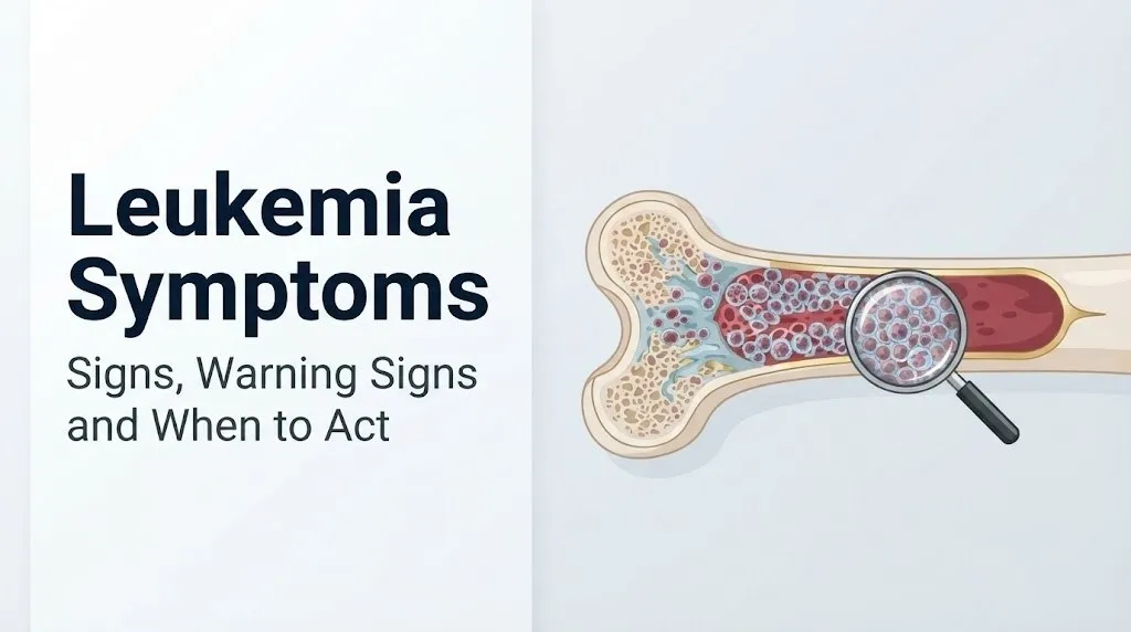
Leukemia causes symptoms primarily because the expanding population of malignant leukemic blasts physically crowds out normal hematopoietic cells within the bone marrow, a process called bone marrow failure or marrow replacement, which disrupts the production of all three major blood cell types simultaneously.
Think of the bone marrow like a factory floor with a fixed amount of production space. Normal blood cell production fills every station efficiently: some stations make red blood cells, others make platelets, others make immune cells. When leukemic blasts begin multiplying without stopping, they progressively occupy more and more floor space. The red blood cell stations are displaced first in many leukemias, then platelet production, then normal immune cell production. The output from normal stations drops toward zero while the blast population grows unchecked.
The three specific production failures each produce a distinct symptom set:
- Failure of erythropoiesis (red blood cell production): produces anemia, which causes the fatigue, pallor, shortness of breath on minimal exertion, and racing heart rate (tachycardia) that many leukemia patients report as their first noticeable symptoms
- Failure of thrombopoiesis (platelet production): produces thrombocytopenia, which causes easy bruising, pinpoint skin hemorrhages called petechiae, bleeding from the gums, nosebleeds, and in severe cases, life-threatening hemorrhage
- Failure of normal leukopoiesis (white blood cell production): produces neutropenia (dangerously low functional neutrophil count), which leaves the body unable to mount an effective defense against bacterial, fungal, and viral pathogens, leading to frequent, severe, or unusual infections and fever
According to research published in Blood (the journal of the American Society of Hematology), the degree of marrow replacement correlates directly with symptom severity: patients with bone marrow blast percentages above 70 to 80% typically present with more severe pancytopenia and more acute symptoms than those with lower blast infiltration percentages.
Pregnant women who develop leukemia face a compounded physiological challenge: pregnancy itself causes physiological anemia (dilutional, from plasma volume expansion) and relative immune suppression, which can mask or modify leukemia symptoms and delay diagnosis. Any pregnant woman with unexplained severe fatigue, fever, or bruising beyond what is expected for gestational age should have an urgent complete blood count evaluated by her obstetrician-gynecologist with hematology consultation.
Leukemia Symptoms: The Complete Clinical Picture
Leukemia symptoms arise from three distinct pathological processes: bone marrow failure producing blood cell deficiencies, leukemic cell infiltration of organs outside the marrow, and cytokine release from malignant and immune cells producing constitutional systemic effects.
The full symptom inventory recognized by clinical hematology, organized by mechanism:
Bone marrow failure symptoms:
- Persistent fatigue, weakness, and reduced exercise tolerance (anemia-driven)
- Pallor (pallor of the skin, conjunctiva, and nail beds from reduced hemoglobin)
- Shortness of breath with minimal exertion (reduced oxygen-carrying capacity)
- Resting or exertional tachycardia (cardiac compensation for anemia)
- Easy bruising with minimal trauma
- Petechiae (pinpoint flat red or purple hemorrhagic spots, typically on the legs, feet, and inside the mouth)
- Spontaneous nosebleeds or gum bleeding
- Heavy or prolonged menstrual bleeding in females
Immune failure symptoms:
- Recurrent bacterial infections (skin, lung, urinary tract)
- Fever that recurs or does not resolve with standard antibiotic therapy
- Oral thrush or other fungal infections suggesting cellular immune failure
- Delayed wound healing
Constitutional and cytokine-driven symptoms:
- Drenching night sweats (IL-1 and TNF-alpha driven)
- Unintended weight loss of more than 10% body weight over six months
- Intermittent fever without identified infection source
Extramedullary infiltration symptoms:
- Left upper quadrant abdominal pain or early satiety from splenomegaly
- Painless swelling of lymph nodes in the neck, armpit, or groin
- Bone pain, particularly in the long bones, sternum, and lower back
- Headache, visual changes, or facial numbness from CNS involvement
The American Cancer Society notes that the presence of three or more of these symptoms simultaneously, particularly when they are new, persistent, and unexplained by a known prior condition, warrants prompt medical evaluation rather than watchful waiting.
Key Takeaway: Leukemia symptoms are not random. They fall into three mechanistic groups (marrow failure, immune failure, and organ infiltration), and recognizing which group a symptom belongs to helps you understand whether it reflects a mild or a medically urgent clinical picture.
Early Warning Signs of Leukemia
The earliest warning signs of leukemia are often dismissed as non-specific because they overlap with ordinary conditions like fatigue from overwork, bruising from minor injury, or mild fever from a routine viral illness. The clinical distinction is not the individual symptom but the pattern, persistence, and combination.
The earliest symptoms that most often bring patients to their first evaluation include:
- Fatigue that does not improve with adequate sleep and rest, lasting more than two to four weeks
- Bruising appearing on body areas not typically exposed to trauma (upper arms, torso, upper thighs)
- Tiny flat red or purple pinpoint spots on the lower legs or inside the mouth (petechiae), often mistaken for a rash
- Fever above 38 degrees Celsius (100.4 degrees Fahrenheit) that recurs more than twice within a month without a diagnosed cause
- Recurrent respiratory infections that resolve incompletely or take longer than expected to clear
- Night sweats severe enough to require changing bedclothes or bed linens
In chronic leukemia, particularly CLL and early-phase CML, the first sign may not be a symptom at all. It may be an abnormal finding on a routine complete blood count (CBC) ordered for an unrelated reason, such as a pre-operative workup. According to the NCI, a substantial proportion of CLL diagnoses are made incidentally from routine blood work in adults over age 60 who have no symptoms at the time of diagnosis.
For older adults over 70, the clinical picture of early leukemia is further complicated by the common presence of anemia from other causes (iron deficiency, vitamin B12 deficiency, or chronic kidney disease) and fatigue from comorbid conditions. The distinguishing feature is that leukemia-related anemia typically does not respond to oral iron supplementation, and its CBC pattern (presence of blasts, elevated or very low white cell count, low platelet count) is distinct from nutritional anemia.
Quick Tip:
- Keep a written symptom log with dates, frequency, and severity. A physician can distinguish a leukemia pattern from coincidental findings far more accurately with a timeline than without one.
- Take photographs of any skin changes (petechiae, bruising, rash) to bring to your appointment with specific dates.
- For older adults: request that any CBC ordered for fatigue evaluation specifically note the differential count (breakdown of white cell types) and platelet count, not just hemoglobin.
Fatigue and Anemia in Leukemia
Leukemia-related fatigue is produced by anemia from suppressed erythropoiesis in the bone marrow, and it is clinically distinct from fatigue caused by poor sleep, depression, or hypothyroidism in its severity, persistence, and associated physical signs.
The mechanism is direct: as leukemic blasts displace the erythroid progenitor cells (CFU-E and BFU-E) in the bone marrow, the production of mature red blood cells falls below the rate of normal red cell senescence and destruction. Hemoglobin, the iron-containing protein in red blood cells that carries oxygen from the lungs to every tissue in the body, falls below the normal range. The body compensates initially by increasing heart rate (tachycardia) and redistributing cardiac output toward the heart and brain. When compensation fails, the tissues begin functioning under chronic oxygen deficiency.
The physical experience of leukemia anemia is described by patients as a deep, bone-level exhaustion that differs qualitatively from normal tiredness. Walking up a flight of stairs may produce breathlessness. Concentrating on a conversation or work task becomes effortful. This reflects both the reduced oxygen delivery to the cerebral cortex and the metabolic stress of the compensatory cardiac tachycardia.
According to research published in JAMA Oncology, hemoglobin levels at leukemia diagnosis range widely, from mildly reduced (below 12 g/dL in females, below 13.5 g/dL in males) to severely reduced (below 7 g/dL), with the most acute presentations typically seen in AML and early ALL where marrow replacement is aggressive and rapid.
Adolescent females are particularly vulnerable to misattribution of leukemia-related anemia as menstrual iron-deficiency anemia, especially if they have heavy menstrual periods, which may themselves be worsening due to thrombocytopenia. Any adolescent female with severe fatigue plus heavy menstrual bleeding plus easy bruising should have a full CBC with differential rather than empirical iron supplementation alone.
Fever, Infections, and Immune Failure in Leukemia
Fever and recurrent infections in leukemia occur because the malignant proliferation of abnormal white cell precursors replaces normal functional immune cells, leaving the body defenseless against pathogens that a healthy immune system would eliminate without difficulty.
The specific mechanism is neutropenia: a dangerously low count of mature neutrophils, the primary white blood cells that engulf and destroy bacteria through phagocytosis and oxidative killing. In leukemia, the displacement of normal myeloid progenitor cells by leukemic blasts prevents adequate neutrophil maturation. The result is an absolute neutrophil count (ANC) that may fall below 500 cells per microliter, defining severe neutropenia. Below this threshold, even bacteria that colonize skin and mucous membranes normally (Staphylococcus aureus, Escherichia coli, Klebsiella species) can produce life-threatening bloodstream infections (bacteremia and sepsis) with extraordinary speed.
Fever in leukemia has two distinct origins. The first is infection-driven: a temperature above 38.3 degrees Celsius (101 degrees Fahrenheit) in a neutropenic patient is a neutropenic fever, a hematological emergency requiring immediate intravenous antibiotic therapy. The second origin is cytokine-driven: leukemic cells and reactive immune cells release interleukin-1 (IL-1) and tumor necrosis factor-alpha (TNF-alpha), which act on the hypothalamic thermoregulatory center to produce fever in the absence of active infection.
Clinicians generally distinguish these two fever patterns by clinical context, blood cultures, and response to antibiotics, but in the early diagnostic phase before a leukemia diagnosis is established, any recurring fever without a clear source should prompt a CBC rather than simply another course of antibiotics.
Patients receiving chemotherapy for leukemia face the most extreme form of this vulnerability. Any patient currently undergoing leukemia treatment who develops a fever above 38.3 degrees Celsius must go to the emergency room immediately, regardless of the time of day, for blood cultures, IV antibiotics, and infectious disease or oncology evaluation. This is not a situation for home management or waiting until the morning.
Bruising, Bleeding, and Petechiae in Leukemia
Easy bruising, spontaneous bleeding, and petechiae in leukemia are produced by thrombocytopenia: a platelet count low enough that normal hemostasis (the process of stopping blood vessel injuries) cannot be maintained.
Platelets are produced by megakaryocytes, large bone marrow cells that shed fragments of their cytoplasm as circulating platelets. When leukemic blasts crowd out megakaryocyte progenitors in the marrow, platelet production falls. A normal platelet count is 150,000 to 400,000 platelets per microliter. At counts below 50,000, easy bruising and prolonged bleeding after minor cuts become clinically apparent. At counts below 20,000, spontaneous bleeding from mucous membranes (gums, nasal passages, gastrointestinal tract) becomes likely. At counts below 10,000, the risk of spontaneous intracranial hemorrhage becomes a medical emergency.
Petechiae are one of the most specific and underrecognized visible signs of thrombocytopenia. They appear as flat, non-blanching (pressing the skin does not cause them to temporarily disappear) pinpoint red or purple spots, typically on dependent areas of the body: the lower legs, the soles of the feet, and the soft palate and inner cheeks. They are often mistaken for small bruises, insect bites, or an allergic rash. The blanching test is simple: press a glass firmly against the spot. A petechiae will not disappear. A rash from inflammation or allergic reaction will.
Ecchymoses (bruises) in leukemia appear in areas not normally exposed to trauma, are often larger than expected from the mechanism of injury, and take longer than normal to resolve because the platelet plug that limits bruise spread forms inadequately.
A specific and particularly dangerous bleeding pattern occurs in acute promyelocytic leukemia (APL), a subtype of AML caused by the t(15;17) chromosomal translocation. APL leukemic cells release procoagulant substances that trigger disseminated intravascular coagulation (DIC), a condition in which clotting factors are consumed system-wide, producing simultaneous clotting and bleeding. Undiagnosed APL with DIC is a genuine oncological emergency.
Key Takeaway: Petechiae on the lower legs combined with easy bruising and fatigue are one of the most specific leukemia symptom combinations, and any adult or child presenting with this triad without a clear alternative explanation needs a same-day complete blood count, not reassurance.
Bone Pain and Joint Pain in Leukemia
Bone pain is among the most diagnostically telling symptoms of leukemia, particularly in children, and it occurs because rapidly expanding leukemic blasts within the marrow cavity create pressure against the periosteum (the richly nerve-innervated outer membrane of bone) and cause localized inflammatory and vascular changes in the bone itself.
The periosteum contains dense sensory nerve fibers supplied by branches of regional somatic nerves. When the medullary space of a bone becomes overcrowded with proliferating leukemic blasts, intramedullary pressure rises and presses outward against the periosteum. This periosteal pressure produces a deep, aching pain that is often described as worse at night and poorly localized compared to the sharp, point-specific pain of a fracture.
In children with ALL, bone pain is present in approximately 25 to 30% of cases at diagnosis, according to research published in Blood. It most commonly affects the long bones of the legs (femur, tibia), the lower back (lumbar vertebral bodies), and the sternum. A child who begins limping, refusing to bear weight, or complaining of persistent bone pain in the legs without a documented injury should be evaluated promptly. This symptom is frequently misattributed to growing pains, sports injuries, or viral arthralgia, causing delays in ALL diagnosis that have direct prognostic consequences in a disease where early treatment response determines long-term outcome.
In adults with AML, bone pain tends to be more diffuse, affecting the lower back, hips, and sternum. Sternal tenderness on direct palpation (pressing on the breastbone) is a clinical sign that hematologists specifically check, because the sternum is a primary site of active adult bone marrow and its tenderness reflects marrow expansion under pressure.
Older adults with CML can develop bone pain at the onset of blast crisis, the transformation from chronic-phase CML to an acute leukemia-like state that signals disease acceleration and requires immediate re-evaluation by a hematologist-oncologist.
Leukemia Skin Symptoms and Lymph Node Changes
Leukemia produces skin changes and lymph node swelling through two distinct mechanisms: thrombocytopenia-driven hemorrhagic lesions (petechiae and ecchymoses, discussed above) and direct infiltration of the skin and lymphatic tissue by leukemic cells.
Leukemia cutis is the specific clinical term for malignant leukemic cell infiltration of the dermis and subcutaneous tissue, producing visible skin lesions. These appear as firm, non-tender nodules or papules with a characteristic violaceous (purple-red) or flesh-colored appearance, most commonly on the trunk, extremities, and face. They are most associated with AML (particularly the monocytic subtypes M4 and M5), CLL, and ALL with T-cell phenotype. According to research published in the Journal of the American Academy of Dermatology, leukemia cutis is present at initial leukemia diagnosis in approximately 3 to 5% of AML cases and up to 30% of specific AML monocytic subtypes.
Gingival hyperplasia, an abnormal overgrowth of the gum tissue, is a hallmark presentation of AML-M5 (acute monocytic leukemia), produced by monocytic leukemic cell infiltration of the gingival connective tissue. Patients often present to dentists rather than physicians initially, which creates a diagnostic opportunity: a dentist who sees unexplained, rapidly progressive gingival swelling in an adult without periodontal disease or drug exposure should consider referral for a CBC with differential.
Lymphadenopathy (swollen lymph nodes) occurs in lymphoid leukemias when malignant lymphoid cells populate and expand the germinal centers and paracortex of lymph nodes. In CLL, bilateral, non-tender cervical, axillary, and inguinal lymph node enlargement is among the earliest clinical findings. In ALL, lymphadenopathy may be asymmetrical and is more often associated with mediastinal lymph node enlargement (anterior mediastinal mass), which can cause cough, shortness of breath, and superior vena cava syndrome if the mass compresses thoracic vascular structures.
Leukemia CNS Symptoms and Extramedullary Disease
Central nervous system (CNS) involvement in leukemia occurs when leukemic blasts cross the blood-brain barrier and populate the leptomeninges (the arachnoid and pia mater membranes surrounding the brain and spinal cord), the cerebrospinal fluid (CSF), and occasionally the brain parenchyma itself.
CNS leukemia is most common in ALL, where it occurs at diagnosis in approximately 5 to 10% of adult cases and at higher rates in high-risk pediatric ALL, according to the Leukemia and Lymphoma Society. It is less frequent but clinically serious when it occurs in AML. CLL and CML rarely produce CNS involvement except at advanced stages.
The symptoms of CNS leukemia reflect meningeal irritation and elevated intracranial pressure:
- Persistent or severe headache, often bifrontal or generalized
- Nausea and vomiting without gastrointestinal cause
- Visual disturbances including blurred vision, double vision, or transient visual loss
- Cranial nerve palsies producing facial drooping, eyelid ptosis, or hearing changes
- Neck stiffness (meningismus)
- In severe cases: altered consciousness, seizure activity, or focal neurological deficits
Splenomegaly (enlargement of the spleen from leukemic cell infiltration of splenic red and white pulp) produces left upper quadrant abdominal discomfort, a sensation of fullness after only small amounts of food (early satiety), and occasionally left shoulder pain from diaphragmatic irritation by the enlarged spleen. The spleen can become massively enlarged in CML (often palpable below the umbilicus) and CLL.
Hepatomegaly from leukemic portal tract infiltration produces right upper quadrant fullness and pressure but is less commonly painful than splenomegaly.
Any patient with a known or suspected leukemia diagnosis who develops new neurological symptoms, including severe headache, visual changes, facial weakness, or confusion, requires emergency evaluation by an emergency medicine physician with immediate oncology and neurology consultation.
Key Takeaway: CNS symptoms in a patient with leukemia, particularly new severe headache, vision changes, or facial weakness, are not migraine or stress; they are leptomeningeal leukemia until proven otherwise, and they require emergency evaluation the same day.
Acute Leukemia Symptoms Versus Chronic Leukemia Symptoms
The temporal pattern of symptom onset is one of the most clinically important distinctions in leukemia, because acute and chronic leukemias produce dramatically different timelines of symptom development, requiring different levels of urgency in the response.
Acute leukemias (ALL and AML) produce symptoms over days to weeks. The rapid uncontrolled proliferation of poorly differentiated blasts overwhelms normal marrow function quickly. Patients with AML, for example, may feel completely normal in early June and present in acute medical distress by late June with severe anemia, febrile neutropenia, and uncontrolled mucosal bleeding. This compression of symptom development is a clinical emergency, and the interval between the first symptom and diagnosis in acute leukemia should be measured in days, not weeks.
Chronic leukemias (CLL and CML) develop over months to years. The malignant cells in CLL are relatively mature, long-lived B-lymphocytes that accumulate gradually rather than proliferating explosively. Many CLL patients are diagnosed on a routine blood test without a single symptom. CML in chronic phase produces a slow-building pattern of fatigue, early satiety from splenomegaly, and night sweats over months before accelerating to a more symptomatic phase.
| Feature | Acute Leukemia (ALL, AML) | Chronic Leukemia (CLL, CML) |
|---|---|---|
| Symptom onset | Days to weeks | Months to years |
| Symptom severity at diagnosis | Often severe | Often mild or absent |
| Common discovery method | Symptomatic presentation to ED or clinic | Incidental abnormal CBC |
| Marrow blast percentage at diagnosis | Greater than 20% (WHO 2022) | Typically below 20% initially |
| Urgency of treatment initiation | Immediate, within days | Often days to weeks for planning |
| Febrile neutropenia risk at presentation | High | Low initially |
Patients with CML who transition from chronic phase to blast crisis experience a rapid clinical deterioration that mirrors acute leukemia in symptom severity: sudden worsening of bone pain, new fevers, new bleeding, and rapidly rising blast counts. This transformation requires immediate re-evaluation and urgent treatment escalation by the managing hematologist-oncologist.
Leukemia Symptoms by Subtype: ALL, AML, CLL, and CML
Each leukemia subtype has a characteristic symptom fingerprint shaped by the specific cell of origin, the speed of disease progression, and the organs preferentially infiltrated by malignant cells.
Acute Lymphoblastic Leukemia (ALL):
ALL presents acutely and aggressively. Classic features include marked bone pain (particularly in children), lymphadenopathy, splenomegaly, and a mediastinal mass (more common in T-cell ALL) that can cause cough and respiratory distress. CNS involvement at diagnosis is more common in ALL than in other subtypes. Testicular leukemia (painless testicular enlargement) occurs specifically in males with ALL and represents a leukemic sanctuary site due to the blood-testis barrier limiting chemotherapy penetration.
Acute Myeloid Leukemia (AML):
AML presents with abrupt onset of pancytopenia-related symptoms (severe fatigue, fever, bleeding). Gingival hyperplasia is specific to monocytic AML subtypes. Leukemia cutis is most common in AML. The t(15;17) APL subtype is distinguished by severe coagulopathy and DIC risk and requires immediate recognition because treatment with all-trans retinoic acid (ATRA) changes the clinical course dramatically. Skin, gum, and CNS infiltration are more characteristic of AML than of CLL or CML.
Chronic Lymphocytic Leukemia (CLL):
CLL is most often diagnosed in adults over 65 during routine blood work. Symptoms when present include painless bilateral lymph node swelling, fatigue from anemia, recurrent infections (because CLL B-cells are dysfunctional and normal antibody production falls over time), night sweats, and splenomegaly. Richter transformation, the conversion of CLL to a diffuse large B-cell lymphoma, presents with suddenly accelerating lymph node growth, fever, and rapid clinical deterioration, and constitutes a diagnostic emergency.
Chronic Myeloid Leukemia (CML):
CML is driven by the BCR-ABL1 fusion oncogene resulting from the Philadelphia chromosome t(9;22) translocation. The chronic phase produces insidious onset fatigue, massive splenomegaly (sometimes extending below the umbilicus), early satiety, and left upper quadrant discomfort. Night sweats and low-grade fever are present in many patients. CML progresses through an accelerated phase before blast crisis, and each phase transition brings worsening constitutional symptoms and new bone pain.
According to data from the NCI SEER Program, five-year relative survival rates vary substantially by subtype and age: pediatric ALL has a five-year survival above 90% with modern therapy, while AML in adults over 60 has a five-year survival below 30%, reflecting both disease biology and treatment tolerance differences.
Leukemia Symptoms in Children Versus Adults
Leukemia presents differently in children than in adults, and those differences reflect not just which subtype predominates in each age group but also the physiological context in which the disease develops.
In children, ALL accounts for approximately 75 to 80% of all pediatric leukemia cases, according to the American Cancer Society. Children with ALL typically present with an acute onset of symptoms over days to weeks, including:
- Pronounced bone and joint pain (particularly in the legs), often severe enough to produce a limp or refusal to walk
- Pallor that parents describe as the child looking “washed out” or gray
- Enlarged, non-tender lymph nodes in the neck or armpit
- Abdominal distension from splenomegaly and hepatomegaly
- Recurrent fever without identified infection
- Unusual bruising or petechiae on the shins and feet
Children rarely present with the insidious, gradual symptom onset more characteristic of chronic adult leukemias. The acute nature of pediatric ALL often drives a relatively rapid path from symptom onset to diagnosis, but the symptoms are still frequently attributed to viral illness, sports injuries, or growing pains in initial clinical encounters.
In adults over 60, the predominant form is CLL, often asymptomatic at diagnosis. When symptoms occur in elderly adults with CLL, they include progressive fatigue, recurrent respiratory tract infections from hypogammaglobulinemia (declining antibody levels as normal B-cells are crowded out), and gradual painless lymph node enlargement. The pace of disease is slow enough that many elderly CLL patients are managed with watchful waiting (active surveillance) rather than immediate treatment.
Adults in middle age who develop AML face the most acute diagnostic picture of any adult leukemia: rapid onset, severe symptoms, and a clinical urgency that requires same-day hematology evaluation once an abnormal CBC is identified. Any adult between 40 and 70 with unexplained anemia, thrombocytopenia, and leukocytosis on a CBC requires urgent hematology referral, not a follow-up appointment in two to three weeks.
Leukemia Symptoms Often Mistaken for Other Conditions
Several leukemia symptoms are so non-specific that they mimic common benign conditions, and this overlap is a primary reason why leukemia diagnoses are sometimes delayed by weeks to months after first presentation.
| Leukemia Symptom | Commonly Misattributed To | Key Distinguishing Feature |
|---|---|---|
| Fatigue and pallor | Iron deficiency anemia, depression, hypothyroidism | Does not respond to iron; CBC shows pancytopenia or blasts |
| Recurrent fever without source | Viral upper respiratory infection | Fever recurs more than twice monthly; no clear viral prodrome |
| Bone pain in children | Growing pains, sports injury | Night-predominant, bilateral, at unusual sites; no injury history |
| Petechiae on lower legs | Allergic rash, insect bites | Non-blanching; does not itch; associated with bruising |
| Gum bleeding and swelling | Gingivitis, periodontal disease | Non-plaque driven; rapid onset in previously healthy gums |
| Left upper quadrant fullness | Gastritis, constipation, IBS | Associated with early satiety; spleen palpable on exam |
| Persistent lymphadenopathy | Reactive lymphadenopathy from infection | Nodes persist beyond 4 to 6 weeks; non-tender; bilateral |
| Night sweats | Anxiety, menopause, medication side effects | Associated with fatigue, weight loss; not hormonally explained |
The most clinically dangerous misattribution is the combination of bone pain, fever, and pallor in a child being labeled as a viral illness or juvenile arthritis, delaying ALL diagnosis. Research published in The Lancet Oncology has identified that delays in pediatric ALL diagnosis beyond four weeks from first symptom presentation are associated with higher disease burden at diagnosis, though not consistently with worse long-term outcomes given the sensitivity of pediatric ALL to chemotherapy.
Any patient whose symptoms persist beyond four weeks, do not respond to the expected treatment for the presumed benign diagnosis, or worsen progressively should have a CBC with differential as a minimum first step, regardless of prior reassurance.
If you are experiencing a persistent symptom cluster that matches multiple leukemia patterns and you are in acute psychological distress about a potential cancer diagnosis, please know that help is available. If you are in crisis or experiencing thoughts of self-harm or suicide, contact the 988 Suicide and Crisis Lifeline by calling or texting 988 at any time. This service is free, confidential, and available 24 hours a day, 7 days a week.
Key Takeaway: Leukemia symptoms are frequently misattributed to benign causes for weeks to months before diagnosis, and the single most effective safety net for earlier detection is a CBC with differential ordered promptly for any persistent, unexplained combination of fatigue, fever, bruising, or bone pain.
How Leukemia Is Diagnosed
Leukemia is diagnosed through a stepwise clinical and laboratory evaluation that begins with a blood test and typically requires bone marrow examination for definitive characterization.
To understand how a leukemia diagnostic workup typically unfolds:
- Complete blood count (CBC) with differential: the first and most accessible test. It measures the number of red blood cells, white blood cells (broken down by type: neutrophils, lymphocytes, monocytes, eosinophils, basophils), and platelets. In acute leukemia, the CBC often shows pancytopenia (low red cells, low platelets, low or elevated white cell count) and may show blasts (immature precursor cells not normally present in peripheral blood). In CLL, the CBC shows an elevated lymphocyte count. In CML, the CBC shows a very high white cell count with a characteristic distribution across maturity stages.
- Peripheral blood smear: a blood sample is spread on a glass slide and examined under a microscope by a hematologist or hematopathologist. This allows direct visualization of cell morphology: blast cells in acute leukemia have characteristic nuclear-to-cytoplasmic ratio changes, and Auer rods (needle-like cytoplasmic inclusions) are pathognomonic for AML.
- Bone marrow aspiration and biopsy: a needle inserted into the posterior iliac crest (hip bone) or occasionally the sternum extracts both liquid marrow (for aspiration, flow cytometry, and cytogenetics) and a core of solid marrow tissue (for biopsy and histological analysis). This is the definitive diagnostic step for all leukemia subtypes. The WHO Classification of Haematolymphoid Tumours (5th edition, 2022) defines acute leukemia as the presence of 20% or more blasts in the bone marrow or peripheral blood.
- Flow cytometry and immunophenotyping: determines which surface proteins the malignant cells express, identifying the specific lineage (B-cell, T-cell, myeloid) and maturation stage. This differentiates ALL from AML and identifies specific subtypes.
- Cytogenetic analysis and FISH (fluorescence in situ hybridization): identifies chromosomal abnormalities including the Philadelphia chromosome t(9;22) in CML, t(15;17) in APL, and t(12;21) in pediatric ALL. These findings have direct implications for prognosis and treatment selection.
- PCR (polymerase chain reaction) for specific gene fusions: detects BCR-ABL1 in CML and MRD (minimal residual disease) monitoring in ALL treatment response assessment.
- Lumbar puncture (spinal tap): performed to evaluate for CNS leukemia in ALL and high-risk AML by examining cerebrospinal fluid cytology and flow cytometry.
A board-certified hematologist-oncologist should direct this diagnostic process once an abnormal CBC raises clinical concern for leukemia. The primary care physician who identifies the abnormal CBC should refer urgently, typically with a phone call to the hematology practice rather than a routine referral letter, given the time-sensitive nature of acute leukemia management.
When Leukemia Symptoms Require Emergency Evaluation
Certain leukemia symptoms and clinical situations require immediate emergency evaluation. Do not wait to see if these resolve on their own.
Call 911 or go to the nearest emergency room immediately if you or someone you are with experiences:
- Fever above 38.3 degrees Celsius (101 degrees Fahrenheit) in a patient with known or suspected leukemia, particularly one currently receiving chemotherapy: this is neutropenic fever until proven otherwise, a medical emergency requiring immediate IV antibiotics, blood cultures, and urgent oncology evaluation
- Uncontrolled bleeding from any site: gum bleeding that does not stop after 10 minutes of direct pressure, heavy nosebleeds lasting more than 20 minutes, or signs of gastrointestinal bleeding (vomiting blood, black tarry stools), all of which indicate severe thrombocytopenia and potential DIC requiring emergency platelet transfusion
- Sudden severe headache, vision changes, confusion, facial drooping, or weakness in an arm or leg in a patient with known leukemia: these are signs of intracranial hemorrhage or CNS leukemia requiring emergency neurological evaluation and imaging
- Shortness of breath at rest or with minimal movement, particularly if associated with pallor and rapid heart rate: severe anemia with hemoglobin below 7 g/dL can produce hemodynamic instability requiring emergency transfusion
- Sudden deterioration in a patient with CML or CLL: rapid worsening of symptoms, sudden new bone pain, or rapidly enlarging lymph nodes may signal blast crisis or Richter transformation, both of which require urgent hematology evaluation the same day
- Bruising on the trunk or internal hemorrhage signs (abdominal pain with rigidity, flank discoloration) in a patient with thrombocytopenia: suggests internal bleeding requiring immediate imaging and hematology assessment
- New or worsening confusion, extreme drowsiness, or seizure in any patient being treated for leukemia: may indicate CNS disease progression, metabolic derangement, or treatment-related neurotoxicity requiring emergency evaluation
These presentations can indicate neutropenic sepsis, intracranial hemorrhage, DIC, blast crisis, or severe hemodynamic compromise from anemia. All require emergency hospital evaluation with full laboratory assessment, IV access, and specialist consultation. Do not manage these at home or at an urgent care center.
Frequently Asked Questions About Leukemia Symptoms
What are the first signs of leukemia?
The first signs of leukemia most commonly include persistent unexplained fatigue, easy bruising or petechiae (pinpoint red or purple spots on the skin), recurring fever without an identified infection, and bone or joint pain, particularly in the legs.
These initial symptoms reflect bone marrow failure producing anemia, thrombocytopenia, and immune deficiency before any other clinical sign is apparent.
Because these symptoms overlap with common benign conditions, a complete blood count (CBC) with differential is the essential first diagnostic step for anyone experiencing this symptom combination persistently for more than two to four weeks.
Can you have leukemia with no symptoms?
Yes, a substantial proportion of chronic leukemia patients, particularly those with CLL, have no symptoms at the time of diagnosis and are identified only through an abnormal white blood cell count on a routine CBC.
According to the NCI, CLL is frequently discovered incidentally in adults over 60 during blood work ordered for unrelated reasons, and many patients remain symptom-free for years after diagnosis.
Acute leukemias (ALL and AML), by contrast, almost universally produce symptoms because the rapid marrow replacement produces acute anemia, thrombocytopenia, and immune failure within weeks of disease onset.
What does leukemia fatigue feel like?
Leukemia-related fatigue is typically described as a profound, unremitting exhaustion that does not improve with rest or adequate sleep, and is accompanied by physical symptoms of anemia including pallor, shortness of breath with minimal exertion, and a racing heartbeat.
It is qualitatively different from the fatigue of overwork or poor sleep: patients report that climbing stairs or walking short distances feels effortful in ways they have not previously experienced, reflecting reduced oxygen delivery from anemia rather than simple tiredness.
This fatigue that does not resolve with rest and is accompanied by pallor, bruising, or unusual infections warrants a CBC from a primary care physician promptly rather than empirical reassurance or sleep improvement strategies.
How is leukemia different from lymphoma in terms of symptoms?
Leukemia primarily affects the blood and bone marrow, producing symptoms of marrow failure (fatigue, bruising, infections) and circulating leukemic cells; lymphoma primarily involves the lymph nodes and lymphatic system, with painless lymph node enlargement as the most characteristic presenting feature.
Both conditions can cause fever, night sweats, and weight loss (the “B symptoms” used in lymphoma staging), and both can involve the spleen and bone marrow at advanced stages, creating symptom overlap.
The definitive distinction between leukemia and lymphoma requires blood counts, bone marrow biopsy, and lymph node biopsy interpreted by a hematopathologist, and cannot be made from symptoms alone.
At what point should you go to the ER for leukemia symptoms?
Any fever above 38.3 degrees Celsius (101 degrees Fahrenheit) in a patient with known or suspected leukemia, uncontrolled bleeding from any site, new neurological symptoms (severe headache, vision changes, confusion, focal weakness), or severe shortness of breath with pallor requires immediate emergency room evaluation, not an urgent care visit or a phone call to a clinic.
Patients currently undergoing chemotherapy for leukemia should treat any fever as a neutropenic emergency and go directly to the emergency room without waiting for an outpatient callback.
Home management is not appropriate for any of these presentations: they require IV access, laboratory assessment, and specialist evaluation available only in a hospital emergency setting.
Can a regular blood test detect leukemia?
A standard complete blood count (CBC) with differential can provide the first indication of leukemia by showing abnormalities such as very high or very low white blood cell counts, low red blood cell counts, low platelet counts, or the presence of immature blast cells in the peripheral blood.
However, a CBC alone cannot diagnose the specific type or confirm leukemia; that requires a peripheral blood smear review by a hematopathologist and typically a bone marrow aspiration and biopsy with flow cytometry and cytogenetic analysis.
Any primary care physician who receives a CBC showing blasts, marked pancytopenia, or extreme leukocytosis should make an urgent same-day referral to a board-certified hematologist-oncologist rather than scheduling a routine follow-up appointment.
Closing
Every leukemia symptom from the fatigue that makes a flight of stairs feel insurmountable to the petechiae on a child’s shins has a specific cellular explanation rooted in what happens to the bone marrow when malignant blast cells take over. That mechanistic understanding is what converts a vague worry into a clinically useful action: scheduling a CBC now rather than in six weeks, going to the emergency room rather than waiting for morning, or asking a hematologist the right diagnostic question at the right appointment.
If you or someone you care for has experienced unexplained persistent fatigue, easy bruising, recurring fever, and bone pain for more than two to three weeks without a clear alternative explanation, request a complete blood count with differential from a primary care physician. That single test is the gateway to diagnosis.
The difference between watchful waiting and prompt action in acute leukemia is measured in days, not months. You now have the clinical framework to act decisively on what your body is telling you