What Causes High Eye Pressure? Causes, Risks, and Signs
High pressure in the eye, called ocular hypertension, occurs when intraocular pressure (IOP) rises above the clinically accepted normal range of 10 to 21 millimeters of mercury (mmHg). The most common reason this happens is that the eye’s internal drainage system fails to move fluid out of the eye as fast as it enters.
According to the National Eye Institute, an estimated 3 to 6 million Americans have elevated IOP without a diagnosis of glaucoma, and another 2.7 million have glaucoma, many of whom went undetected for years because elevated eye pressure often produces no symptoms at all. That silent quality is exactly what makes understanding its causes so clinically important.
This article covers every major cause of high pressure in the eye, from the most common drainage failure to steroid use, eye injuries, systemic diseases, and genetic risk factors. You’ll also find out which presentations are ophthalmic emergencies and what specific IOP thresholds should prompt you to contact a board-certified ophthalmologist.
What Causes High Pressure in the Eye?
High pressure in the eye is caused by an imbalance between the rate at which the eye produces aqueous humor and the rate at which it drains. When production exceeds drainage, fluid accumulates in the anterior chamber of the eye, raising IOP above the normal range.
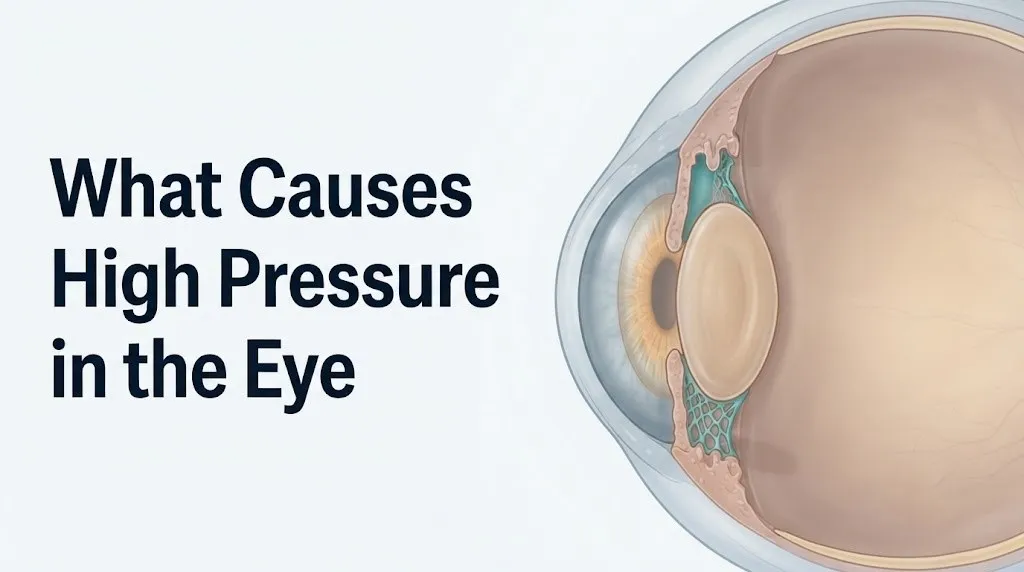
The ciliary body, a ring-shaped structure sitting just behind the iris, continuously produces aqueous humor through its ciliary processes. This fluid flows from the posterior chamber of the eye through the pupil into the anterior chamber, then exits primarily through the trabecular meshwork at the drainage angle, passes into Schlemm’s canal, and eventually rejoins the body’s venous circulation through aqueous veins.
Think of this system like a sink with the tap running at a constant rate. If the drain is partially blocked, water levels rise. The tap doesn’t have to run faster for the sink to overflow. The trabecular meshwork is that drain, and in the majority of people with elevated IOP, the drain is the problem.
The causes that follow all act through one of three final pathways: impaired outflow through the trabecular meshwork, elevated pressure in the episcleral veins (which reduces the gradient needed to pull fluid out), or, in rare cases, abnormally increased aqueous production by the ciliary body.
What Is Normal Eye Pressure and What Counts as High?
Normal intraocular pressure ranges from 10 to 21 mmHg, measured in millimeters of mercury, with the clinical threshold for ocular hypertension set at any reading above 21 mmHg on repeated measurement. A single elevated reading can reflect measurement variability, corneal thickness differences, or testing technique; diagnosis of true ocular hypertension requires confirmed elevation across multiple measurements.
IOP is not static. It follows a diurnal variation pattern, typically peaking in the early morning hours and declining through the afternoon. A 2021 study published in the British Journal of Ophthalmology found that IOP can fluctuate by 3 to 6 mmHg across a single day in healthy individuals, and by even wider margins in people with glaucoma. This means a single midday reading may underestimate a person’s true peak IOP.
| IOP Level (mmHg) | Clinical Classification | Clinical Action |
|---|---|---|
| 10 to 21 | Normal range | Routine monitoring per age and risk |
| 22 to 24 | Mildly elevated | Repeat measurement, risk factor assessment |
| 25 to 29 | Moderately elevated | Ophthalmologist evaluation for glaucoma workup |
| 30 or above | High; urgent evaluation | Same-day or next-day ophthalmologist visit |
| Sudden severe elevation with symptoms | Possible acute angle-closure | Emergency room or emergency ophthalmology immediately |
Central corneal thickness (CCT) affects IOP readings directly. Thicker corneas produce artificially high readings; thinner corneas produce falsely low ones. The Ocular Hypertension Treatment Study (OHTS) identified CCT below 555 microns as an independent risk factor for conversion from ocular hypertension to glaucoma, separate from the IOP reading itself.
Older adults and people with high myopia (nearsightedness greater than 6 diopters) often have lower scleral rigidity, which can also affect measured IOP. A board-certified ophthalmologist will interpret IOP readings alongside pachymetry (corneal thickness measurement) for accurate clinical assessment.
How Aqueous Humor Drainage Controls Eye Pressure
Eye pressure is controlled almost entirely by the rate of aqueous humor outflow, not by the rate of production, because aqueous production by the ciliary body remains remarkably constant under normal physiological conditions. When drainage slows, pressure rises with near mathematical predictability.
Aqueous humor exits the eye through two parallel pathways. The conventional pathway, which handles approximately 70 to 90 percent of outflow, moves fluid from the anterior chamber through the trabecular meshwork into Schlemm’s canal and then into episcleral veins. The uveoscleral pathway handles the remaining 10 to 30 percent, moving fluid through the ciliary body and out across the sclera.
Each of these pathways can be disrupted by different causes. Trabecular meshwork blockage is the mechanism behind primary open-angle glaucoma and most secondary glaucomas. Uveoscleral pathway disruption is the mechanism targeted by prostaglandin analog eye drops, which increase uveoscleral outflow to lower IOP therapeutically.
Episcleral venous pressure matters too. This is the pressure in the small veins on the surface of the eye into which Schlemm’s canal drains. When episcleral venous pressure rises (as it can in certain vascular conditions, thyroid eye disease, or carotid cavernous fistulas), the pressure gradient pulling aqueous out of Schlemm’s canal shrinks, and IOP rises even when the trabecular meshwork itself is undamaged.
Key Takeaway: The root cause of high eye pressure in most people is a drainage problem in the trabecular meshwork, not too much fluid production. This distinction directly shapes every treatment approach used by ophthalmologists.
Trabecular Meshwork Obstruction as the Primary Cause
Trabecular meshwork obstruction is the primary cause of elevated IOP in primary open-angle glaucoma, the most common form of glaucoma worldwide, and is responsible for the majority of cases of ocular hypertension seen in clinical practice. The trabecular meshwork is a sponge-like tissue located at the drainage angle between the cornea and iris, and its gradual dysfunction is often the central event in IOP elevation.
The trabecular meshwork contains specialized cells that actively maintain the extracellular matrix (ECM) composing the meshwork’s filtering structure. In primary open-angle glaucoma, the aqueous drainage outflow resistance through this tissue increases due to accumulation of abnormal ECM proteins, loss of trabecular cells, and stiffening of the meshwork structure. Research published in Investigative Ophthalmology and Visual Science has documented progressive changes in trabecular meshwork gene expression and cytoskeletal organization that reduce its compliance and outflow capacity over time.
The meshwork doesn’t close visibly. The anterior chamber angle (the space where the iris and cornea meet, containing the meshwork) remains anatomically open, which is why this form is called open-angle. Think of it like a shower drain that’s gradually clogged with debris rather than physically blocked by a stopper. Water still enters the drain, but flow rate is progressively reduced.
Pseudoexfoliation syndrome, a condition where abnormal fibrillar protein material deposits throughout the anterior segment, is one of the most potent forms of trabecular meshwork obstruction. According to the American Academy of Ophthalmology, pseudoexfoliation is present in approximately 30 percent of open-angle glaucoma cases worldwide and is significantly more common in people of Scandinavian descent.
Pigment dispersion syndrome produces a similar obstruction through a different material. In this condition, pigment granules from the posterior iris epithelium are released into the aqueous humor and accumulate in the trabecular meshwork, progressively reducing outflow. It affects primarily young, myopic adults, and men are affected roughly twice as often as women.
What Causes Ocular Hypertension from Steroid Use?
Corticosteroids cause ocular hypertension by activating glucocorticoid receptors in trabecular meshwork cells, triggering accumulation of abnormal extracellular matrix proteins that stiffen the meshwork and reduce aqueous outflow. This effect is well-established by controlled human clinical research and applies regardless of the route of steroid administration.
Approximately 5 to 6 percent of the general population are “high responders” to corticosteroids, meaning their IOP rises markedly (by 15 mmHg or more) with prolonged steroid use. Another 30 percent show a moderate IOP response of 6 to 15 mmHg. People with pre-existing primary open-angle glaucoma or a family history of glaucoma are disproportionately likely to be high responders.
Routes of corticosteroid administration that can raise IOP include:
- Topical ophthalmic steroid drops (highest IOP risk due to direct ocular contact)
- Periocular injections (sub-Tenon or intravitreal corticosteroid)
- Inhaled corticosteroids (nasal and pulmonary, with modest but real risk at high doses and prolonged use)
- Systemic oral or intravenous corticosteroids (lower but measurable IOP risk, especially at high doses)
- Topical skin creams applied near the eye (documented IOP elevating effect with prolonged periocular use)
Steroid-induced IOP elevation typically reverses within weeks of discontinuing the corticosteroid, provided optic nerve damage has not already occurred. However, chronic steroid use in unmonitored patients can produce permanent trabecular meshwork damage and glaucoma. Any patient on long-term systemic, inhaled, or topical corticosteroids should have baseline IOP measured by an ophthalmologist and monitored every 6 to 12 months during treatment.
Eye Injuries and Conditions That Cause Increased Eye Pressure
Eye injuries and specific anterior segment conditions can raise IOP by physically blocking aqueous outflow, triggering inflammation that obstructs drainage, or releasing cellular material that clogs the trabecular meshwork. These are classified as secondary causes of ocular hypertension because they arise from a named event or condition rather than primary trabecular dysfunction.
Hyphema, the accumulation of blood in the anterior chamber following blunt trauma to the eye, raises IOP through two mechanisms: the physical presence of red blood cells in the trabecular meshwork reduces outflow directly, and the metabolic products released from lysing red blood cells further compromise trabecular cell function. Traumatic hyphema typically resolves with management, but severe or repeated cases can cause permanent trabecular damage.
Uveitis (inflammation of the uveal tract, which includes the iris, ciliary body, and choroid) raises IOP primarily by releasing inflammatory cells, proteins, and debris into the aqueous humor. These inflammatory materials clog the trabecular meshwork. Uveitic glaucoma can also develop from posterior synechiae (adhesions between the iris and lens) that block the pupil, preventing aqueous from reaching the drainage angle at all. The American Uveitis Society notes that IOP management in uveitis is complicated by the fact that treating inflammation often requires corticosteroids, which themselves can raise IOP.
Other injury-related causes include:
- Chemical burns to the anterior segment, which disrupt trabecular cell viability
- Lens dislocation (subluxation) that physically blocks aqueous flow
- Ghost cell glaucoma, where degenerated red blood cell remnants obstruct the trabecular meshwork weeks after a vitreous hemorrhage
Key Takeaway: Any blunt injury to the eye followed by pain, light sensitivity, or vision change in the days or weeks afterward warrants evaluation by a board-certified ophthalmologist, as delayed IOP elevation is a recognized complication of ocular trauma.
Systemic Diseases That Cause Eye Pressure to Rise
Several systemic medical conditions can raise IOP by elevating episcleral venous pressure, increasing blood viscosity, promoting neovascularization of the iris, or directly compressing orbital and ocular structures. These connections between body-wide disease and intraocular pressure are frequently missed in primary care settings.
Thyroid eye disease (Graves’ orbitopathy) raises IOP through two mechanisms: first, proptosis (forward displacement of the eyeball) compresses the episcleral veins and impairs venous drainage; second, restriction of the inferior rectus muscle increases resistance to upward gaze, artificially elevating IOP in the upgaze position by 5 to 10 mmHg in some patients.
Diabetes mellitus is associated with elevated IOP through several pathways. Observational data published in JAMA Ophthalmology in 2019 found an association between type 2 diabetes and increased IOP, with proposed mechanisms including increased episcleral venous pressure, glycation of trabecular meshwork proteins, and neovascular glaucoma (where new blood vessels formed in diabetic retinopathy grow over the iris and drainage angle, physically blocking aqueous outflow). Neovascular glaucoma can produce IOP values of 40 to 70 mmHg and represents one of the most aggressive forms of secondary glaucoma.
Obstructive sleep apnea (OSA) has been associated with elevated IOP in multiple observational studies. The proposed mechanisms include nocturnal hypoxia, elevated arterial carbon dioxide levels, and increased venous pressure from repeated apneic episodes. A 2022 review published in Sleep Medicine Reviews found a statistically meaningful association between moderate-to-severe OSA and glaucoma risk. People with diagnosed OSA who also have any glaucoma risk factors should discuss IOP screening with their ophthalmologist.
Medications That Cause Eye Pressure to Increase
Several medication classes raise intraocular pressure by impairing aqueous outflow, dilating the pupil (which can narrow the drainage angle), or elevating episcleral venous pressure. This is a clinically important and frequently overlooked cause of elevated IOP, particularly in older adults on multiple medications.
Patients taking any of the following medication classes should disclose them to their ophthalmologist at every visit:
- Corticosteroids (any route, as detailed in the previous section)
- Anticholinergic medications (including some antihistamines, antispasmodics, bladder medications, and tricyclic antidepressants): these dilate the pupil, which in people with naturally shallow anterior chamber angles can precipitate angle-closure by pushing the iris root against the trabecular meshwork
- Sulfa-based medications (including topiramate used for migraines and epilepsy, and hydrochlorothiazide): can cause supraciliary effusion that pushes the iris and lens forward, narrowing the angle
- Selective serotonin reuptake inhibitors (SSRIs): associated with IOP elevation in some observational studies, with proposed mechanism involving serotonin receptor activity affecting aqueous humor dynamics
- Sympathomimetics (including pseudoephedrine in decongestants and some eye drops marketed for redness relief): pupil dilation can precipitate angle-closure in susceptible individuals
| Medication Class | Mechanism of IOP Elevation | Risk Level | Population at Greatest Risk |
|---|---|---|---|
| Corticosteroids (all routes) | Trabecular ECM accumulation | High | Glaucoma family history; prior diagnosis |
| Anticholinergics | Pupil dilation, angle narrowing | Moderate | Narrow-angle anatomy, older adults |
| Topiramate | Supraciliary effusion, angle closure | Moderate | Women, patients on rapid dose escalation |
| SSRIs | Aqueous dynamics via serotonin receptors | Low to moderate | Older patients, those with elevated baseline IOP |
| Decongestant eye drops (vasoconstrictors) | Sympathomimetic pupil dilation | Low to moderate | Pre-existing narrow angle |
Anyone starting a new medication who notices eye pain, halos, or blurring should contact a board-certified ophthalmologist promptly and mention the new medication by name.
Key Takeaway: Topiramate (Topamax) and anticholinergic medications are among the most clinically significant drug causes of elevated IOP, and neither is commonly flagged during routine prescribing in non-eye care settings.
What Causes Eye Pressure to Rise Suddenly?
Sudden IOP elevation is most often caused by acute angle-closure glaucoma, a condition where the peripheral iris physically blocks the trabecular meshwork drainage angle, trapping aqueous humor in the anterior chamber and causing IOP to spike dramatically, sometimes within minutes to hours. This is a genuine ophthalmic emergency.
In acute angle-closure, the iris is pushed or pulled forward until it contacts the peripheral cornea at the drainage angle. This contact seals the trabecular meshwork entirely. IOP can rise from a normal value to 50 or even 70 mmHg within hours. At these pressures, the optic nerve can be irreversibly damaged in a matter of hours. This is not a situation to monitor at home overnight.
Other causes of rapid IOP elevation include:
- Inflammatory uveitis with pupillary block: inflammation can bind the iris to the lens, blocking aqueous from passing through the pupil to reach the drainage angle
- Neovascular glaucoma from acute vascular events: sudden retinal vein occlusion can trigger rapid neovascularization of the iris within weeks, leading to rapid IOP rise
- Spontaneous hyphema: bleeding into the anterior chamber without trauma can occur in patients on anticoagulants or with iris neovascularization
- Lens-particle glaucoma: a sudden cataract rupture can release lens material into the anterior chamber, causing rapid trabecular blockage
People with hyperopia (farsightedness) have anatomically shallower anterior chambers, making them structurally predisposed to angle-closure. Women are affected by acute angle-closure approximately three times more often than men, largely due to anterior chamber depth differences related to biological sex and lens size. East Asian populations have a substantially higher prevalence of narrow-angle anatomy compared to European-ancestry populations.
Risk Factors That Make High Eye Pressure More Likely
The Ocular Hypertension Treatment Study, a landmark multicenter randomized controlled trial published in JAMA Ophthalmology, identified five independent risk factors that predict conversion from ocular hypertension to glaucoma: older age, elevated baseline IOP, larger cup-to-disc ratio, thinner central corneal thickness, and pattern standard deviation on visual field testing. Each of these adds predictive power beyond IOP alone.
Additional risk factors for developing elevated IOP in the first place include:
- Family history: First-degree relatives of people with primary open-angle glaucoma have a 2 to 5 times greater risk of developing elevated IOP and glaucoma
- African ancestry: Research published in the Archives of Ophthalmology found that primary open-angle glaucoma affects African American individuals 3 to 4 times more often than European American individuals, with more aggressive presentation and earlier onset. The physiological basis involves differences in trabecular meshwork morphology, scleral biomechanics, and optic nerve susceptibility to pressure-related damage
- East Asian ancestry: People of East Asian descent have higher rates of narrow anterior chamber angles, predisposing them to angle-closure glaucoma rather than the open-angle form
- High myopia (nearsightedness above 6 diopters): associated with elongated axial length, altered trabecular morphology, and optic nerve characteristics that increase glaucoma risk
- Low ocular perfusion pressure: people with low systemic blood pressure relative to IOP may develop optic nerve damage at IOP levels that would be tolerated by someone with higher blood pressure
Systemic hypertension does not directly cause ocular hypertension. However, it does affect ocular perfusion pressure, and the relationship between systemic blood pressure and IOP is nonlinear. Age is a consistent independent risk factor, with IOP tending to rise modestly across decades, partly due to increasing trabecular meshwork dysfunction with cellular aging.
What Causes High Pressure in One Eye Only?
Unilateral IOP elevation (high pressure in one eye only) strongly suggests a secondary cause affecting that eye specifically, including previous trauma, unilateral uveitis, unilateral pigment dispersion, pseudoexfoliation, or angle-closure affecting one eye before the other. Bilateral IOP elevation more commonly reflects primary open-angle glaucoma, systemic causes, or medication effects.
According to the American Academy of Ophthalmology, any asymmetric IOP difference of more than 3 to 4 mmHg between the two eyes warrants thorough investigation, even if both eyes fall within the normal range. The pressure asymmetry itself is a clinical signal. A clinician will check for evidence of previous trauma (even old trauma the patient may have forgotten), signs of uveitis, pigment in the trabecular meshwork, pseudoexfoliation material on the lens capsule, and angle configuration differences.
Certain causes of unilateral IOP elevation carry specific clinical significance:
- Fuchs’ heterochromic iridocyclitis: a chronic low-grade uveitis that typically affects one eye, causes heterochromia (different iris color between the two eyes), and is associated with elevated IOP and cataract formation. Patients are often unaware of the underlying uveitis because it produces no pain.
- Previous penetrating eye injury or surgery: scar tissue from old injury or surgery can distort the drainage angle unilaterally
- Intraocular tumors: choroidal melanoma and iris tumors can impede aqueous outflow in one eye through direct mechanical obstruction or exudative changes
- Carotid cavernous fistula: an abnormal connection between the carotid artery and the cavernous sinus can raise episcleral venous pressure on one side, causing unilateral IOP elevation with a characteristic reddish, dilated appearance of episcleral vessels
Key Takeaway: A difference of more than 3 mmHg between the IOP of your two eyes is a clinically meaningful finding, not a normal measurement variation, and warrants a focused ophthalmological examination even if both readings appear normal in isolation.
What Causes Increased Eye Pressure in Older Adults?
IOP tends to rise modestly with age due to progressive trabecular meshwork dysfunction, increased aqueous outflow resistance from ECM accumulation in the meshwork, and a higher prevalence of pseudoexfoliation syndrome, which increases steeply in incidence after age 60. The National Eye Institute reports that glaucoma, which elevated IOP can drive, is the second leading cause of blindness worldwide and disproportionately affects people over age 60.
Several age-specific mechanisms compound this risk. Trabecular cells decline in number with age, reducing the active maintenance of the meshwork’s filtering structure. The remaining cells produce more transforming growth factor-beta (TGF-beta), a cytokine that promotes ECM accumulation and trabecular stiffening. Simultaneously, the crystalline lens continues to grow throughout life, gradually reducing the space available in the anterior chamber and narrowing the drainage angle, a process that can eventually precipitate angle-closure in eyes that were previously safe.
Older adults are also more likely to be using corticosteroids for arthritis, asthma, chronic obstructive pulmonary disease, or autoimmune conditions, and anticholinergics for bladder overactivity or other conditions. Both drug classes raise IOP. This polypharmacy reality makes regular IOP screening especially important for anyone over age 60, regardless of eye symptoms.
The American Academy of Ophthalmology recommends a comprehensive eye examination including IOP measurement every 1 to 2 years for adults aged 40 and above, and annually for those over 65 or those with any known risk factors. Many older adults skip these examinations because elevated IOP is silent. By the time peripheral vision loss from glaucoma is noticeable, significant optic nerve damage has typically already occurred.
What Does High Eye Pressure Feel Like?
In the vast majority of cases, elevated intraocular pressure from chronic open-angle conditions produces no symptoms whatsoever, which is one of the most clinically important facts about ocular hypertension: you cannot feel it accumulating over years. This is why the condition is sometimes compared to hypertension of the blood vessels, silent at first, destructive over time.
When IOP does produce symptoms, this usually indicates either a very rapid pressure spike (as in acute angle-closure glaucoma) or secondarily induced corneal changes from chronically elevated pressure. Symptoms that can accompany elevated IOP include:
- Blurred vision (due to corneal edema from high pressure affecting the corneal endothelium’s fluid pumping mechanism)
- Colored halos around lights, particularly at night (produced by corneal edema diffracting light abnormally)
- A sensation of eye fullness or pressure behind the eye (not always reliable, as many people with very high IOP report no fullness sensation)
- Headache localized to the brow or temple on the affected side
- Nausea and vomiting in acute angle-closure (due to the vagal reflex triggered by severe intraocular pain)
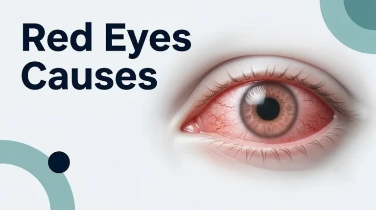
- Eye redness (from conjunctival vessel dilation)
The absence of symptoms does not mean pressure is normal. Many people with IOP in the 25 to 30 mmHg range for years feel nothing until an ophthalmologist measures their pressure. Conversely, some people with normal IOP report significant pressure and fullness sensations from entirely unrelated causes (eye muscle strain, sinus congestion, dry eye). Subjective eye pressure sensation is not a reliable indicator of actual IOP.
People with migraine, cluster headache, or trigeminal neuralgia may describe eye pressure symptoms that have nothing to do with IOP. A board-certified ophthalmologist can differentiate these presentations with a direct pressure measurement.
How High Eye Pressure Is Measured and Diagnosed
Intraocular pressure is measured clinically using tonometry, with Goldmann applanation tonometry considered the gold standard for accuracy in a clinical setting. A comprehensive evaluation for elevated IOP goes well beyond a single pressure reading and includes corneal thickness measurement, optic nerve assessment, and drainage angle examination.
The diagnostic process for someone with elevated IOP typically unfolds as follows:
- IOP measurement by tonometry: Goldmann applanation tonometry uses a calibrated probe to flatten a small area of the cornea while the patient is at the slit lamp. Non-contact tonometry (“air puff test”) is less accurate but used for screening. Rebound tonometry (iCare) is increasingly used for monitoring.
- Pachymetry (central corneal thickness): measured by ultrasound or optical coherence techniques. CCT directly corrects IOP reading interpretation and identifies glaucoma conversion risk independently.
- Gonioscopy: a contact lens examination that allows the ophthalmologist to directly visualize the trabecular meshwork and drainage angle, classifying it as open, narrow, or closed, and identifying secondary causes such as pigment deposition, pseudoexfoliation material, or angle recession from prior trauma.
- Optic nerve evaluation: slit-lamp biomicroscopy and/or optical coherence tomography (OCT) of the optic nerve head and retinal nerve fiber layer to detect early structural damage from elevated IOP.
- Visual field testing (perimetry): automated threshold perimetry detects functional vision loss from optic nerve damage. Loss is typically peripheral first and is detectable before the patient notices it subjectively.
- Diurnal IOP curve: multiple IOP readings across a single day to capture peak pressure, particularly important when a single reading appears borderline.
Key Takeaway: A single normal eye pressure reading does not rule out clinically significant IOP elevation, because pressure fluctuates across the day and peaks in the early morning before most clinic visits occur.
The Relationship Between High Eye Pressure and Glaucoma
Elevated intraocular pressure is the primary modifiable risk factor for glaucoma, but having high IOP does not mean you have glaucoma, and having glaucoma does not require IOP to be elevated at all. The relationship between the two conditions is probabilistic, not deterministic.
Ocular hypertension is defined as IOP above 21 mmHg without evidence of optic nerve damage or visual field loss. Primary open-angle glaucoma is defined by characteristic optic nerve damage and visual field loss, which often but not always occurs in the context of elevated IOP. Normal tension glaucoma, a form that affects an estimated 30 to 40 percent of glaucoma patients in the United States and higher proportions in East Asian populations, involves progressive glaucomatous optic nerve damage at IOP levels that fall within the statistically normal range.
The Ocular Hypertension Treatment Study found that lowering IOP by 20 percent from baseline with medication reduced the risk of developing glaucoma over 5 years by approximately 50 percent in people with ocular hypertension. This remains one of the most actionable findings in glaucoma clinical research. It also confirmed that not everyone with elevated IOP progresses to glaucoma: in the untreated group, only approximately 9.5 percent developed glaucoma over 5 years.
Risk of conversion from ocular hypertension to glaucoma increases with higher IOP, thinner corneas, larger cup-to-disc ratio, and older age. The optic nerve head is not equally susceptible across all people at a given IOP level. Some optic nerves tolerate elevated pressure for decades; others sustain damage at pressures that most people would never notice. This variability is why a single IOP measurement should always be evaluated in the context of optic nerve appearance and risk factors, not treated as a standalone diagnosis.
When to Seek Care for High Eye Pressure
Anyone whose IOP is measured above 21 mmHg on repeated assessment should schedule an evaluation with a board-certified ophthalmologist within two to four weeks, not an optometrist screening alone, for full optic nerve and drainage angle assessment. Acute or sudden onset symptoms require same-day or emergency evaluation.
| Situation | Urgency | Action |
|---|---|---|
| IOP 22 to 24 mmHg, no symptoms, no optic nerve changes | Routine | Ophthalmologist evaluation within 2 to 4 weeks |
| IOP 25 to 29 mmHg, no symptoms | Prompt | Ophthalmologist within 1 to 2 weeks |
| IOP 30+ mmHg, no symptoms | Urgent | Ophthalmologist same day or next day |
| Any IOP with early visual field changes | Urgent | Ophthalmologist within 24 to 48 hours |
| Sudden eye pain, halos, blurred vision, headache, nausea | Emergency | Emergency room or emergency ophthalmology immediately |
| Eye injury followed by persistent pain or vision change | Urgent | Ophthalmologist within 24 hours |
People with a first-degree relative diagnosed with glaucoma should begin IOP screening and comprehensive eye examinations by age 35 rather than the standard age 40. African American individuals face higher risk with earlier onset and are recommended by the American Academy of Ophthalmology to begin regular comprehensive eye examinations no later than age 40 and ideally in their mid-30s.
Pregnant women represent a special case. IOP typically decreases during pregnancy, likely due to increased outflow facility from progesterone-related effects on the trabecular meshwork and reduced episcleral venous pressure. Women on IOP-lowering eye drops who become pregnant should discuss medication safety with both their ophthalmologist and obstetrician-gynecologist, as some prostaglandin analogs are contraindicated in pregnancy due to uterotonic effects.
Emergency Symptoms: When to Call 911 or Go to the ER
Certain symptoms associated with high pressure in the eye require immediate emergency evaluation. Do not wait to see if these resolve on their own.
Call 911 or go to the nearest emergency room immediately if you experience:
- Sudden severe pain in one or both eyes, described as intense pressure or aching
- Halos or rainbow-colored rings around lights, particularly combined with eye pain
- Sudden blurred or foggy vision in one eye that does not improve within minutes
- Headache over the brow or temple on one side accompanied by any eye symptom
- Nausea or vomiting associated with eye pain (a vagal sign of severe acute IOP elevation)
- Sudden loss of peripheral or central vision in any portion of the visual field
- A reddened, hard-feeling eye with any accompanying visual disturbance
These presentations can indicate acute angle-closure glaucoma or another cause of rapid, severe IOP elevation that can produce permanent optic nerve damage and blindness within hours. This requires emergency ophthalmology or emergency room assessment, not an urgent care appointment or a phone call.
Frequently Asked Questions About High Pressure in the Eye
What is the most common cause of high pressure in the eye?
The most common cause of high pressure in the eye is impaired aqueous humor drainage through the trabecular meshwork, the primary drainage structure located at the anterior chamber angle.
This mechanism is responsible for primary open-angle glaucoma and most secondary glaucomas, and it typically develops slowly over years without producing any symptoms until optic nerve damage is established.
A board-certified ophthalmologist can evaluate trabecular meshwork function indirectly through IOP measurement, gonioscopy, and optic nerve imaging.
Can stress or anxiety cause eye pressure to increase?
Acute psychological stress can cause a temporary, modest rise in IOP through sympathetic nervous system activation and increased blood pressure, but chronic stress alone is not established as a primary cause of clinically elevated IOP.
The evidence for a direct stress-IOP relationship is observational and preliminary, not confirmed by controlled clinical trials as of 2026.
If you are under significant chronic stress and have any glaucoma risk factors, mentioning this to your ophthalmologist during a routine IOP evaluation is reasonable, but stress reduction alone should not be relied on as an IOP management strategy.
What does high eye pressure feel like?
In most people with chronically elevated IOP, high eye pressure produces no sensation at all, which is why it often goes undetected for years without regular eye examinations.
Symptoms only tend to appear when IOP rises rapidly (as in acute angle-closure) or when the pressure is high enough to affect the cornea, causing blurred vision or halos around lights.
A subjective sense of pressure, fullness, or tightness behind the eye can occur but is not a reliable indicator of actual IOP, as many people with very high pressure feel nothing while others with normal pressure report significant discomfort from unrelated causes.
Can high blood pressure in the body cause high pressure in the eye?
Systemic hypertension (high blood pressure) does not directly cause elevated IOP through a simple pressure transmission pathway, because the eye regulates its own pressure independently of blood pressure.
However, systemic hypertension affects ocular perfusion pressure and episcleral venous pressure in ways that can indirectly influence IOP, and uncontrolled hypertension is associated with increased glaucoma risk in some observational studies.
Anyone with diagnosed systemic hypertension should inform their ophthalmologist, particularly if they are over age 50 or have other glaucoma risk factors.
What medications cause eye pressure to rise?
Corticosteroids are the most clinically significant medication class known to raise IOP, through glucocorticoid receptor activation in trabecular meshwork cells that reduces aqueous outflow.
Anticholinergic medications, topiramate, and some decongestants can also raise IOP, primarily by narrowing the drainage angle through pupil dilation or anterior segment anatomical changes.
Patients taking any of these drug classes should tell their ophthalmologist at every visit and should not discontinue prescribed medications without discussing the IOP implications with both their prescribing physician and their ophthalmologist.
At what eye pressure level should I be worried?
Any IOP reading above 21 mmHg on repeated measurement warrants evaluation by a board-certified ophthalmologist, as this is the clinically accepted threshold for ocular hypertension.
IOP above 30 mmHg requires same-day or next-day ophthalmologist evaluation, and any IOP elevation accompanied by eye pain, halos, or vision changes requires emergency evaluation the same hour.
The number alone doesn’t tell the whole story: a 22 mmHg reading in someone with a thin cornea, large optic nerve cup, and family history of glaucoma carries different risk than the same reading in a 30-year-old with a thick cornea and no risk factors.
High pressure in the eye is rarely felt before it has already begun doing damage. That’s the clinical reality that makes understanding the causes and risk factors genuinely useful, not just interesting. If you have been told your IOP is above 21 mmHg, the single most important next step is scheduling a comprehensive evaluation with a board-certified ophthalmologist, not just an optometry screening, for gonioscopy, pachymetry, optic nerve imaging, and visual field testing.
If you use corticosteroids in any form, have a family history of glaucoma, are over age 40, or have African or East Asian ancestry, baseline IOP screening and regular monitoring are medically indicated regardless of symptoms. Ask your ophthalmologist specifically about your cup-to-disc ratio and corneal thickness, because these two measurements tell you more about your personal glaucoma risk than your IOP number alone.
The good news: when elevated IOP is caught before optic nerve damage occurs, treatment with pressure-lowering eye drops or minimally invasive procedures can prevent vision loss with a high degree of effectiveness. What you cannot do is catch it by waiting for symptoms that, in most cases, will never arrive on their own.