Heel Spur Symptoms: What They Feel Like and When to Act
Heel spur symptoms typically begin as sharp, stabbing pain under or behind the heel with the first few steps of the morning, easing somewhat as you move but returning after prolonged standing or sitting. Not every calcaneal spur causes pain, but when it does, the symptom pattern is distinctive enough that most people who have one describe the experience in nearly identical terms before they have any diagnosis at all.
According to the National Institute of Arthritis and Musculoskeletal and Skin Diseases (NIAMS), heel pain is one of the most common musculoskeletal complaints in adults, and calcaneal spurs are present on imaging in a substantial proportion of adults who report chronic heel pain. Research published in the Journal of the American Podiatric Medical Association found that approximately 50% of people with plantar fasciitis have a detectable calcaneal spur on X-ray, yet an equally notable proportion of people with visible spurs on imaging have no symptoms at all.
This article covers every recognized symptom of heel spurs with the physiological mechanism behind each one, the difference between inferior (plantar) and dorsal (posterior) spur presentations, how symptoms change over time, how heel spur pain differs from plantar fasciitis, which populations are most affected, when to stop self-managing and see a provider, and the specific clinical thresholds that require same-day or emergency evaluation.
Heel Spur Symptoms: The Clinical Overview
Heel spur symptoms are produced by a combination of bony calcaneal osteophyte formation and the soft tissue inflammation that either caused the spur to develop or is perpetuated by its presence at the enthesis.
The calcaneus is the largest bone in the foot. Its inferior surface is the attachment point for the plantar fascia, a thick band of connective tissue that supports the arch. Its posterior surface is where the Achilles tendon inserts. Both attachment sites are called entheses, and both can become sites of chronic traction-driven bone spur formation.
The key clinical distinction that most symptom descriptions miss: the spur itself is often not the direct source of pain. Instead, the inflamed periosteum (the fibrous membrane covering bone), the irritated enthesis tissue, and the compressed or irritated soft tissue structures around the spur are what generate the pain signal. This is why removing a spur surgically does not always resolve pain, and why the symptom pattern mirrors plantar fasciitis so closely.
Key symptoms at a glance:
- Sharp stabbing pain with first steps in the morning or after rest
- Tenderness directly under or behind the heel on palpation
- Aching pain that worsens with prolonged standing or walking
- Pain that briefly improves with movement but worsens again with extended activity
- Localized swelling or warmth at the heel in flare states
- Altered gait (walking on the outer edge of the foot to avoid heel contact)
What Is a Heel Spur and How Does It Form
A heel spur is an abnormal bony projection, clinically called an osteophyte, that forms on the calcaneus as a response to repetitive mechanical stress at the site where soft tissue attaches to bone.
The formation process follows a predictable sequence. Repetitive traction at the plantar fascia enthesis or Achilles tendon insertion creates microscopic tears in the fibrous tissue where it meets the calcaneal periosteum. The periosteum responds to this chronic low-grade injury by activating osteoblasts, the bone-forming cells. Over months to years, calcium hydroxyapatite is deposited at the site of repeated injury, building into a bony projection that can range from a few millimeters to over a centimeter in length.
Think of it like a callus, but inside the foot instead of on the skin. Just as a callus forms where the skin experiences repeated friction, a bone spur forms where the periosteum experiences repeated traction. The structure is the body’s attempt to reinforce a chronically stressed attachment point.
Two anatomical locations produce two distinct spur types. An inferior calcaneal spur grows from the bottom of the heel, at or near the plantar fascia attachment. A dorsal calcaneal spur (also called a posterior spur) grows from the back of the heel, at or near the Achilles tendon insertion. These two types have different symptom profiles, different aggravating factors, and different clinical implications, which this article covers separately below.
The American Academy of Orthopaedic Surgeons notes that heel spurs are associated with plantar fasciitis but are not synonymous with it. Many people have spurs without fasciitis, and many people have fasciitis without a visible spur.
Bone Spur in Heel Symptoms: The Core Pain Pattern
The defining bone spur in heel symptoms are centered on pain that behaves in a specific, recognizable pattern tied to the mechanics of weight-bearing and the inflammatory cycle at the calcaneal enthesis.
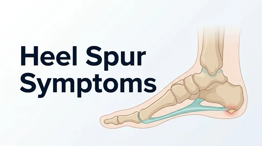
The most reported first symptom is sharp, stabbing pain under the heel with the very first steps after waking or after any prolonged period of rest. This happens because the plantar fascia shortens during non-weight-bearing rest. The contracted fascia puts the already-inflamed enthesis under acute tensile load the moment the foot bears weight. The inflammatory mediator prostaglandin E2, produced at the periosteum via the COX-2 enzyme, is at its highest local concentration after a period of inactivity, which amplifies the pain signal during that initial contact.
Core symptoms by location:
| Symptom | Location | Typical Timing |
|---|---|---|
| Sharp stabbing pain | Under the heel (inferior spur) | First steps after rest |
| Dull aching pain | Through the arch | After prolonged standing |
| Tenderness on pressure | Directly over the spur site | With palpation or firm footwear |
| Localized warmth/swelling | Heel border | During or after a flare |
| Altered gait / toe-walking | Whole foot | When pain is severe |
An important note for people who take anti-inflammatory medications regularly for arthritis or other chronic pain conditions: those medications (specifically COX-2 inhibitors and non-steroidal anti-inflammatory drugs) may partially suppress the inflammatory component of heel spur pain, which can obscure the symptom pattern and make the presentation seem milder than the underlying structural change actually is. A primary care physician should know about all medications when evaluating heel pain.
Symptoms of a Heel Spur: The Full Clinical Picture
The symptoms of a heel spur extend beyond the classic morning pain that most descriptions focus on, and understanding the full clinical picture helps explain why so many people misattribute their symptoms or delay appropriate evaluation.
Pain character ranges from sharp and stabbing during acute phases to a deep, persistent ache during chronic phases. During a flare, some people describe a sensation of a stone or pebble pressing into the heel from inside, even when standing on a smooth, flat surface. This sensation is produced by pressure on the inflamed periosteum over the bony projection.
Beyond pain, the full symptom spectrum includes:
- Tenderness on direct pressure: Applying finger pressure to the specific point on the heel where the spur is located typically produces sharp, localized pain. This is the single most consistent physical examination finding.
- Arch pain: Because the plantar fascia runs from the calcaneus to the metatarsal heads, enthesis irritation at the heel often produces referred aching along the medial arch of the foot.
- Morning stiffness: Beyond the sharp first-step pain, many people experience 5 to 15 minutes of significant stiffness before the foot loosens up.
- End-of-day fatigue pain: Pain returns or worsens after several hours on the feet, as the inflamed tissue is progressively loaded through the day.
- Antalgic gait: People with significant heel spur pain frequently walk on the ball of the foot or the outer edge of the heel to reduce contact with the painful area, which can secondarily produce hip, knee, or lower back pain from altered biomechanics.
Research published in Foot and Ankle International found that gait alteration in response to chronic heel pain significantly increases the mechanical load on the contralateral (opposite) limb, making bilateral lower extremity complaints a documented downstream consequence of untreated unilateral heel spur pain.
Key Takeaway: The full symptom picture of a heel spur includes not just heel pain but arch aching, morning stiffness, end-of-day pain recurrence, and altered gait that can create secondary pain in the knee, hip, and lower back if left unmanaged.
Calcaneal Spur Symptoms: What the Research Shows
Calcaneal spur symptoms vary considerably between individuals, and understanding why requires knowing something important: the imaging finding of a spur and the experience of symptoms are not reliably correlated.
A cross-sectional study published in the Journal of the American Podiatric Medical Association found calcaneal spurs present on plain radiographs in 16 to 27% of the general adult population, depending on age and imaging protocol. Of those with visible spurs, roughly half reported no heel pain at the time of imaging. This does not mean the spur is irrelevant to the painful group; it means the pain is driven primarily by the state of the soft tissue at the enthesis rather than by the mechanical size or presence of the bony projection alone.
When symptoms do occur, their severity correlates more closely with the degree of plantar fascia enthesopathy than with spur size on imaging. A 2-centimeter spur in a person with minimal fascia inflammation may be nearly painless. A smaller spur in someone with significant enthesopathy and periosteal irritation can cause debilitating pain.
Symptom factors most closely associated with clinical severity:
- Duration of the inflammatory cycle at the enthesis (chronic cases hurt differently from acute cases)
- Degree of mechanical loading from body weight, footwear, and daily activity demands
- Presence of associated retrocalcaneal bursitis or plantar fasciitis
- Individual pain sensitization level, which varies by nervous system factors not specific to the foot
- Concurrent systemic inflammatory conditions such as rheumatoid arthritis, reactive arthritis, or ankylosing spondylitis, which can amplify enthesis inflammation beyond what mechanical factors alone would produce
For people with a known inflammatory arthropathy, a board-certified rheumatologist should be part of the evaluation, as the enthesitis in these conditions responds to different treatment approaches than mechanical heel spur pain.
Dorsal Heel Spur Symptoms: The Back-of-Heel Presentation
Dorsal heel spur symptoms are distinct from inferior heel spur symptoms because they involve a different anatomical structure, a different location on the calcaneus, and different aggravating activities.
A dorsal (posterior) calcaneal spur develops at the superior and posterior aspect of the calcaneus, where the Achilles tendon inserts. The key soft tissue structures affected are the Achilles tendon itself, the retrocalcaneal bursa (a fluid-filled sac between the Achilles tendon and the calcaneal bone), and the superficial calcaneal bursa (located between the tendon and the skin).
The primary symptom of a dorsal calcaneal spur is pain at the back of the heel, not the bottom. This is the single most important distinguishing feature. People with dorsal spurs typically report:
- Pain and tenderness directly at the back of the heel, often visible as a prominent bony bump
- Pain that worsens with activities that load the Achilles tendon: running, jumping, climbing stairs, or walking uphill
- Pain aggravated by the back edge of a rigid shoe counter pressing against the bony prominence
- Localized swelling and redness at the back of the heel, particularly after activity
- Morning stiffness at the back of the heel that is relieved partially by gentle calf stretching
The bony prominence associated with dorsal calcaneal spurs is sometimes called a Haglund deformity when the bone of the posterior-superior calcaneal angle is particularly prominent. Haglund deformity and dorsal calcaneal spur often co-occur and together can compress the retrocalcaneal bursa, producing retrocalcaneal bursitis, which adds a burning, swollen quality to the back-of-heel pain.
Women who wear rigid low-backed dress shoes or pumps are disproportionately affected by Haglund-related symptoms, which is why this presentation is sometimes informally called “pump bump.” The rigid shoe counter directly compresses the posterior calcaneal prominence.
Bone Spur on Back of Heel Symptoms: Haglund and Achilles Factors
Bone spur on back of heel symptoms frequently involve the interaction between the calcaneal osteophyte, the Achilles tendon, and the retrocalcaneal bursa in a way that produces a complex of overlapping pain patterns that can be difficult to separate clinically without imaging.
The retrocalcaneal bursa sits in a tight anatomical space between the posterior calcaneal surface and the Achilles tendon. When a dorsal spur is present, this space is mechanically compromised. Each step compresses the bursa, triggering an inflammatory cycle that produces prostaglandin E2 locally. Over time, the bursa can become chronically thickened and inflamed, a condition called retrocalcaneal bursitis, which adds to the pain burden of the underlying spur.
Simultaneously, the Achilles tendon itself may develop enthesopathy at its calcaneal insertion: microscopic tearing, disorganized collagen, and local neovascularization that shows up as increased blood flow on Doppler ultrasound. This is a form of tendinopathy, not true tendinitis, and it responds differently to treatment.
Symptoms that suggest involvement of both the spur and the Achilles insertion include:
- Pain that is worse with passive dorsiflexion of the ankle (pulling the foot upward), which stretches the Achilles against the spur
- Pain that is sharp with the first steps but also returns with a burning quality after sustained running or walking
- A palpable nodule or tender thickening along the lower Achilles tendon
- Visible swelling at the back of the heel that does not resolve completely between activity sessions
Key Takeaway: Dorsal heel spur symptoms involve the back of the heel and worsen with Achilles-loading activities like stair climbing and running, while inferior calcaneal spurs produce bottom-of-heel pain worst with the first steps of the morning after rest.
What Does a Heel Spur Feel Like Day to Day
What a heel spur feels like on a daily basis follows a pattern that most people describe with remarkable consistency, and that pattern gives clinicians useful diagnostic information even before imaging is ordered.
The typical daily pain arc looks like this: severe, sharp pain with the first five to ten steps out of bed or after sitting for more than 20 to 30 minutes. That initial sharpness gives way to a milder, more tolerable ache as the fascia and surrounding soft tissue warm up and the prostaglandin load disperses slightly with movement. After several hours of sustained activity, pain returns, often as a deep, burning ache that is worse toward the end of the workday or after a long walk.
Sitting down for a lunch break and then standing up again can produce a second sharp episode, almost as intense as the morning pain. This is the same nocturnal contracture mechanism compressed into a shorter rest period. Some people describe it as “my heel resets to painful” every time they stop moving for more than 15 minutes.
To describe this pattern accurately to a provider:
- Note the exact time of day when pain is worst and what you were doing immediately before.
- Rate pain on a scale of 0 to 10 using the Visual Analog Scale for pain approach: 0 is no pain, 10 is the worst pain imaginable.
- Record whether pain improves after 10 to 15 minutes of walking or stays constant.
- Note whether pain has changed over weeks or months: better, worse, or the same.
- Document what makes it worse: specific footwear, specific activities, going barefoot on hard floors.
- Note any changes in how you walk (favoring one side, avoiding heel contact).
People who are on their feet for work (teachers, healthcare workers, retail workers, construction workers) often report that the end-of-day pain becomes the dominant complaint as the condition progresses, sometimes overtaking the morning pain in severity. This shift suggests progression from an acute inflammatory phase to a more chronic degenerative enthesopathy pattern.
Heel Bone Spur Symptoms by Severity
Heel bone spur symptoms range from an occasional, barely noticeable twinge to pain severe enough to substantially alter walking, work capacity, and daily function, and understanding where on that spectrum your symptoms fall has real practical implications.
The American Academy of Orthopaedic Surgeons classifies heel pain broadly by functional impact, and podiatrists typically use the clinical severity of symptom interference with daily life to guide treatment escalation. Here is how calcaneal spur symptoms typically map across a severity spectrum:
| Severity Level | Symptom Description | Functional Impact | Clinical Action |
|---|---|---|---|
| Mild | Brief morning pain, resolves within minutes, no gait change | Minimal; tolerable | Self-care: stretching, footwear modification, cushioning |
| Moderate | Morning pain lasting 15 to 30 min, recurs with prolonged standing, tender to touch | Noticeable; interferes with work or activity | Primary care or podiatry evaluation; physical therapy |
| Severe | Pain constant throughout the day, altered gait, prevents normal walking | Significant; limits occupation and daily tasks | Podiatrist or orthopedic specialist; imaging; advanced conservative or procedural care |
| Emergency-Qualifying | Sudden complete inability to bear weight, fever with heel swelling, or tearing sensation | Complete functional loss | Emergency evaluation: go to the emergency room |
Pain severity also varies by time within the condition’s natural history. Early-stage calcaneal spur symptoms tend to be more acutely inflammatory and more responsive to anti-inflammatory strategies. Long-standing cases often transition to a degenerative enthesopathy pattern where inflammation is less prominent but structural tissue disorganization produces chronic mechanical pain.
Older adults with a BMI over 30 tend to present at a higher baseline severity because both age-related reduction in plantar fat pad thickness and increased mechanical loading from body weight compound the enthesis stress. A primary care physician should assess for metabolic contributors in this population before attributing all symptoms to mechanical spur alone.
Symptoms of Heel Spurs That Worsen Over Time
Symptoms of heel spurs that worsen over time are clinically important to recognize because they signal a shift from a condition that typically responds well to conservative care into one that requires structured medical management.
The natural history of untreated calcaneal spur symptoms is variable, but some patterns consistently indicate progression. Pain that initially occurred only in the morning and now persists throughout the day is a progression marker. Gait alteration that was previously absent and has now become habitual is a sign that the condition has moved beyond mild. Night pain, meaning pain that wakes you from sleep or is present at rest without weight-bearing, is not typical of a mechanical heel spur and should prompt evaluation for an alternative or additional diagnosis.
Worsening symptom patterns to watch for:
- Pain now present during seated rest, not just with standing or walking
- Swelling that remains visible at the heel even on rest days
- Tingling, numbness, or burning that radiates from the heel into the arch or toward the toes (suggests tarsal tunnel syndrome or medial calcaneal nerve involvement)
- Second heel beginning to develop similar symptoms (bilateral cases suggest a systemic contributing factor rather than pure mechanical cause)
- Significant loss of ankle range of motion alongside heel pain (suggests Achilles tendon involvement beyond the spur itself)
According to the American Orthopaedic Foot and Ankle Society, most patients with plantar heel pain who receive consistent conservative care (stretching, footwear modification, orthotic support) show improvement within 6 to 12 months. However, approximately 10% of cases do not resolve with conservative measures and require procedural or surgical intervention. Persistent worsening beyond three to six months of consistent conservative care is the threshold that should trigger a structured evaluation by a podiatrist or orthopedic foot and ankle specialist.
Key Takeaway: Night pain, radiating numbness or tingling into the foot, and bilateral onset of heel spur symptoms are three specific warning signs that the condition has progressed or changed character and warrants professional evaluation rather than continued self-management.
Bone Spur Foot Symptoms vs. Plantar Fasciitis
Bone spur foot symptoms and plantar fasciitis symptoms are so closely intertwined that many people have both conditions simultaneously, which is why separating them in everyday symptom descriptions is genuinely difficult.
Plantar fasciitis is an inflammatory and degenerative condition of the plantar fascia itself, involving microtearing along the length of the fascia and enthesopathy at its calcaneal origin. An inferior calcaneal spur forms at the exact same site: the calcaneal enthesis. The two conditions share the same triggering mechanism, often the same risk factors, and a nearly identical morning pain pattern.
Here is how the symptom picture differs in practical terms:
| Feature | Calcaneal Spur | Plantar Fasciitis |
|---|---|---|
| Pain location | Directly under the heel bone | Heel plus arch, often extends toward toes |
| Tenderness point | Single point directly over spur site | Medial calcaneal tubercle + along fascia |
| Arch pain | Secondary, mild | Often prominent; fascia-length aching |
| Imaging finding | Bony spur visible on X-ray | No bony abnormality; thickened fascia on ultrasound |
| Both can coexist | Yes, very commonly | Yes |
| Night pain | Not typical | Not typical; if present, evaluate further |
The practical clinical truth: because these two conditions coexist so frequently, most clinicians treat the soft tissue pathology rather than the spur, regardless of which diagnosis is leading. This is why imaging that confirms a spur does not automatically change the initial treatment approach from what is used for plantar fasciitis.
A person with isolated spur and no fascia pathology may have no symptoms at all. The fascia pathology tends to drive the pain. This is the reason that imaging a spur is most useful for ruling out other causes of heel pain (stress fracture, tarsal tunnel syndrome, calcaneal cyst) rather than for confirming the specific pain generator.
What Are Heel Spur Symptoms in Different Populations
Heel spur symptoms present differently across sex, age group, activity level, and pre-existing condition status, and recognizing these differences helps identify who is at greatest risk and who may need earlier or more aggressive evaluation.
Women are diagnosed with heel spurs and plantar fasciitis at higher rates than men, with some epidemiological studies suggesting a ratio of approximately 2:1. This disparity is partly attributed to footwear choices (high heels load the plantar fascia differently than flat shoes), but hormonal factors also play a role. Estrogen affects collagen quality in connective tissue; the reduction in estrogen after menopause is associated with decreased enthesis resilience and a higher prevalence of enthesopathy. Postmenopausal women presenting with new-onset heel pain should have their foot type, footwear history, and hormone status noted in any clinical evaluation.
Runners and athletes experience calcaneal enthesopathy and spur formation through repetitive impact loading rather than through body weight alone. In runners, symptoms often begin insidiously after a sudden increase in training volume or a change in running surface. Dorsal heel spurs are proportionally more common in athletes who perform jumping activities. A sports medicine physician or physiatrist is an appropriate provider for athletes with heel spur symptoms, particularly when return to sport is a time-sensitive concern.
Older adults over 60 tend to have a thinner plantar fat pad (the natural cushioning on the sole of the heel), which means the calcaneal spur may produce symptoms more easily and at lower activity levels than in younger adults. The National Institutes of Health notes that plantar fat pad atrophy with aging is a documented contributor to heel pain independent of spur formation.
People with flat feet (pes planus) or high-arched feet (pes cavus) have abnormal tension distribution across the plantar fascia. Flat feet increase tension at the medial calcaneal enthesis; high-arched feet create a rigid fascia with poor shock absorption. Both structural types are associated with elevated spur risk, and both benefit from custom orthotic evaluation by a podiatrist.
People with rheumatoid arthritis, reactive arthritis, or ankylosing spondylitis can develop enthesitis (systemic inflammation at entheses) that mimics or compounds mechanical heel spur symptoms. These cases require rheumatological management alongside any local foot treatment.
How Heel Spurs Are Diagnosed
Heel spurs are diagnosed through a combination of clinical history, physical examination, and imaging, and the sequence of these steps is important because not every person with heel pain needs advanced imaging as a first step.
The clinical evaluation a primary care physician or podiatrist typically performs:
- History taking: Location of pain, timing pattern (morning vs. throughout day), aggravating and relieving factors, footwear assessment, activity level, weight history, and any prior episodes.
- Physical examination: Palpation of the calcaneal enthesis (inferior and posterior), assessment of ankle range of motion, foot arch type evaluation, gait observation, and the Windlass test (dorsiflexion of the big toe to assess plantar fascia tension and reproduce pain).
- Plain radiograph (X-ray): The standard first imaging tool. Shows bony spur morphology, size, and location. Does not show soft tissue pathology.
- Diagnostic ultrasound: Increasingly used to assess plantar fascia thickness (more than 4 mm is associated with pathology), enthesis integrity, and the presence of local neovascularization. Real-time imaging allows guided injection if indicated.
- Magnetic resonance imaging (MRI): Reserved for cases where soft tissue pathology (plantar fascia tear, stress fracture, calcaneal cyst, tarsal tunnel pathology) needs to be evaluated. MRI shows structures that X-ray cannot.
- Bone scan: Occasionally used to assess for calcaneal stress fracture when a stress reaction is suspected and MRI is not available.
The American Academy of Orthopaedic Surgeons recommends that imaging be correlated with clinical findings; the presence of a spur on X-ray alone does not confirm that the spur is the pain source, and clinical examination findings must guide treatment decisions.
Blood tests (including inflammatory markers such as C-reactive protein, erythrocyte sedimentation rate, and rheumatoid factor) are appropriate when systemic inflammatory arthropathy is suspected as a contributing factor.
Bone Spur on Heel Symptoms That Need a Doctor
Certain bone spur on heel symptoms clearly warrant professional medical evaluation rather than home management, and identifying the specific threshold matters.
Most early heel spur symptoms can be managed initially with footwear modification, stretching, and cushioning. But the following specific presentations require evaluation by a primary care physician or podiatrist:
- Pain that persists beyond six to eight weeks of consistent home care without improvement
- Pain that is severe enough to cause you to limp or change how you walk
- Swelling visible at the heel that persists between activity sessions
- Heel pain that woke you from sleep or is present at rest
- Numbness or tingling radiating from the heel into the arch or toes (suggests nerve involvement at the tarsal tunnel or medial calcaneal nerve)
- Bilateral heel pain developing in both feet within a short period (suggests systemic inflammatory condition)
- Heel pain following an ankle injury or fall (requires imaging to rule out calcaneal fracture)
- Inability to bear full weight on the affected foot without severe pain
Key Takeaway: Heel spur symptoms that produce gait changes, persist beyond six to eight weeks of home care, involve nerve-type sensations (numbness, tingling), or develop bilaterally are clear signals for professional evaluation, not reasons to continue self-managing.
According to the American Orthopaedic Foot and Ankle Society, delayed presentation to clinical care is one of the factors most strongly associated with prolonged recovery in calcaneal heel pain conditions, because untreated chronic enthesopathy undergoes structural changes (collagen disorganization, local calcification) that make recovery more complex.
Foot Spurs Symptoms and the Limits of Self-Care
Foot spurs symptoms in early stages do genuinely respond to self-care measures, but self-care has clear limits that are important to understand before assuming that home management is sufficient.
The self-care measures with the strongest evidence base for calcaneal spur and associated plantar fasciitis symptoms:
- Plantar fascia stretching: Stretching the plantar fascia and calf muscles before the first step of the morning reduces the acute contracture load on the enthesis. The AOFAS and AAOS both support stretching as a first-line conservative measure. Specific protocols include seated towel stretches and standing calf stretches held for 30 seconds, performed three times each morning before weight-bearing.
- Footwear modification: Shoes with adequate arch support, a slightly elevated heel (to reduce plantar fascia tension), and a cushioned heel counter address the mechanical loading component. Going barefoot or wearing unsupportive flat footwear on hard floors consistently worsens symptoms.
- Heel cushion or orthotic inserts: Over-the-counter silicone heel cups provide direct cushioning at the calcaneal contact point. Custom orthotics from a podiatrist address underlying arch mechanics more precisely but are more costly.
- Activity modification: Reducing the high-impact activities that directly load the enthesis (running on hard surfaces, jumping, prolonged standing on concrete) during an acute phase gives the inflamed tissue a chance to partially recover.
- Ice application: Applying ice to the heel for 15 to 20 minutes after activity can reduce local prostaglandin-mediated swelling. Do not apply ice directly to bare skin.
What self-care cannot do: self-care cannot correct an anatomical foot type problem (severe flat foot, rigid high arch) without an orthotic, cannot treat a calcaneal stress fracture, cannot address retrocalcaneal bursitis that has become chronic and thickened, cannot manage Achilles tendon tears, and cannot treat systemic inflammatory arthropathy contributing to enthesitis.
People who are pregnant should avoid self-medicating heel pain with NSAIDs, particularly in the third trimester. Any medication decision during pregnancy should involve the managing obstetrician or midwife.
When Heel Spur Symptoms Require Specialist Evaluation
Heel spur symptoms that require specialist evaluation rather than primary care fall into several clear categories, and knowing which type of specialist is appropriate for which situation saves time and ensures the right diagnostic approach.
- Podiatrist (Doctor of Podiatric Medicine): First specialist of choice for most heel spur presentations. Performs biomechanical assessment, gait analysis, prescribes custom orthotics, performs diagnostic ultrasound, administers corticosteroid injections, and manages conservative care escalation.
- Board-certified orthopedic surgeon with foot and ankle subspecialty: Appropriate when imaging suggests structural pathology requiring surgical evaluation, when conservative care has failed after six months, or when Achilles tendon pathology is severe enough to require surgical consideration.
- Physiatrist (Physical Medicine and Rehabilitation specialist): Appropriate for athletes, people with complex biomechanical contributors, or anyone where rehabilitation and return to function rather than surgical consideration is the priority.
- Rheumatologist: Appropriate when bilateral enthesitis, systemic inflammatory markers, or clinical features suggest rheumatoid arthritis, reactive arthritis, or a spondyloarthropathy is contributing to the heel symptoms.
- Neurologist or neurosurgeon: Appropriate when imaging or clinical examination suggests tarsal tunnel syndrome, sciatic nerve involvement, or lumbar radiculopathy is contributing to heel and foot pain (lumbar nerve root compression at L5 or S1 can produce heel pain that mimics a calcaneal spur).
What to bring to your specialist appointment:
- A written pain diary noting the daily timing and severity of pain over two to three weeks
- All footwear you currently use (work shoes, exercise shoes, casual footwear)
- Any X-rays or imaging already performed
- A list of all medications, including any NSAIDs or supplements you have been taking
- A description of all self-care measures you have tried and their effect
Emergency Symptoms: When to Call 911 or Go to the ER
Certain symptoms associated with heel and calcaneal bone conditions require immediate emergency evaluation. Do not wait to see if these resolve on their own.
Call 911 or go to the nearest emergency room immediately if you experience:
- Sudden, severe inability to bear any weight on the heel combined with a tearing or snapping sensation at the back of the ankle (suggests Achilles tendon rupture, a surgical emergency requiring time-sensitive evaluation)
- Heel pain accompanied by high fever (above 101.5°F or 38.6°C), significant warmth, swelling, and redness of the heel (suggests septic arthritis or calcaneal osteomyelitis, a serious bone infection requiring urgent medical imaging and IV antibiotic therapy)
- Sudden severe heel or foot pain following a fall from height or a direct impact to the heel (suggests calcaneal fracture, which requires emergency imaging)
- Heel pain with calf swelling, redness, and warmth in the calf above it (suggests possible deep vein thrombosis, a blood clot that can travel to the lungs)
- Heel or foot pain with loss of sensation or sudden weakness in the foot or ankle (suggests acute nerve compression or vascular emergency)
These presentations can indicate severe structural injury, bone infection, or vascular emergency and require emergency medical assessment, not an urgent care appointment or a phone call to a nurse line.
Frequently Asked Questions About Heel Spur Symptoms
What does a heel spur feel like first thing in the morning?
A heel spur typically feels like a sharp, stabbing pain directly under or behind the heel with the very first steps after waking, often described as stepping on a nail or a sharp stone.
This pain is caused by the plantar fascia contracting during non-weight-bearing sleep and then abruptly re-stretching the inflamed calcaneal enthesis upon first weight-bearing.
Most people find the pain eases somewhat after 10 to 15 minutes of walking as the tissue warms and the inflammatory mediator concentration disperses, though it often returns after prolonged activity.
Can a heel spur cause pain without plantar fasciitis?
A heel spur can cause pain without concurrent plantar fasciitis, though this is less common because both conditions typically share the same mechanical origin at the calcaneal enthesis.
In cases where a dorsal (posterior) spur is present without inferior spur formation, the pain involves the back of the heel and is associated with Achilles tendon and retrocalcaneal bursa irritation rather than plantar fascia pathology.
A podiatrist can use diagnostic ultrasound and physical examination to determine which structures are inflamed and driving the pain in your specific case.
How do I know if I have a heel spur or plantar fasciitis?
You cannot reliably distinguish between a heel spur and plantar fasciitis based on symptoms alone, because both conditions produce nearly identical morning heel pain from nearly identical anatomical locations.
An X-ray will show a calcaneal spur if one is present, but the presence of a spur does not confirm it is the pain generator, since approximately half of people with visible calcaneal spurs report no symptoms.
A podiatrist or primary care physician can order the appropriate imaging and perform a physical examination to clarify which structures are involved and guide treatment accordingly.
Do heel spurs go away on their own?
The bony osteophyte of a heel spur does not dissolve or disappear without intervention; once calcium is deposited at the calcaneal enthesis, the structural change is permanent without surgical removal.
However, the inflammatory soft tissue component that drives the pain can resolve with appropriate conservative care, which is why many people with confirmed calcaneal spurs on imaging eventually become pain-free without surgery.
According to the American Orthopaedic Foot and Ankle Society, the majority of patients with calcaneal heel pain improve with six to twelve months of consistent conservative management.
What happens if a heel spur is left untreated?
An untreated painful heel spur typically follows a progressive course: the inflammatory enthesopathy becomes chronic, structural tissue changes at the fascia and bursa accumulate, and the compensatory gait alterations produce secondary pain in the knee, hip, or lower back.
In a minority of cases, the plantar fascia can partially or fully tear at its calcaneal insertion, which produces a sudden change in the quality of pain and requires separate evaluation.
Leaving neurological symptoms such as numbness or tingling untreated risks progression to more sustained nerve compression injury at the tarsal tunnel.
Which doctor should I see for heel spur symptoms?
A podiatrist (Doctor of Podiatric Medicine) is the most appropriate first specialist for most cases of heel spur symptoms, as they specialize in foot biomechanics, perform on-site diagnostic ultrasound, prescribe custom orthotics, and administer local injections when indicated.
A primary care physician is an appropriate first contact if you are unsure of the diagnosis or if you need a referral, and they can order initial imaging and rule out systemic conditions contributing to the pain.
If conservative care fails after six months, a board-certified orthopedic surgeon with foot and ankle subspecialty training is the appropriate specialist to evaluate surgical options.
Closing
The most practical thing to take away from everything covered here is this: the symptom pattern of a heel spur is recognizable, the physiological reasons behind each symptom are well understood, and there is a clear, evidence-grounded sequence from home management to professional evaluation to specialist care that works for the majority of people who deal with this condition.
If your heel pain matches the morning-onset, rest-to-activity pattern described in this article, start with footwear modification, daily plantar fascia stretching before your first step, and heel cushioning. If pain persists beyond six to eight weeks, produces gait changes, involves numbness or tingling, or develops in both heels simultaneously, contact a podiatrist or primary care physician. Bring your shoes, your symptom history, and any imaging you already have.
Heel spur pain responds well to the right treatment at the right time. The structure causing the problem is small, the mechanism is understood, and the path to managing it is clear. You now have the clinical context to take the next appropriate step.