Sacroiliitis Symptoms: Pain, Stiffness, and Warning Signs (2026)
Sacroiliitis symptoms include deep pain in the sacral region, buttock aching, morning stiffness lasting more than 45 minutes, referred pain down one or both legs, and groin discomfort, and they arise from inflammation of the sacroiliac joint, the large, load-bearing joint connecting the base of the spine to the pelvis on each side. The condition can be the first visible sign of a systemic inflammatory disease like ankylosing spondylitis, or it can result from pregnancy, trauma, or in rare cases, a bacterial joint infection requiring emergency treatment.
According to the National Institute of Arthritis and Musculoskeletal and Skin Diseases (NIAMS), sacroiliitis is among the most common causes of chronic lower back pain in adults under age 45, yet it is frequently misdiagnosed as lumbar disc disease or nonspecific low back pain for years before the correct diagnosis is established. The Assessment of SpondyloArthritis International Society (ASAS) estimates that the average delay from first symptom onset to confirmed axial spondyloarthropathy diagnosis has historically ranged from 6 to 8 years, largely because sacroiliac pain is not recognized as a specific clinical entity by many primary care providers.
This guide covers every clinically recognized sacroiliitis symptom with its anatomical and physiological explanation, how the inflammatory pattern differs from mechanical sacroiliac pain, how to distinguish sacroiliitis from sciatica, which symptoms require emergency evaluation, and which specialist to involve based on your specific presentation.
Sacroiliitis Symptoms Overview
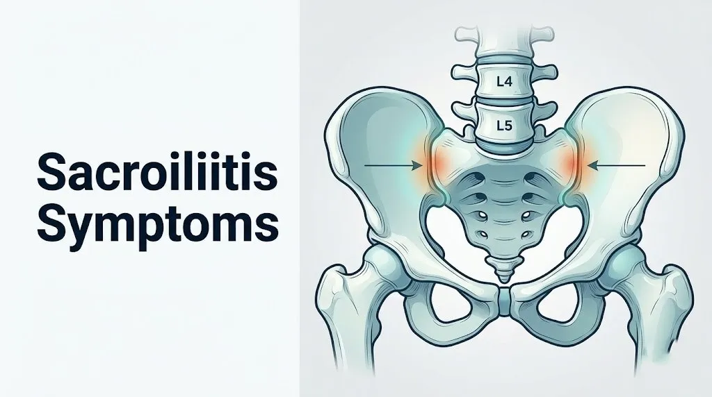
Sacroiliitis symptoms are produced when inflammation disrupts the sacroiliac joint, a weight-bearing joint formed by the auricular surface of the sacrum and the auricular surface of the ilium, held in place by one of the strongest ligament complexes in the human body.
The sacroiliac joint is not a typical freely moving joint. Its articular surfaces are covered by a hybrid cartilage layer: hyaline cartilage on the sacral side and fibrocartilage on the iliac side. The joint is surrounded by a dense ligamentous complex including the posterior sacroiliac ligament (the strongest ligament in the body by weight-to-strength ratio), the ventral sacroiliac ligament, the interosseous sacroiliac ligament, the sacrotuberous ligament, and the sacrospinous ligament. These structures together allow only 2 to 4 degrees of rotation and minimal translational movement, which is why sacroiliac joint inflammation produces a qualitatively different pain than a freely moveable joint like the knee or shoulder.
The joint is innervated primarily by the posterior rami of spinal nerves L4, L5, S1, S2, and S3. These posterior rami supply both the posterior joint capsule and the overlying skin via the superior cluneal nerves (derived from L1-L3). When inflammatory mediators activate these nerve endings, pain is perceived in the lower back, sacral region, buttock, and, through central sensitization, referred into the posterior thigh and calf along non-dermatomal pathways that frequently mimic sciatic nerve distribution.
| Sacroiliitis Symptom | Anatomical Mechanism | Typical Severity |
|---|---|---|
| Sacral and lower back pain | Posterior rami nociceptor activation by inflammatory mediators | Moderate to severe |
| Buttock pain | L5-S2 posterior rami referral, piriformis irritation | Moderate to severe |
| Morning stiffness over 45 minutes | Overnight IL-17 and TNF-alpha accumulation in immobile joint | Often most disabling symptom |
| Leg pain (non-dermatomal) | Central sensitization of lumbosacral posterior horn neurons | Mild to moderate |
| Groin and hip pain | Anterior joint capsule and iliopsoas muscle involvement | Variable, often unilateral |
| Fever (septic form only) | Systemic inflammatory response to pyogenic joint infection | High, acute onset |
People with obesity experience sacroiliac joint symptoms more intensely than those of normal weight because elevated body mass increases shear stress across the joint surface, amplifying mechanical irritation on top of any inflammatory component.
Sacrum and Lower Back Pain in Sacroiliitis
Sacrum and lower back pain in sacroiliitis is typically described as a deep, aching discomfort centered in the region just below the beltline on one or both sides, precisely over the posterior sacroiliac joint, and it is consistently worsened by positions that load the joint asymmetrically.
The pain originates where the inflammatory process is most active: at the synovial membrane lining the inferior third of the sacroiliac joint (the joint is partly synovial and partly ligamentous), and at the entheses (the insertion points of the posterior sacroiliac ligaments into the iliac and sacral bone surfaces). Enthesitis, inflammation at the ligament-bone interface, is a hallmark of HLA-B27-associated sacroiliitis and produces a qualitatively different pain from pure synovitis: it is often described as sharp with movement, dull at rest, and exquisitely tender to firm palpation directly over the posterior superior iliac spine (the bony prominence felt at the dimples of the lower back).
Activities that consistently provoke sacroiliitis-related lower back pain include: prolonged standing on one leg, climbing stairs, rolling over in bed, rising from a seated position, and any sustained static posture (prolonged sitting or prolonged standing with weight shifted to one side). Pain typically improves with walking and gentle movement, a clinical feature that distinguishes inflammatory sacroiliac pain from mechanical lumbar disc pain, which often worsens with prolonged walking.
According to the American College of Rheumatology (ACR), inflammatory lower back pain that improves with exercise but not with rest, begins before age 40, and is associated with morning stiffness lasting more than 30 minutes should prompt clinical evaluation for axial spondyloarthropathy.
Older adults (over 60) presenting with sacroiliac region pain are more likely to have degenerative sacroiliac joint disease (osteoarthritic changes, not inflammatory synovitis) than HLA-B27-associated sacroiliitis, and the two conditions require different management approaches. Degenerative sacroiliac pain tends to be more constant, less responsive to movement, and not associated with elevated inflammatory markers.
Buttock Pain as a Sacroiliitis Symptom
Buttock pain is present in the vast majority of patients with sacroiliitis and is produced by two distinct mechanisms operating simultaneously: direct referred pain from the L5, S1, and S2 posterior rami supplying the posterior sacroiliac joint capsule, and irritation of the piriformis muscle, which lies directly posterior to the joint.
The piriformis muscle originates from the anterior surface of the sacrum (S2-S4) and inserts on the greater trochanter of the femur. When sacroiliac joint inflammation extends to the periarticular soft tissue, the piriformis, which passes directly over the posterior capsule of the joint, can develop reactive muscle spasm. This spasm has two consequences: it deepens the buttock pain, and in approximately 17 percent of anatomical variants where the sciatic nerve passes through (rather than below) the piriformis, it compresses the sciatic nerve, producing additional leg symptoms on top of the already-referred sacroiliac pain.
A distinguishing clinical feature of sacroiliitis-related buttock pain is its location. The pain tends to concentrate in the medial buttock (close to the sacral midline) rather than the deep lateral buttock characteristic of piriformis syndrome or the posterior thigh distribution of gluteal tendinopathy. Many patients can point to one spot with a single finger: this clinical sign is called the Fortin finger sign, and it reliably localizes pain to the posterior sacroiliac joint region when the finger points to the area just medial to the posterior superior iliac spine.
Quick Tip:
- If you can point to your pain location with one finger, and that location is just below and medial to the dimple of your lower back, describe this precisely to your provider. It is one of the most clinically useful localizing signs for sacroiliac joint pathology.
- Buttock pain that worsens with sitting on a hard surface for more than 30 minutes and improves with standing suggests sacroiliac joint loading as the provocation mechanism.
- People who sit asymmetrically (always crossing the same leg, always leaning to one side) place chronic unilateral shear load on the sacroiliac joint, amplifying both inflammatory and mechanical symptoms.
Key Takeaway: Sacroiliitis lower back and buttock pain is produced by L4-S3 posterior rami activation at the inflamed sacroiliac joint capsule and entheses; the characteristic inflammatory pattern of pain that improves with movement but not with rest is the most clinically useful early differentiating feature from lumbar disc disease.
Sacroiliitis Leg Pain and Referred Pain Patterns
Sacroiliitis can cause leg pain extending from the buttock into the posterior thigh and occasionally into the calf, and this referred pain arises through a non-dermatomal central sensitization mechanism that is fundamentally different from the nerve root compression producing true radiculopathy.
Think of the spinal cord’s dorsal horn like a telephone switchboard with worn connectors. When the L4-S3 posterior rami are continuously bombarded by pain signals from the inflamed sacroiliac joint, the second-order neurons at those spinal cord levels become hyperexcitable. They begin responding to lower-intensity inputs from adjacent dermatomes, projecting pain perception into the leg even though no nerve root is compressed. The result is a deep, aching leg pain that travels into the posterior thigh, occasionally the knee, and rarely the calf, but does not follow a precise dermatomal strip the way L5 or S1 disc herniation radiculopathy does.
This distinction has direct clinical importance. Sacroiliitis-related leg pain does not typically produce the sharp, electric, shooting quality of true radiculopathy. It does not follow the strict L4, L5, or S1 dermatomal maps that disc herniation produces. It rarely extends below the knee as a primary symptom (though it may in severe cases with high levels of central sensitization). And critically, it is not associated with objective neurological deficits (weakness, absent reflexes, or positive straight leg raise test) unless another concurrent pathology is present.
According to research published in the Journal of Rheumatology, sacroiliac joint pain referral patterns documented by intra-articular injection studies demonstrate that the majority of confirmed sacroiliac joint pain refers to the buttock, followed by the posterior thigh, with knee and calf involvement less frequent and occurring primarily in patients with long-duration untreated inflammation.
Young adults (20 to 35) with HLA-B27-positive sacroiliitis who present with buttock and posterior thigh pain without a clear disc herniation on MRI should have sacroiliac joint evaluation specifically requested in their imaging order, because standard lumbar spine MRI does not include the sacroiliac joint field of view.
Morning Stiffness in Sacroiliitis
Morning stiffness lasting more than 45 minutes after waking is the most diagnostically specific symptom of inflammatory sacroiliitis, and it arises from the overnight accumulation of inflammatory cytokines in a joint that has been relatively immobile during sleep.
The mechanism is precise. During sleep, the sacroiliac joint undergoes minimal movement. The synovial membrane continues to produce TNF-alpha (tumor necrosis factor-alpha) and interleukin-17 (IL-17) in an HLA-B27-activated immune state, but without joint movement to distribute synovial fluid and clear inflammatory mediators, these cytokines accumulate in the joint space and periarticular tissue. By morning, the concentration of pain-sensitizing prostaglandins and inflammatory cytokines in the joint reaches its daily peak. The first movements of the day stretch an already-inflamed capsule and entheses, producing intense stiffness and pain that gradually resolves as movement promotes synovial fluid circulation and partial cytokine clearance.
This is why inflammatory sacroiliitis stiffness classically improves with 30 to 60 minutes of movement and worsens again after prolonged inactivity during the day. Mechanical sacroiliac dysfunction produces a qualitatively different pattern: stiffness that lasts fewer than 30 minutes and tends to be present more consistently throughout the day rather than being concentrated in the morning-to-mid-morning window.
The Assessment of SpondyloArthritis International Society (ASAS) includes morning stiffness lasting more than 30 minutes, along with age of onset before 40 and improvement with exercise, as part of the inflammatory back pain criteria used to screen for axial spondyloarthropathy. Meeting four of five of these criteria establishes a high clinical probability of inflammatory rather than mechanical back pain.
Adolescents and young adults with ankylosing spondylitis report morning stiffness as the symptom that most affects school and work attendance, because the stiffest period of the day coincides precisely with the morning hours of activity. This population should be counseled to begin gentle mobilization exercises before rising from bed to reduce the duration and severity of morning stiffness.
Sacroiliitis Groin and Hip Pain
Groin and hip pain in sacroiliitis is produced by the anterior joint capsule of the sacroiliac joint, which is in anatomical proximity to the iliopsoas muscle, and by referred pain patterns from the L4 and L5 posterior rami into the anterior hip and groin region.
The sacroiliac joint’s anterior surface is covered by the ventral sacroiliac ligament and sits in direct anatomical relation to the iliopsoas muscle (formed by the psoas major and iliacus, which originate from the lumbar vertebrae and iliac fossa respectively and merge to insert on the lesser trochanter of the femur). When sacroiliac joint inflammation involves the anterior capsule and ventral ligament, the adjacent iliopsoas can develop reactive irritation, producing a deep groin ache that patients frequently describe as a hip problem rather than a back or pelvis problem.
This anterior pain pattern leads to a common diagnostic error: sacroiliitis patients are evaluated for hip joint pathology (femoroacetabular impingement, hip labral tear) before the sacroiliac joint is assessed. A hip joint problem typically produces groin pain that is provoked by the FADIR test (Flexion, ADduction, Internal Rotation of the hip), while sacroiliac joint pain is typically provoked by the FABER test (Flexion, ABduction, External Rotation, also called the Patrick test), which stresses the sacroiliac joint rather than the hip joint.
Research published in the Annals of the Rheumatic Diseases has documented that groin and hip pain as part of the sacroiliitis presentation is more frequent in women than in men, likely because women’s wider pelvic anatomy creates a different force vector across the sacroiliac joint, more often engaging the anterior capsule and the iliopsoas attachment.
Women with sacroiliitis who report predominantly groin and anterior hip pain should tell their rheumatologist or primary care physician specifically about this pattern, because it may lead to unnecessary hip joint investigation if the examining clinician does not perform a systematic sacroiliac joint provocation examination.
Key Takeaway: Morning stiffness lasting more than 45 minutes that improves with movement is the most diagnostically specific symptom of inflammatory sacroiliitis, driven by overnight cytokine accumulation in an immobile joint, and meeting the ASAS inflammatory back pain criteria is the appropriate clinical screening step for any patient with this pattern.
Sacroiliitis Flare Symptoms
A sacroiliitis flare is an acute worsening of baseline inflammatory joint activity triggered by identifiable physiological events, and it produces a clinical picture more intense than the patient’s typical daily symptoms.
Flares occur when the immune system’s inflammatory output at the sacroiliac joint temporarily escalates. Known triggers include: intercurrent infections (viral upper respiratory infections can trigger systemic TNF-alpha and IL-17 surges that worsen joint inflammation), psychological stress (which activates the hypothalamic-pituitary-adrenal (HPA) axis, temporarily amplifying systemic inflammatory cytokine production), dietary factors in patients with concurrent inflammatory bowel disease (specific food antigens triggering gut-associated lymphoid tissue activation), and stopping or missing doses of anti-inflammatory medication.
During a flare, patients typically experience:
- Sacral and buttock pain intensity escalating to 7 or above on a Visual Analog Scale where 10 is worst imaginable pain
- Morning stiffness extending beyond 60 to 90 minutes instead of the usual 45 minutes
- Difficulty performing activities that were manageable between flares: walking distances greater than one block, rising from a chair without arm support, or rolling over in bed
- Return or intensification of leg referred pain even if it had been well-controlled
- Sleep disruption from pain during position changes, specifically turning over, which loads the sacroiliac joint
- Increased fatigue, which is a direct systemic effect of elevated circulating IL-6 (a cytokine that crosses the blood-brain barrier and activates central fatigue pathways)
Patients on biologic disease-modifying antirheumatic drugs (DMARDs) who experience a sudden severe flare should contact their prescribing rheumatologist promptly, because an intercurrent infection, which may be the flare trigger, may also require that biologic therapy be temporarily paused depending on the type and severity of the infection.
Bilateral vs Unilateral Sacroiliitis Symptoms
Whether sacroiliitis affects one sacroiliac joint (unilateral) or both (bilateral) is not merely a matter of severity: it is a clinically meaningful finding that strongly suggests specific underlying diagnoses.
Bilateral symmetric sacroiliitis, where both sacroiliac joints show equivalent degrees of inflammation on imaging, is the hallmark of ankylosing spondylitis and most forms of HLA-B27-associated axial spondyloarthropathy. The immune dysregulation in this condition affects both joints equally because the initiating mechanism (HLA-B27-mediated immune activation, likely in response to gut microbial antigens) is systemic rather than local. Symptoms are felt on both sides simultaneously or alternate sides in a pattern that patients describe as “switching.”
Unilateral sacroiliitis, where only one joint is affected or one joint is dramatically more inflamed than the other, is more characteristic of psoriatic arthritis (which tends to cause asymmetric joint involvement), reactive arthritis (triggered by preceding gastrointestinal or genitourinary infection), mechanical sacroiliac dysfunction from a leg length discrepancy or hip joint abnormality, and most importantly, septic sacroiliitis (which is almost always unilateral because it is caused by a localized bacterial infection).
According to the Arthritis Foundation, the pattern of unilateral versus bilateral involvement is one of the factors rheumatologists evaluate when determining which subtype of inflammatory arthritis a patient has, because the treatment approach differs meaningfully between symmetric spondylitis and asymmetric psoriatic or reactive arthritis.
| Involvement Pattern | Most Associated Conditions | Clinical Significance |
|---|---|---|
| Bilateral symmetric | Ankylosing spondylitis, HLA-B27 axSpA | High probability of systemic spondyloarthropathy |
| Bilateral asymmetric | Psoriatic arthritis, IBD-associated sacroiliitis | Investigate for skin, bowel, or eye symptoms |
| Unilateral, gradual onset | Reactive arthritis, mechanical dysfunction | Investigate preceding infection, leg length asymmetry |
| Unilateral, acute severe onset | Septic sacroiliitis | Emergency evaluation for infection |
People who notice that their lower back or buttock pain consistently alternates from side to side on different days should describe this pattern precisely to their physician, because alternating buttock pain is one of the ASAS inflammatory back pain criteria and increases clinical suspicion for axial spondyloarthropathy.
Sacroiliitis Symptoms in Ankylosing Spondylitis and Spondyloarthropathy
Sacroiliitis in ankylosing spondylitis and related spondyloarthropathies is the defining feature of an entire family of systemic inflammatory diseases, and its symptoms extend beyond the joint itself to include extra-articular manifestations that affect the eyes, skin, gut, and peripheral joints.
Ankylosing spondylitis (AS), now classified under the broader category of axial spondyloarthropathy (axSpA) by the ASAS criteria, is driven by HLA-B27 antigen-mediated immune dysregulation. Approximately 90 to 95 percent of people with ankylosing spondylitis carry the HLA-B27 antigen, compared to approximately 8 percent of the general white population in the United States, according to data from the National Institute of Arthritis and Musculoskeletal and Skin Diseases (NIAMS). This dramatic enrichment confirms HLA-B27 as a major predisposing factor, though not all HLA-B27-positive individuals develop the disease.
The sacroiliitis of ankylosing spondylitis begins with subchondral bone marrow edema and synovitis, visible on MRI as high-intensity signal on STIR (Short Tau Inversion Recovery) sequences at the sacroiliac joint margins. Over years, untreated inflammation progresses through cartilage erosion, sclerosis, and finally ankylosis, the complete bony fusion of the joint. Once the sacroiliac joint fuses, paradoxically, the pain from that joint often diminishes, but the disease simultaneously advances up the lumbar and thoracic spine.
Extra-articular symptoms that frequently accompany sacroiliitis in this context include:
- Uveitis (acute anterior uveitis): sudden unilateral eye redness, photophobia, and blurred vision, occurring in approximately 25 to 40 percent of AS patients
- Psoriatic skin plaques: present in psoriatic arthritis-associated sacroiliitis
- Inflammatory bowel disease symptoms: cramping, diarrhea, and rectal bleeding in Crohn’s disease or ulcerative colitis-associated sacroiliitis
- Peripheral arthritis: asymmetric swelling of large joints (knee, ankle, shoulder) occurring alongside the axial symptoms
- Enthesitis at other sites: heel pain from Achilles tendon enthesitis, chest pain from costovertebral or manubriosternal enthesitis
Septic Sacroiliitis Symptoms
Septic sacroiliitis is a bacterial infection of the sacroiliac joint that produces a clinical presentation distinct from all other forms of sacroiliitis, and it constitutes a medical emergency requiring same-day evaluation and treatment.
The condition is caused most commonly by Staphylococcus aureus spreading to the joint through the bloodstream from a primary infection site (skin abscess, intravenous drug use, urinary tract infection, or respiratory infection), a process called hematogenous seeding. Less commonly, gram-negative organisms such as Enterobacteriaceae species are responsible, particularly in patients with recent urological procedures or immunosuppression. The sacroiliac joint is particularly vulnerable to hematogenous bacterial seeding because of its rich vascular supply and the complex sinusoidal blood flow through the subchondral bone.
The symptoms of septic sacroiliitis are:
- High fever, typically 38.5 degrees Celsius (101.3 degrees Fahrenheit) or above, appearing rapidly over 24 to 72 hours
- Severe unilateral sacroiliac pain that is abrupt in onset and dramatically more intense than any previous back pain
- Inability to weight-bear on the affected side: patients with septic sacroiliitis frequently cannot walk normally because any loading of the infected joint produces immediate sharp pain
- Severe restriction of hip movement on the affected side, because the adjacent iliopsoas muscle develops a reactive spasm or abscess (psoas abscess is a recognized complication of untreated septic sacroiliitis)
- Systemic illness: sweating, rigors (shaking chills), malaise, and nausea consistent with bacteremia or early sepsis
This condition is frequently misdiagnosed as a lumbar disc herniation, hip flexor strain, or non-specific lower back pain for days to weeks. The diagnostic delay allows progressive joint destruction and, in severe cases, the development of psoas abscess, osteomyelitis of adjacent sacral or iliac bone, and septicemia.
Immunocompromised individuals, people who inject drugs, patients with diabetes mellitus, and those who have recently had invasive procedures are at the highest risk. Any sacroiliac pain with fever, even low-grade, should prompt same-day emergency evaluation rather than home monitoring.
Key Takeaway: Bilateral symmetric sacroiliitis strongly indicates HLA-B27-associated ankylosing spondylitis requiring rheumatological evaluation, while unilateral acute-onset sacroiliac pain with fever indicates septic sacroiliitis, which requires same-day emergency assessment, not a routine appointment.
Sacroiliitis in Pregnancy
Sacroiliac joint pain in pregnancy is among the most frequent musculoskeletal complaints of the second and third trimesters, affecting an estimated 20 to 25 percent of pregnant women according to data in the Journal of Rheumatology, and it is caused by a specific, well-defined hormonal mechanism rather than inflammatory joint disease.
Relaxin, a peptide hormone produced primarily by the corpus luteum of the ovary in early pregnancy and subsequently by the placenta, binds to relaxin receptors expressed on sacroiliac joint ligament fibroblasts. This receptor activation reduces collagen cross-linking in the posterior sacroiliac ligament complex, increasing joint laxity beyond the normal physiological range. The joint, designed to allow only 2 to 4 degrees of movement, now experiences greater shear stress with each step, loading the posterior capsule and activating nociceptors in a way that closely mimics inflammatory sacroiliitis symptomatically.
Relaxin-mediated sacroiliac pain in pregnancy is specifically called pelvic girdle pain (PGP) in the clinical literature, to distinguish it from inflammatory sacroiliitis. The pain has these distinguishing characteristics compared to HLA-B27 sacroiliitis: it is frequently bilateral and symmetric, it is directly related to weight-bearing activities (walking, stair climbing, turning in bed), and morning stiffness, while present, typically resolves within 30 minutes, shorter than the 45-plus-minute morning stiffness of inflammatory disease.
Management of pregnancy-related sacroiliac pain is handled primarily by a physical therapist specializing in pelvic girdle dysfunction, working in coordination with the patient’s obstetrician-gynecologist. Sacroiliac joint belts (pelvic support belts) worn around the posterior pelvis can reduce joint shear and provide pain relief without pharmacological intervention, and current evidence from several Cochrane Reviews supports their use for symptom management in pelvic girdle pain.
Women who have pre-existing HLA-B27-associated sacroiliitis should discuss their inflammatory disease management with their rheumatologist before and during pregnancy, because some biologic medications used to treat inflammatory sacroiliitis have specific safety considerations in pregnancy.
Sacroiliitis Symptoms in Women vs Men
Sacroiliitis does not present identically across sexes: women and men with the same underlying condition frequently describe different symptom patterns, experience different delays to diagnosis, and have different risk profiles for specific subtypes of the disease.
In ankylosing spondylitis, men historically outnumbered women in diagnosed cases by approximately 2 to 3:1, but this ratio has narrowed as recognition of female-specific presentations has improved. Women with AS are more likely to have predominantly peripheral joint involvement (knees, hips, ankles) alongside their axial sacroiliac symptoms, and their axial disease tends to progress more slowly and less commonly to complete spinal fusion than men’s. The result is that women’s AS has historically been misclassified as seronegative rheumatoid arthritis, fibromyalgia, or non-specific low back pain at rates substantially higher than men’s.
Men with HLA-B27 sacroiliitis are more likely to present with the classic bilateral symmetric sacroiliac pain, pronounced morning stiffness, and progression to visible spinal radiographic changes within 5 to 10 years. Their symptom picture more closely matches textbook descriptions, which partly explains why their diagnosis is made sooner on average.
The Arthritis Foundation has explicitly highlighted that women with axial spondyloarthropathy face diagnostic delays averaging 1 to 2 years longer than men with equivalent disease activity, contributing to delayed treatment access and potentially greater cumulative joint damage.
Women who are told their sacroiliac pain is “just from pregnancy” or “probably fibromyalgia” without HLA-B27 testing and sacroiliac MRI should ask their primary care physician specifically for these evaluations if their morning stiffness lasts more than 45 minutes, their pain improves with exercise rather than rest, and they have any family history of ankylosing spondylitis, psoriasis, or inflammatory bowel disease.
Chronic Sacroiliitis Symptoms and Progression
Chronic sacroiliitis that goes undiagnosed or inadequately treated follows a predictable progression through recognizable stages, each producing a distinct symptom pattern that clinicians use to gauge disease activity and structural damage.
Early chronic inflammatory sacroiliitis produces intermittent sacral and buttock pain, fluctuating morning stiffness, and occasional leg referred pain. On MRI during this phase, active inflammation appears as bone marrow edema (STIR-sequence hyperintensity) at the sacroiliac joint margins without visible structural change on plain radiographs. This is classified as non-radiographic axial spondyloarthropathy (nr-axSpA) and is a recognized clinical entity in the ASAS classification system.
As inflammation continues without adequate suppression, structural changes appear. The cartilage thins, small erosions develop at the joint margins, and the subchondral bone undergoes reactive sclerosis (thickening and hardening visible on CT and plain radiographs). Patients at this stage often notice that their pain has become more constant and less clearly triggered by specific activities. They may report that over-the-counter NSAIDs, which were previously effective, no longer provide adequate relief.
In advanced disease, the sacroiliac joint undergoes ankylosis: complete bony fusion replaces the normal joint space. Interestingly, the sacroiliac pain from an ankylosed joint often diminishes because the nociceptive tissue (synovium, cartilage, entheses) has been replaced by bone. However, the disease simultaneously tends to ascend the spine, producing thoracolumbar rigidity, chest expansion limitation (from costovertebral joint involvement), and a characteristic stooped posture as the lumbar lordosis flattens.
People with longstanding untreated sacroiliitis who develop new-onset chest pain, shortness of breath, or reduced chest expansion with deep breathing should be evaluated by their rheumatologist promptly for costovertebral or manubriosternal joint involvement, which can restrict respiratory mechanics in advanced axial spondyloarthropathy.
Key Takeaway: Chronic inflammatory sacroiliitis progresses from intermittent MRI-visible bone marrow edema (nr-axSpA) through structural erosion and sclerosis to eventual joint ankylosis, and the window for most effective treatment intervention is the early inflammatory phase before structural damage is established.
Sacroiliitis vs Sciatica: How to Tell the Difference
Sacroiliitis and sciatica produce overlapping leg and buttock pain, but they arise from completely different anatomical sites and have specific distinguishing features that clinical examination can often separate before imaging is performed.
Sciatica is produced by compression or irritation of the sciatic nerve (formed from the L4, L5, S1, S2, and S3 nerve roots) as it exits the lumbar spine, most commonly from a herniated disc at L4-L5 or L5-S1. The pain of true sciatica is typically sharp, burning, or electric in quality, follows a precise dermatomal map (the outer calf and top of the foot for L5, the heel and sole for S1), and is associated with objective neurological findings: weakness, reduced reflexes, and a positive straight leg raise test (pain reproduced by passively raising the leg to 30 to 70 degrees with the knee straight).
Sacroiliitis-related leg pain is referred through central sensitization of posterior rami and lumbosacral plexus neurons, not through sciatic nerve root compression. The pain quality is typically aching and dull rather than sharp and electric, it does not follow a precise dermatomal pattern, it rarely extends below the knee as a primary complaint, and the straight leg raise test is usually negative (does not reproduce the leg pain) in sacroiliitis without concurrent disc herniation.
| Feature | Sacroiliitis Leg Pain | Sciatica (Disc Herniation) |
|---|---|---|
| Pain quality | Deep aching, non-electric | Sharp, burning, electric |
| Dermatomal distribution | Non-dermatomal, diffuse posterior thigh | Precise dermatomal (L4, L5, or S1 strip) |
| Extension below knee | Uncommon, usually proximal | Common; extends to foot |
| Straight leg raise test | Usually negative | Positive at 30 to 70 degrees |
| Neurological deficits | Absent | May be present (weakness, reflex loss) |
| Morning stiffness pattern | Over 45 minutes, improves with movement | Not a feature; worsens with movement |
| Response to walking | Improves with gentle walking | Worsens with prolonged walking |
Clinical examination by a physician or physiotherapist can often distinguish these two conditions before MRI is obtained, but imaging of both the lumbar spine and the sacroiliac joints may be required in patients with overlapping features.
Sacroiliitis Red Flag Symptoms
Red flag symptoms in sacroiliitis indicate either a more serious underlying diagnosis, a complication of inflammatory disease, or a presentation that requires emergency rather than routine evaluation.
The following symptoms appearing alongside sacroiliac or sacral pain should prompt immediate clinical action:
- Fever above 38.5 degrees Celsius (101.3 degrees Fahrenheit) combined with severe unilateral sacroiliac pain and inability to walk normally: this presentation indicates septic sacroiliitis until proven otherwise and requires emergency evaluation the same day
- Bowel or bladder incontinence, or urinary retention: these symptoms alongside lower sacral pain constitute a cauda equina syndrome presentation, where the cauda equina nerve roots within the spinal canal are compromised, representing a surgical emergency
- Saddle anesthesia (numbness in the inner thighs, perineum, and buttocks, the skin area that would contact a saddle): this is the defining neurological sign of cauda equina syndrome
- Rapidly progressive lower limb weakness: leg weakness that develops over hours to days alongside sacral pain is a neurological emergency, not a musculoskeletal pain complaint
- Sudden severe eye pain, redness, and photophobia: acute anterior uveitis is the most common extra-articular emergency associated with HLA-B27 sacroiliitis and requires same-day ophthalmological evaluation to prevent vision loss from secondary glaucoma
- Unexplained weight loss, drenching night sweats, or persistent fever without a localized infection: this triad alongside sacroiliac pain raises concern for malignancy (primary bone tumor, metastatic disease, or lymphoma affecting the sacrum or iliac bone) and requires urgent imaging and oncological referral
- New-onset incapacitating chest pain with deep breathing: costovertebral joint involvement in advanced spondylitis can restrict chest expansion significantly, but acute-onset pleuritic chest pain in this population also warrants evaluation to exclude pulmonary embolism
These red flag symptoms change the clinical picture from an inflammatory musculoskeletal condition to a potential emergency, and none of them should be monitored at home while waiting for a routine appointment.
Emergency Symptoms: When to Call 911 or Go to the ER
Certain symptoms associated with sacroiliitis or sacrum pain require immediate emergency evaluation. Do not wait to see if these resolve on their own.
Call 911 or go to the nearest emergency room immediately if you experience:
- Bowel or bladder incontinence, or inability to urinate, alongside lower back or sacral pain: these are the defining signs of cauda equina syndrome, a compression of the spinal nerve roots requiring emergency surgical decompression; every hour of delay increases the risk of permanent neurological damage
- Numbness in the perineum, inner thighs, or buttocks (saddle anesthesia): this neurological finding indicates cauda equina compression and is a surgical emergency
- Sudden, severe unilateral sacroiliac pain with fever and inability to walk: this presentation indicates septic sacroiliitis, which can progress to psoas abscess, osteomyelitis, and septicemia without same-day antibiotic treatment
- Rapidly progressive leg weakness developing over hours to a day or two: this is not musculoskeletal pain; it indicates a neurological emergency requiring immediate imaging
- Sudden eye pain, vision loss, or severe photophobia in a patient with known inflammatory sacroiliitis: acute anterior uveitis associated with HLA-B27 disease can cause permanent vision damage within hours without treatment from an ophthalmologist
Call 911 immediately if bowel or bladder function is lost or if leg weakness is rapidly progressive. For fever with severe unilateral sacroiliac pain, go to the nearest emergency room without delay. These presentations can indicate cauda equina syndrome, septic sacroiliitis, or spinal cord compromise and require emergency medical assessment, not an urgent care appointment or a call to a rheumatologist’s office.
How Sacroiliitis Is Diagnosed and Which Specialist to See
Sacroiliitis is diagnosed through a combination of clinical history, physical examination, laboratory testing, and imaging, with MRI of the sacroiliac joints representing the gold standard for detecting active inflammation before structural changes are visible on plain radiographs.
The clinical examination includes specific sacroiliac joint provocation tests performed by a physician or physiotherapist:
To describe your symptoms effectively for a sacroiliitis evaluation appointment:
- Note whether your pain is worst in the morning and how many minutes it takes to improve after you start moving.
- Record whether the pain is unilateral (one side), bilateral (both sides), or alternates between sides.
- Tell the clinician whether you can point to the pain with one finger, and specifically where: just below and medial to the dimple of the lower back (Fortin sign) or more lateral.
- Describe whether walking and gentle movement relieve the pain or worsen it.
- List any concurrent symptoms: eye redness, skin plaques, bowel symptoms, heel pain, or peripheral joint swelling.
- Bring a complete medication list and disclose any family history of psoriasis, inflammatory bowel disease, or ankylosing spondylitis.
| Diagnostic Test | What It Evaluates | When It Is Ordered |
|---|---|---|
| MRI of sacroiliac joints (STIR sequence) | Active bone marrow edema, synovitis, early inflammation | First-line for suspected inflammatory sacroiliitis; detects pre-radiographic disease |
| CT scan of sacroiliac joints | Structural changes: erosion, sclerosis, ankylosis | When structural damage needs detailed bony assessment |
| Plain radiograph (AP pelvis) | Advanced structural changes only | Initial screening; misses early disease |
| HLA-B27 blood test | Genetic marker for spondyloarthropathy risk | Ordered alongside imaging for inflammatory suspicion |
| CRP and ESR | Systemic inflammatory activity | Elevated in active inflammatory sacroiliitis; normal in mechanical dysfunction |
| Complete blood count (CBC) | White cell count elevation in septic sacroiliitis | Essential when infection is suspected |
The appropriate specialist depends on your clinical picture. A board-certified rheumatologist should evaluate suspected inflammatory sacroiliitis, especially if morning stiffness exceeds 45 minutes, HLA-B27 testing is positive, or systemic symptoms are present. Pregnancy-related sacroiliac pain is best managed by a pelvic floor physical therapist working alongside the patient’s obstetrician-gynecologist. Any presentation with fever, neurological signs, or acute severe unilateral symptoms requires an emergency medicine physician as the first point of contact.
Frequently Asked Questions About Sacroiliitis Symptoms
What does sacroiliitis feel like?
Sacroiliitis feels like a deep, aching pain centered in the sacral region (just below the beltline), often extending into one or both buttocks and occasionally into the posterior thigh.
The pain is typically worst first thing in the morning (lasting more than 45 minutes before improving with movement) and is provoked by prolonged sitting, rolling over in bed, rising from a chair, and stair climbing.
Unlike a pulled muscle, sacroiliitis pain is not sharply localized to one muscle but instead feels like it is coming from inside the pelvic girdle, and it tends to improve with gentle walking rather than worsening with activity.
How do I know if my lower back pain is sacroiliitis or a herniated disc?
Sacroiliitis typically presents with pain below the beltline that improves with movement and is associated with morning stiffness lasting over 45 minutes, while a herniated disc produces sharper, more electric pain that often worsens with walking and is associated with leg numbness or weakness following a specific dermatomal pattern.
A positive FABER test (pain provoked by flexing and rotating the hip outward) on physical examination suggests sacroiliac joint involvement, while a positive straight leg raise test suggests lumbar nerve root compression.
MRI of both the lumbar spine and the sacroiliac joints (two separate imaging sequences) is needed to differentiate the two conditions definitively, since standard lumbar spine MRI does not include the sacroiliac joints in its field of view.
Can sacroiliitis cause leg pain and numbness?
Sacroiliitis can cause referred leg pain, typically a deep aching in the posterior thigh that does not follow a precise dermatomal pattern, through central sensitization of the L4-S3 posterior rami.
True numbness, tingling, or weakness in the leg following a specific dermatomal map (outer calf, top of foot, or heel) is more characteristic of sciatic nerve root compression from a disc herniation than sacroiliac joint inflammation.
Leg weakness or bowel and bladder changes alongside sacral pain require emergency evaluation to exclude cauda equina syndrome, regardless of whether sacroiliitis is the suspected underlying diagnosis.
Is sacroiliitis the same as sciatica?
Sacroiliitis and sciatica are not the same condition, though they produce overlapping symptoms in the buttock and leg.
Sciatica is caused by compression of the sciatic nerve or its contributing nerve roots (L4-S1) producing sharp, dermatomal leg pain that typically extends to the foot, while sacroiliitis is joint inflammation producing referred buttock and proximal thigh aching through non-dermatomal central sensitization pathways.
A physician can often distinguish the two through clinical examination using the straight leg raise test (positive in sciatica, negative in isolated sacroiliitis) and the FABER provocation test (positive in sacroiliitis, negative in isolated disc herniation).
Does sacroiliitis ever go away on its own?
Mechanical sacroiliac joint dysfunction related to pregnancy or minor trauma can resolve over weeks to months with appropriate physical therapy and activity modification.
Inflammatory sacroiliitis caused by HLA-B27-associated spondyloarthropathy does not resolve without treatment and progresses in most untreated patients through stages of increasing joint damage over years to decades.
Septic sacroiliitis never resolves on its own and progresses to psoas abscess and septicemia without antibiotic treatment initiated in an emergency or inpatient setting.
Which doctor should I see for sacroiliitis symptoms?
Suspected inflammatory sacroiliitis with morning stiffness exceeding 45 minutes, alternating buttock pain, or improvement with exercise warrants a referral to a board-certified rheumatologist who can order sacroiliac MRI, HLA-B27 testing, and inflammatory markers.
Pregnancy-related sacroiliac pain is best initially addressed with a referral to a physical therapist specializing in pelvic girdle dysfunction, working in coordination with your obstetrician-gynecologist.
Sacroiliac pain accompanied by fever, inability to walk, bowel or bladder changes, or rapidly progressive leg weakness requires immediate evaluation by an emergency medicine physician, not a scheduled outpatient appointment.
Closing
Sacroiliitis is not generic back pain. It has specific anatomical mechanisms, specific symptom patterns, and specific clinical thresholds that separate inflammatory joint disease from mechanical dysfunction and urgent emergencies from manageable chronic conditions. The morning stiffness pattern, the non-dermatomal leg referral, the Fortin finger sign pointing to the posterior superior iliac spine: each of these is a piece of clinical information that a rheumatologist needs.
If your lower back or sacral pain has been present for more than six weeks, improves with movement rather than rest, and causes stiffness for more than 45 minutes every morning, ask your primary care physician for sacroiliac joint MRI and HLA-B27 testing specifically. Bring a written record of when the pain is worst, how long morning stiffness lasts, and whether it is unilateral, bilateral, or alternating.
If you develop fever alongside sudden severe sacroiliac pain, or if you experience any bowel or bladder changes with lower sacral pain, treat these as emergencies. The anatomy and the evidence are clear: getting the right diagnosis early is what separates effective treatment from years of inadequate management.