Carotid Artery Symptoms: Warning Signs to Know in 2026
Carotid artery symptoms range from a brief, painless episode of vision loss in one eye to sudden one-sided limb weakness, slurred speech, or facial drooping — and every one of them can represent a stroke in progress or a transient ischemic attack warning that a stroke is imminent. Recognizing these symptoms accurately and acting on them immediately is not optional; it is time-critical in a way that few medical situations are.
According to the Centers for Disease Control and Prevention (CDC), stroke is the fifth leading cause of death in the United States and a leading cause of long-term disability, with carotid artery disease responsible for approximately 10 to 15 percent of all ischemic strokes. The American Heart Association states that individuals who experience a transient ischemic attack have approximately a 10 percent risk of stroke within 90 days, with the highest risk concentrated in the first 48 hours.
This article covers every clinically recognized carotid artery symptom with the precise anatomical and physiological reason each one occurs, the distinction between a TIA warning and an active stroke, what a carotid bruit means when a doctor finds it, how stenosis severity maps to symptom risk, and exactly which symptoms require calling 911 versus scheduling a same-week appointment.
Carotid Artery Symptoms: What They Are and Why They Matter
Carotid artery symptoms are neurological warning signs produced when narrowing, blockage, or unstable plaque in one or both carotid arteries reduces or interrupts blood flow to the brain, retina, or both.
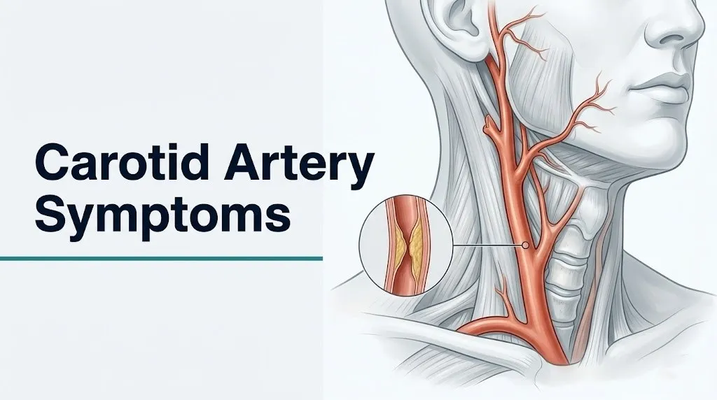
The carotid arteries are the primary conduits for oxygenated blood traveling from the heart to the brain. Each side of the neck contains a common carotid artery (CCA) that divides at approximately the level of the C3 to C4 vertebrae into the internal carotid artery (ICA) and the external carotid artery (ECA). The ICA supplies the brain, the retina, and the anterior pituitary. The ECA supplies the face and scalp. When the ICA is compromised, the neurological and visual consequences are specific, predictable, and often sudden.
Symptoms matter because their appearance, even briefly and even if they resolve completely within minutes, indicates active cerebrovascular pathology. A symptom that resolves is not a symptom that was harmless.
| Symptom Category | Vascular Territory Involved | Clinical Significance |
|---|---|---|
| Transient vision loss in one eye | Ophthalmic artery (ICA branch) | High: indicates ipsilateral ICA disease |
| Contralateral face or arm weakness | Middle cerebral artery (ICA territory) | High: indicates cortical ischemia |
| Speech difficulty (production or comprehension) | Left MCA territory (dominant hemisphere) | High: indicates hemispheric ischemia |
| Sudden severe headache | Variable; may indicate hemorrhagic complication | Emergency |
| Brief loss of consciousness | Bilateral cerebral or brainstem involvement | Emergency |
People taking antiplatelet medications such as aspirin or clopidogrel, or anticoagulants such as warfarin or direct oral anticoagulants, may have suppressed thrombotic activity that partially reduces symptom intensity. This does not mean their carotid disease is less dangerous. It means the pharmacological protection may create a false sense of security if a new or worsening symptom is attributed to medication rather than disease progression.
What Is Carotid Artery Disease and How Does It Develop?
Carotid artery disease is the progressive narrowing of the internal carotid artery caused primarily by atherosclerosis, a disease process in which inflammatory plaques accumulate within the tunica intima (the innermost layer of the arterial wall), reducing the lumen diameter available for blood flow.
Atherosclerosis in the carotid artery preferentially targets the carotid bifurcation because of a specific hemodynamic phenomenon: at arterial branch points, blood flow becomes turbulent and oscillatory rather than smooth and laminar. This turbulence creates regions of low wall shear stress, which injures the endothelial cells lining the arterial wall. Injured endothelium becomes permeable to oxidized low-density lipoprotein (oxLDL), which accumulates in the tunica intima and triggers macrophage infiltration. Those macrophages engulf the oxLDL and transform into foam cells, the cellular hallmark of early atherosclerotic plaque.
Think of the carotid bifurcation like a highway interchange where traffic slows, changes direction, and creates turbulence. Arterial endothelium is designed for smooth, unidirectional flow. The turbulence at that fork is precisely where the damage accumulates over decades.
Over years, foam cells accumulate into a fatty streak, then a fibrous plaque with a lipid-rich necrotic core surrounded by a fibrous cap of collagen and smooth muscle cells. When inflammatory cytokines, particularly matrix metalloproteinase-9 (MMP-9), degrade the fibrous cap, the plaque becomes unstable. A ruptured cap exposes the lipid core to circulating blood, activating platelets via thromboxane A2 (TXA2) and adenosine diphosphate (ADP), forming a thrombus. Fragments of that thrombus, called microemboli, travel distally into cerebral or retinal arteries and cause focal ischemia.
According to the National Institute of Neurological Disorders and Stroke (NINDS), the process from initial endothelial injury to clinically detectable stenosis typically occurs over decades, which is why carotid artery disease is most prevalent in adults over 65 but can be accelerated significantly by smoking, hypertension, diabetes, and dyslipidemia.
Symptoms of Carotid Artery Blockage: The Complete Picture
The symptoms of carotid artery blockage reflect which specific cerebral or retinal territory has been deprived of blood flow, either transiently (TIA) or persistently (ischemic stroke), and they map with anatomical precision to the arteries supplied by the affected internal carotid artery.
The symptom pattern is predictably ipsilateral for visual symptoms (the eye on the same side as the blocked artery loses vision) and contralateral for motor and sensory symptoms (the arm, leg, or face on the opposite side of the body from the blocked artery becomes weak or numb). This crossed pattern exists because the motor fibers of the corticospinal tract cross at the medullary pyramids in the brainstem before descending into the spinal cord, while the ophthalmic artery branches directly from the ICA before the crossing occurs.
The core symptoms of carotid artery blockage are:
- Sudden, painless, transient loss of vision in one eye (the eye on the same side as the blocked artery), described by patients as a curtain or shade being drawn across the visual field
- Sudden weakness or paralysis of the face, arm, or leg on the side opposite the blocked artery
- Sudden numbness or loss of sensation in the face, arm, or hand on the side opposite the blocked artery
- Sudden difficulty speaking, understanding speech, or finding words (when the dominant hemisphere, usually the left, is affected)
- Sudden severe headache with no prior history of similar headaches
- Sudden confusion or altered consciousness
People who experience symptoms that resolve completely within minutes or hours often do not recognize the event as a medical emergency. This is one of the most consequential misconceptions in cerebrovascular medicine. Resolution of symptoms does not indicate resolution of risk. It often indicates a TIA, which carries a stroke risk of up to 10 percent within 90 days according to American Stroke Association data.
Key Takeaway: Carotid artery blockage produces ipsilateral vision loss and contralateral limb weakness because of the anatomical crossing of motor fibers at the medullary pyramids; any symptom in this pattern, even if it resolves in minutes, is a potential TIA and requires same-day emergency evaluation.
Transient Ischemic Attack and Carotid Artery Symptoms
A transient ischemic attack (TIA) is a brief episode of focal neurological dysfunction caused by temporary focal ischemia without permanent infarction, and it is the most important warning sign that carotid artery disease is at a stage where stroke risk is acutely elevated.
By clinical definition, a TIA produces symptoms that resolve completely, typically within minutes to an hour, leaving no detectable brain damage on imaging. The American Stroke Association updated its guidance to emphasize that the diagnosis is now based on the absence of infarction on diffusion-weighted MRI, not simply on symptom duration. Even a 10-minute episode of arm weakness or speech difficulty qualifies as a potential TIA requiring emergency evaluation.
The ABCD2 score is a validated clinical tool used by emergency physicians to stratify a patient’s 2-day stroke risk after a TIA. It assigns points based on:
| ABCD2 Factor | Scoring Criterion | Points |
|---|---|---|
| Age | 60 years or older | 1 |
| Blood pressure | Systolic at or above 140 mmHg or diastolic at or above 90 mmHg | 1 |
| Clinical features | Unilateral weakness (2 points) or speech disturbance without weakness (1 point) | 1 to 2 |
| Duration | 60 minutes or more (2 points) or 10 to 59 minutes (1 point) | 0 to 2 |
| Diabetes | Present | 1 |
A score of 4 or above indicates high short-term stroke risk. Patients with a score of 6 or 7 have an estimated 2-day stroke risk of approximately 8 percent, according to a landmark validation study published in The Lancet in 2007.
Younger adults, particularly those under 50, may experience TIA from carotid dissection (a tear in the arterial wall) rather than atherosclerosis. The clinical presentation overlaps but may include neck pain and a partial Horner’s syndrome (ptosis, miosis, and anhidrosis on the side of the dissection). Emergency physicians routinely screen for dissection in younger patients presenting with TIA symptoms.
Amaurosis Fugax and Carotid Artery Warning Signs
Amaurosis fugax is transient monocular blindness caused by temporary ischemia of the retina, and it is one of the most specific warning signs of ipsilateral internal carotid artery disease because the retina receives its blood supply through the central retinal artery, which branches from the ophthalmic artery, which is the first major branch of the internal carotid artery.
The episode typically lasts seconds to minutes and is described by patients as a gray or black curtain or shade that descends (or ascends) across one eye, blurring or completely eliminating vision in that eye only, then lifting. The visual loss is painless. Crucially, it affects only the eye on the same side as the carotid lesion, never the opposite eye, because the ophthalmic artery branches before the carotid enters the circle of Willis and has no collateral pathway from the contralateral side at this anatomical level.
A retinal embolus causing amaurosis fugax can sometimes be directly visualized. An ophthalmologist or emergency physician examining the fundus may see a Hollenhorst plaque, a bright, yellowish, cholesterol-containing embolus lodged at a retinal artery bifurcation. Its presence confirms that emboli from an atherosclerotic source, almost certainly the ipsilateral carotid artery, are reaching the ocular circulation.
According to the American Heart Association, amaurosis fugax carries approximately the same short-term stroke risk as hemispheric TIA, meaning it must be treated with the same urgency. A patient who experiences a transient episode of monocular vision loss should receive same-day evaluation in an emergency department or a certified stroke center, not a next-week appointment with an ophthalmologist.
Women over 50 and postmenopausal women should be aware that retinal migraine can produce monocular visual disturbances that superficially resemble amaurosis fugax. The distinction is that migraine-associated visual symptoms are typically accompanied by a positive visual phenomenon (zigzag lines, scintillating scotoma) and often preceded or followed by headache, while amaurosis fugax produces pure, painless visual loss. Only a clinician with access to imaging and vascular history can reliably distinguish these in practice.
Carotid Bruit Symptoms and What They Mean
A carotid bruit is an abnormal whooshing or blowing sound heard through a stethoscope placed over the carotid bifurcation in the neck, produced by turbulent blood flow through a narrowed arterial segment, and its discovery by a physician during a routine physical examination is often the first indication that carotid artery disease is present.
The physics behind a bruit are straightforward: when a fluid is forced through a constriction, its velocity increases (a consequence of the Bernoulli principle) and the flow shifts from laminar to turbulent. That turbulence creates vibrations in the arterial wall, and those vibrations produce an audible sound that propagates through surrounding tissue to the skin surface where a stethoscope can detect it.
The clinical interpretation of a carotid bruit requires nuance. A bruit indicates turbulent flow, which indicates some degree of narrowing, but the loudness of the bruit does not reliably correspond to the severity of stenosis. Moderate stenosis (around 50 to 60% narrowing) often produces the loudest, most audible bruits. Severe stenosis (above 90%) may produce a softer bruit or even no bruit at all, because at extreme narrowing the flow velocity drops rather than continuing to rise, reducing the turbulence that generates the sound.
The presence of a carotid bruit on physical examination has a reported sensitivity of approximately 63 to 76% and a specificity of approximately 61 to 91% for detecting hemodynamically stenosis above 50%, according to data reviewed in clinical vascular medicine guidelines. This means a bruit is a useful screening finding that warrants further investigation, not a diagnostic confirmation, and the absence of a bruit does not exclude severe stenosis.
Quick Tip:
- A carotid bruit found incidentally during a routine physical examination warrants referral for a carotid duplex ultrasound to grade stenosis severity.
- Tell your primary care physician if you experience any neurological symptom, no matter how brief, after a bruit has been discovered, because the clinical urgency changes dramatically.
- Patients with aortic stenosis (narrowing of the aortic heart valve) can transmit a cardiac murmur upward into the neck that may be mistaken for a carotid bruit; a vascular specialist can distinguish these clinically.
Key Takeaway: A carotid bruit heard by a physician is a meaningful vascular finding that requires carotid duplex ultrasound follow-up, but the loudness of the bruit does not reliably indicate how severe the stenosis is.
Blood Clot in Neck Artery Symptoms
Symptoms of a blood clot in the neck artery reflect either a thrombus forming directly on an atherosclerotic plaque in the carotid artery (in situ thrombosis) or a thromboembolus traveling from the carotid artery to a distal cerebral or retinal artery, with the clinical presentation depending on which downstream territory is affected and whether the occlusion is complete or partial.
In situ carotid thrombosis most often occurs when an unstable, lipid-rich plaque ruptures, exposing its necrotic core to circulating blood. Platelets aggregate on the exposed surface via glycoprotein IIb/IIIa receptor binding, and a fibrin-platelet clot forms rapidly. If the clot expands to occlude the ICA completely, the result is acute hemispheric stroke, with sudden onset of dense contralateral hemiplegia, hemisensory loss, gaze deviation toward the side of the lesion, and global or expressive aphasia if the dominant hemisphere is involved.
A carotid dissection represents a distinct mechanism for neck artery clot formation. Dissection occurs when a tear in the tunica intima allows blood to enter and track between the intima and the tunica media, forming an intramural hematoma. This hematoma can narrow the arterial lumen from within. Dissection can occur spontaneously, following neck trauma, or after a chiropractic cervical manipulation. Unlike atherosclerotic disease, carotid dissection most commonly affects adults between 35 and 50 years old.
The distinguishing features of carotid dissection-related symptoms include:
- Ipsilateral neck pain or headache, often described as sharp and sudden, preceding the neurological symptoms
- Ipsilateral partial Horner’s syndrome (drooping of the upper eyelid and constriction of the pupil on the same side as the dissection, caused by compression of the sympathetic fibers that travel adjacent to the ICA)
- Pulsatile tinnitus in the ear on the affected side
- Subsequent TIA or stroke symptoms from emboli or hemodynamic compromise
According to a review published in Stroke journal, carotid artery dissection accounts for approximately 15 to 25% of ischemic strokes in patients under 45, making it the most common identifiable cause of stroke in younger adults.
Symptoms of Clogged Carotid Artery Affecting Speech and Cognition
Speech and cognitive symptoms from a clogged carotid artery occur when ischemia reaches the cerebral cortex of the dominant hemisphere, specifically the language-processing regions supplied by branches of the middle cerebral artery (MCA), which is the primary territory of the internal carotid artery.
Two distinct speech disruptions can occur, and they reflect different cortical areas affected. Broca’s aphasia (expressive aphasia) occurs when ischemia affects Broca’s area in the left inferior frontal gyrus (in right-handed people and approximately 90% of left-handed people). The patient understands language but cannot produce fluent speech. Words come out labored, halting, and reduced in quantity. The person may say “car…hospital…doctor” rather than forming complete sentences.
Wernicke’s aphasia (receptive aphasia) occurs when ischemia affects Wernicke’s area in the left posterior superior temporal gyrus. The patient speaks fluently but produces nonsensical or paraphasic speech and cannot understand language directed at them. A patient with Wernicke’s aphasia may speak in complete sentences that carry no coherent meaning, making it superficially appear as though they are confused or having a psychiatric episode rather than a stroke.
Dysarthria is a third speech symptom that differs from aphasia. In dysarthria, the language content is intact but articulation is impaired because the motor cortex cannot effectively coordinate the muscles of the lips, tongue, and palate. Speech sounds slurred or thick, like speaking with anesthesia still affecting the mouth. Dysarthria can occur with either right or left ICA territory ischemia.
Cognitive symptoms, including sudden confusion, difficulty concentrating, or a feeling of mental “fogging” without clear motor or speech deficits, are less specific but can represent frontal lobe ischemia from MCA branch occlusion. These symptoms are more likely to be dismissed or attributed to fatigue or anxiety, which delays emergency care. According to the National Institute of Neurological Disorders and Stroke, atypical presentations including isolated confusion, without obvious motor or speech signs, account for a meaningful proportion of missed stroke diagnoses in emergency settings.
Symptoms of Blocked Carotid Artery Causing Limb Weakness
Limb weakness from a blocked carotid artery is produced by ischemia of the primary motor cortex (located in the precentral gyrus of the frontal lobe) and the corticospinal tract fibers descending from it, both supplied by the middle cerebral artery in the territory of the internal carotid artery.
The weakness appears on the side of the body opposite to the blocked artery, always. This is not coincidence. It reflects a fundamental principle of human neuroanatomy: the motor neurons originating in the left motor cortex descend through the internal capsule and brainstem, then cross at the medullary pyramids before continuing down the spinal cord to synapse on lower motor neurons controlling the right side of the body. An occlusion of the right ICA, reducing flow through the right MCA to the right motor cortex, therefore produces left-sided weakness.
The degree of weakness ranges from subtle hand clumsiness (a dropped cup, difficulty buttoning a shirt) to dense hemiplegia (complete inability to move the arm or leg on the affected side). The arm is typically more severely affected than the leg in MCA territory strokes because the cortical representation of the arm occupies the lateral convexity of the motor cortex, which is directly in the MCA’s distribution, while the leg representation sits at the medial interhemispheric surface, which receives some supply from the anterior cerebral artery (ACA).
The American Heart Association emphasizes that unilateral limb weakness with sudden onset is one of the most reliable early indicators of stroke or TIA, and its presence should trigger immediate activation of emergency services, not a wait-and-see approach.
To describe limb weakness to an emergency provider accurately:
- Note which side of the body is affected (left arm, right leg, both, etc.) and note when it began.
- Note whether the weakness came on suddenly (seconds to minutes) or gradually (hours).
- Note whether any other symptoms accompanied it (speech difficulty, facial droop, vision change).
- Note whether the weakness has fully resolved, partially resolved, or is ongoing.
- Note any prior similar episodes, even brief ones, that may not have been reported to a physician previously.
Key Takeaway: Limb weakness from carotid artery disease always appears on the opposite side of the body from the blocked artery because motor fibers cross at the medullary pyramids; right-sided arm weakness indicates left carotid or left MCA territory involvement.
Asymptomatic vs. Symptomatic Carotid Artery Blockage Symptoms
The most clinically consequential distinction in carotid artery disease is between asymptomatic stenosis, where narrowing exists but no TIA or stroke has occurred, and symptomatic stenosis, where the stenosis has already produced at least one ischemic event, because the stroke risk profiles and management urgency differ enormously between these two categories.
Symptomatic carotid stenosis refers specifically to stenosis of 50% or greater by NASCET criteria (the method used in the North American Symptomatic Carotid Endarterectomy Trial, published in the New England Journal of Medicine in 1991) in a patient who has experienced an ipsilateral TIA or non-disabling stroke within the preceding six months. The 2-year stroke risk in this group without surgical treatment was approximately 26% in the NASCET trial for stenosis between 70% and 99%.
Asymptomatic carotid stenosis refers to stenosis of any degree in a patient who has had no neurological symptoms attributable to the affected artery. The risk of stroke with optimal medical therapy alone in asymptomatic stenosis is substantially lower, approximately 1 to 2% per year according to contemporary observational data cited by the Society for Vascular Surgery.
| Category | Stenosis Threshold | Stroke Risk (Approximate) | Management Approach |
|---|---|---|---|
| Asymptomatic, mild | Below 50% | Less than 1% per year | Medical therapy; surveillance imaging |
| Asymptomatic, moderate to severe | 50% to 99% | 1 to 2% per year | Medical therapy; consider specialist referral |
| Symptomatic, moderate | 50% to 69% | Approximately 15% at 5 years (NASCET data) | Specialist evaluation; individualized decision |
| Symptomatic, severe | 70% to 99% | Approximately 26% at 2 years (NASCET data) | Specialist evaluation; intervention often considered |
| Complete occlusion | 100% | Variable; depends on collateral circulation | No revascularization possible; medical therapy |
A person with known asymptomatic carotid stenosis who develops any new neurological symptom has, by definition, converted from asymptomatic to symptomatic disease. This conversion requires urgent re-evaluation by a board-certified vascular neurologist or board-certified vascular surgeon the same day the symptom occurs, not at the next scheduled follow-up.
Carotid Artery Symptoms in Women
Women experience carotid artery disease and its symptoms with several important differences from men, driven by hormonal, anatomical, and sociological factors that alter both the likelihood of specific symptoms and the probability that those symptoms will be recognized and treated promptly.
The American Heart Association reports that stroke kills more women than men each year in the United States in absolute numbers, partly because women live longer and partly because women are more likely to experience and report atypical stroke symptoms that emergency providers are less likely to initially recognize as cerebrovascular emergencies. These atypical presentations in women can include sudden onset of fatigue without localizing weakness, altered consciousness or confusion, nausea or vomiting accompanying a neurological symptom, and general disorientation rather than the classic facial droop and arm weakness pattern.
Hormonal factors play a direct role. Before menopause, estrogen exerts vasoprotective effects by promoting endothelial nitric oxide synthase (eNOS) activity, increasing nitric oxide (NO) production, and suppressing inflammatory pathways in the arterial wall. This partially explains why premenopausal women have lower rates of carotid atherosclerosis than age-matched men. After menopause, the loss of this estrogen-mediated protection accelerates atherogenesis, particularly in women with additional risk factors such as hypertension, dyslipidemia, or type 2 diabetes.
Women are also more likely to experience carotid artery dissection as a cause of carotid-related TIA or stroke during pregnancy and the postpartum period. Pregnancy-associated connective tissue changes and hemodynamic stress on arterial walls increase dissection risk. A pregnant or recently postpartum woman presenting with sudden ipsilateral neck pain and contralateral limb weakness should be evaluated for carotid dissection as the primary diagnosis, not atherosclerotic disease, by an emergency medicine physician with access to MRI and MRA imaging.
Carotid Artery Symptoms in Older Adults and People with Diabetes
Older adults and people with diabetes represent two populations in whom carotid artery symptoms are more likely to be present, more likely to be atypical in presentation, and more likely to be missed or attributed to other conditions than in middle-aged adults without metabolic disease.
In adults over 75, the prevalence of carotid stenosis above 50% increases substantially. The USPSTF 2021 Recommendation Statement on Carotid Artery Stenosis Screening notes that while the prevalence of significant stenosis rises with age, the evidence for routine screening in asymptomatic adults remains insufficient to recommend population-wide screening. This means clinicians rely heavily on the presence of symptoms to identify carotid disease in older patients. But older adults are more likely to have concurrent conditions (orthostatic hypotension, vestibular disorders, cervical spondylotic myelopathy) that produce symptoms resembling TIA, making accurate attribution more difficult.
Cognitive baseline is a specific complicating factor in older adults. An older person with pre-existing mild cognitive impairment (MCI) may not register or report a brief episode of confusion, word-finding difficulty, or unsteadiness as unusual, because these symptoms may feel consistent with their daily experience. Family members or caregivers are often the more reliable reporters of acute symptom change in this population.
People with type 2 diabetes mellitus face two compounding problems. First, diabetes accelerates atherogenesis through glycation of LDL particles, endothelial dysfunction mediated by advanced glycation end-products (AGEs), and chronic low-grade inflammation driven by elevated fasting glucose and insulin resistance. Second, diabetic peripheral neuropathy can reduce the sensory acuity that would normally allow a person to clearly perceive and localize numbness or weakness in an affected limb, causing a TIA’s sensory symptoms to be underreported or misinterpreted as neuropathy rather than a cerebrovascular event.
According to a systematic review published in Stroke journal, people with diabetes have approximately double the risk of ischemic stroke compared to non-diabetic individuals, even after adjusting for other cardiovascular risk factors.
Key Takeaway: Older adults with pre-existing cognitive symptoms and diabetic patients with peripheral neuropathy are at the highest risk of having a TIA symptom attributed to another cause, making family member awareness of sudden symptom change the most reliable early detection tool in these groups.
How Carotid Artery Blockage Is Diagnosed
Carotid artery blockage is diagnosed through a combination of clinical history, physical examination (including auscultation for a carotid bruit), and vascular imaging, with the specific choice of imaging modality depending on the clinical urgency and the information needed to guide treatment decisions.
The standard first-line diagnostic test is carotid duplex ultrasound, a non-invasive test that combines B-mode ultrasound imaging of the arterial wall with Doppler flow velocity measurements. The Doppler component measures peak systolic velocity (PSV) and end-diastolic velocity (EDV) in the ICA and CCA. Elevated PSV values in the ICA (typically above 125 cm/s for greater than 50% stenosis and above 230 cm/s for greater than 70% stenosis, per Society of Radiologists in Ultrasound consensus criteria) indicate hemodynamically stenosis. Duplex ultrasound can also detect plaque morphology, including echolucent (soft, lipid-rich) plaques that may be more vulnerable to rupture.
For cases where surgical or interventional planning is required, or where duplex ultrasound provides technically inadequate images, additional imaging options include:
- CT angiography (CTA) of the neck and head vessels: Provides detailed three-dimensional imaging of the carotid and cerebral arteries. Requires iodinated contrast administration. Offers excellent sensitivity and specificity for stenosis grading. Preferred for pre-operative planning.
- Magnetic resonance angiography (MRA): Provides soft-tissue and vascular imaging without ionizing radiation. Can characterize plaque composition (identifying lipid-rich necrotic core and fibrous cap integrity). Preferred in younger patients and those with contrast allergies.
- Digital subtraction angiography (DSA): The historical gold standard for stenosis grading. Invasive (requires arterial catheterization). Reserved for cases where non-invasive imaging is indeterminate or when intervention is planned simultaneously.
- Carotid intima-media thickness (CIMT) measurement: Performed by ultrasound; measures the combined thickness of the tunica intima and tunica media as a marker of subclinical atherosclerosis. Used primarily for cardiovascular risk stratification, not for acute diagnosis.
An evaluation by a board-certified vascular neurologist will typically include diffusion-weighted MRI of the brain to look for evidence of silent infarction, electrocardiography to exclude atrial fibrillation as an alternative source of cardiogenic embolism, and a fasting lipid panel and fasting glucose to characterize the underlying metabolic risk profile.
Carotid Artery Stenosis Grading and Symptom Severity
Carotid artery stenosis is graded by the percentage reduction in the arterial lumen diameter compared to the normal ICA segment distal to the stenosis, using the NASCET method (diameter of the normal distal ICA lumen minus the diameter at the point of maximal stenosis, divided by the normal distal ICA diameter, expressed as a percentage), and this grading directly determines which clinical management pathway applies.
The NASCET grading system was established through the landmark North American Symptomatic Carotid Endarterectomy Trial, which randomized patients with symptomatic carotid stenosis to either carotid endarterectomy plus best medical therapy or best medical therapy alone. The trial’s findings, published in the New England Journal of Medicine in 1991, demonstrated that patients with symptomatic stenosis of 70% to 99% had a dramatically reduced 2-year stroke risk with surgical treatment (9%) compared to medical therapy alone (26%), establishing the basis for surgical intervention thresholds that remain foundational to current clinical guidelines.
Understanding the grading categories helps clarify why the same stenosis percentage means different things for different patients:
| NASCET Stenosis Grade | Lumen Reduction | Typical Hemodynamic Effect | Guideline-Referenced Management Context |
|---|---|---|---|
| Mild | Less than 50% | Minimal flow disruption | Lifestyle modification, risk factor management |
| Moderate | 50% to 69% | Turbulent flow, audible bruit likely | Risk stratification; intervention not uniformly recommended |
| Severe | 70% to 99% | Hemodynamic compromise possible; high embolic risk | Specialist evaluation; intervention often appropriate for symptomatic patients |
| Near-occlusion | Greater than 99%, minimal residual lumen | Flow critically reduced | Complex management; benefit of intervention uncertain |
| Complete occlusion | 100% | No flow through ICA | Revascularization not possible; collateral-dependent circulation |
Stenosis grading interacts with symptom status in determining management. A 75% stenosis in an asymptomatic patient carries a different risk-benefit calculus for surgical intervention than the same 75% stenosis in a patient who experienced a TIA two weeks ago. A board-certified vascular surgeon or vascular neurologist interprets these numbers in the context of the individual patient’s age, cardiovascular comorbidities, and procedural risk before making any intervention recommendation.
Emergency Symptoms: When to Call 911 or Go to the ER
Certain carotid artery symptoms require immediate emergency evaluation. Do not wait to see if they resolve on their own, and do not drive yourself to the hospital.
Call 911 immediately if you or someone near you experiences any of the following:
- Sudden loss of vision in one eye, even if it lasts only seconds or minutes (this is amaurosis fugax and indicates active cerebrovascular embolism until proven otherwise)
- Sudden weakness, heaviness, or inability to move one arm or leg, particularly on one side of the body
- Sudden drooping of one side of the face, or an inability to smile symmetrically
- Sudden difficulty speaking, producing words, or understanding speech
- Sudden numbness or tingling affecting one side of the face, arm, or hand
- Sudden severe headache with no prior history of similar headaches (the “thunderclap” headache can indicate hemorrhagic transformation or cerebral venous thrombosis)
- Sudden confusion, disorientation, or inability to recognize familiar surroundings or people
- Loss of consciousness or collapse without warning
Time is brain tissue. The American Stroke Association states that approximately 1.9 million neurons are lost every minute that a large-vessel ischemic stroke goes untreated. Intravenous thrombolysis with tissue plasminogen activator (tPA) has a treatment window that closes at 4.5 hours from symptom onset in eligible patients. Mechanical thrombectomy can be performed within 6 to 24 hours in selected patients, depending on imaging findings.
These symptoms indicate a potential acute ischemic stroke or TIA requiring emergency neuroimaging, vascular evaluation, and time-critical treatment at a certified stroke center, not an urgent care visit, not a scheduled physician appointment the next morning.
Blocked Artery in Neck Symptoms: What to Tell Your Doctor
Describing blocked artery in neck symptoms accurately to a physician or emergency provider determines how quickly the correct diagnosis is made and how appropriate the immediate treatment decision is, particularly in the critical window after a TIA when stroke risk is highest.
The most common reason a TIA is missed or delayed in diagnosis is that patients describe their experience vaguely or omit details they did not consider relevant. A clinician assessing potential carotid artery disease needs specific information to determine whether the symptom pattern matches a cerebrovascular distribution and warrants urgent imaging.
To describe your symptoms accurately to a provider:
- State the exact onset time: when did the symptom begin, to the nearest minute if possible? For stroke evaluation, the last known time the patient was normal is the clinical reference point for treatment eligibility.
- Describe which side of the body was affected: left arm, right face, left eye, etc. Unilateral symptoms are far more concerning for vascular territory involvement than bilateral or diffuse symptoms.
- Describe what the symptom felt like: weakness (inability to move or reduced strength), numbness (loss of sensation), tingling, visual loss (which eye, what the visual loss looked like, complete or partial), or speech difficulty (could not find words, could not understand others, slurred speech).
- State how long the symptom lasted: seconds, minutes, or hours. Whether symptoms are still present at the time of evaluation changes the immediate management approach.
- Report any associated symptoms: neck pain (dissection consideration), recent trauma or chiropractic treatment, headache character, nausea, or loss of consciousness.
- Bring a list of all current medications, particularly antiplatelet agents, anticoagulants, antihypertensives, and statins, along with their doses.
Things to tell your primary care physician or neurologist at a follow-up appointment after a TIA or newly discovered bruit:
- Any prior brief episodes, even years ago, that were not reported at the time
- Family history of stroke or carotid artery disease before age 65
- History of smoking (pack-years), type 2 diabetes, atrial fibrillation, or prior cardiovascular procedures
- Recent blood pressure readings at home if monitored
Key Takeaway: The single most important piece of information for any emergency physician evaluating a potential TIA is the exact time the symptom began or the last moment the patient was known to be neurologically normal, because this time stamp determines eligibility for time-sensitive stroke treatments.
Frequently Asked Questions About Carotid Artery Symptoms
What are the first warning signs of a blocked carotid artery?
The first warning signs of a blocked carotid artery are typically a brief episode of vision loss in one eye, sudden weakness or numbness on one side of the face or arm, or sudden difficulty speaking or understanding speech.
These symptoms often resolve within minutes and are called a transient ischemic attack (TIA), which the American Stroke Association describes as a medical emergency with a stroke risk of up to 10% in the 90 days following the event.
Any of these symptoms, regardless of how briefly they lasted, requires same-day emergency evaluation by calling 911.
Can a blocked carotid artery cause dizziness or headaches?
Dizziness and headache are not the primary symptoms of carotid artery blockage in the internal carotid artery territory, but they can occur in specific circumstances.
Severe stenosis causing hemodynamic compromise may produce lightheadedness during position changes, and sudden severe headache can accompany a hemorrhagic stroke or cerebral venous thrombosis complicating carotid disease.
Isolated dizziness without any focal neurological symptoms (arm weakness, speech difficulty, vision change) is more commonly caused by vestibular disorders or blood pressure changes than by carotid artery disease, but any sudden severe headache accompanying neurological symptoms warrants immediate emergency evaluation.
What does a carotid bruit sound like and is it dangerous?
A carotid bruit is a whooshing or blowing sound heard through a stethoscope placed over the side of the neck, produced by turbulent blood flow through a narrowed carotid artery segment.
The patient cannot hear a bruit themselves; it is detected only by a clinician during physical examination, which is one reason many people with carotid artery disease are unaware of it until it is found during a routine check.
The finding is clinically meaningful and warrants a carotid duplex ultrasound to grade the degree of stenosis, but the bruit itself does not cause symptoms and its absence does not exclude severe stenosis.
What is the difference between a TIA and a stroke from carotid artery disease?
A transient ischemic attack produces focal neurological symptoms that resolve completely without permanent brain damage, while a stroke produces focal neurological symptoms that persist beyond 24 hours and are associated with permanent infarction of brain tissue.
The distinction is confirmed by diffusion-weighted MRI: a TIA shows no infarction on imaging while a stroke shows a region of permanent ischemic damage.
Both conditions require emergency evaluation because a TIA carries a high short-term risk of conversion to a completed stroke, particularly within the first 48 hours, and the treatments that reduce that risk are time-dependent.
At what percentage of blockage does a carotid artery become dangerous?
Carotid artery stenosis becomes associated with significantly elevated stroke risk when it reaches 50% or greater by NASCET criteria in a patient who has already experienced a TIA or stroke (symptomatic stenosis), according to landmark clinical trial data published in the New England Journal of Medicine.
For asymptomatic stenosis, stroke risk remains relatively low (approximately 1 to 2% per year) even at high stenosis grades under optimal medical therapy, according to data from the Society for Vascular Surgery.
The decision to consider surgical intervention depends on stenosis percentage, symptom status, age, procedural risk, and the clinical judgment of a board-certified vascular surgeon or vascular neurologist.
Can you have a blocked carotid artery with no symptoms at all?
Yes, a carotid artery can be narrowed by 50% to 99% or even develop a complete occlusion without producing any recognized neurological symptoms, a condition called asymptomatic carotid stenosis.
The USPSTF 2021 Recommendation Statement notes that asymptomatic carotid stenosis is found in approximately 0.5% to 1% of adults aged 50 and older, rising to higher rates in older age groups with cardiovascular risk factors, and many of these individuals have no knowledge of the condition.
Asymptomatic stenosis is typically discovered incidentally during imaging performed for another reason, or during vascular screening in patients with multiple cardiovascular risk factors, which is why risk factor management (blood pressure, lipids, smoking cessation) is the first line of protection for people who have not yet had symptoms.
Closing
Carotid artery symptoms are not subtle background noise to track over weeks. A brief episode of vision loss in one eye, sudden arm weakness, or slurred speech that resolves in minutes is the arterial equivalent of a smoke detector going off: the fact that the alarm stopped does not mean the fire is out.
The single most actionable step after any episode of transient neurological symptoms is to call 911 or go to the nearest certified stroke center immediately. Do not wait for a scheduled appointment. Do not drive yourself. Bring the exact time of symptom onset with you to the emergency department, because that timestamp governs what treatments are available.
For anyone who has been told they have a carotid bruit or mild to moderate stenosis without symptoms: schedule follow-up with a primary care physician for a carotid duplex ultrasound if one has not been done, ask specifically about your blood pressure, LDL cholesterol, and smoking status as the three most modifiable risk factors, and make sure the people closest to you know what these symptoms look like. You may not be the one who notices the warning sign first.