L4-L5 Nerve Root Compression Symptoms: Full Guide 2026
L4-L5 nerve root compression symptoms include radiating leg pain, numbness and tingling along a specific track down the leg and foot, muscle weakness in the lower leg or foot, and in severe cases, loss of bowel or bladder control that constitutes a medical emergency. Knowing which nerve root is compressed at this spinal level matters enormously, because the L4 nerve root and the L5 nerve root produce distinct, identifiable patterns of symptoms that point to different anatomical locations within the same disc space.
According to the National Institute of Neurological Disorders and Stroke (NINDS), lumbar disc herniation causing nerve root compression affects an estimated 1 to 3 percent of adults in any given year, with the L4-L5 disc level being one of the two most commonly affected sites in the lumbar spine alongside L5-S1. The landmark SPORT trial, published in the New England Journal of Medicine in 2006 and 2007, remains the largest randomized study of lumbar disc herniation outcomes and showed that most patients improve substantially within three months, but a specific subset with progressive neurological deficit does not, making early recognition of warning signs essential.
This article covers every documented symptom of L4-L5 nerve root compression with the specific anatomical and physiological mechanism behind each one, explains the critical clinical difference between L4 and L5 nerve root presentations, distinguishes disc herniation from spinal stenosis and spondylolisthesis symptoms, identifies the emergency presentation of cauda equina syndrome, and tells you exactly when each type of provider needs to be involved.
L4-L5 Nerve Root Compression Symptoms: What They Are and Why They Occur
L4-L5 nerve root compression symptoms occur when a structural problem at the fourth and fifth lumbar vertebral level physically deforms or chemically irritates either the L4 or L5 nerve root, disrupting the normal electrical signals those nerves carry to and from the lower extremity.
The nerve root is not simply a passive wire. It is a living tissue with its own blood supply, myelin sheath, and metabolic requirements. Compression from a herniated disc or narrowed spinal canal reduces blood flow within the nerve root, causing ischemic neuropathy at the site of compression. Simultaneously, biochemical irritants leaking from torn disc tissue, particularly phospholipase A2 and tumor necrosis factor-alpha (TNF-alpha), trigger an inflammatory response at the nerve root surface that sensitizes nearby pain receptors and amplifies radicular pain beyond what the mechanical compression alone would produce.
This two-mechanism model explains a clinically important phenomenon: why some people have severe leg pain from a small disc herniation on MRI, while others with a large herniation on imaging have surprisingly mild symptoms. The degree of chemical irritation from the nucleus pulposus, not just the size of the mechanical compression, drives much of the pain intensity.
The full symptom profile of L4-L5 nerve root compression includes all of the following:
- Lower back pain, often described as deep and aching, localized to the lumbosacral region
- Radiating pain down the leg, following the specific dermatomal path of the compressed nerve root
- Numbness or tingling in a specific zone of the lower leg or foot corresponding to the affected nerve’s sensory territory
- Muscle weakness in specific muscle groups supplied by the compressed nerve root
- Reflex changes: diminished patellar reflex (L4) or normal patellar reflex with no Achilles reflex change (L5, which does not supply the Achilles reflex)
- In severe cases: loss of bowel or bladder function indicating cauda equina compromise
People with pre-existing type 2 diabetes mellitus and peripheral neuropathy may experience more diffuse, less clearly dermatomal symptoms, because the superimposed neuropathy blurs the specific nerve root distribution. This makes clinical diagnosis significantly more challenging and often requires electromyography (EMG) and nerve conduction velocity (NCV) testing to distinguish radiculopathy from peripheral neuropathy.
The Anatomy of L4-L5: Why This Spinal Level Is So Vulnerable to Nerve Compression
The L4-L5 spinal segment is the most mechanically stressed level of the lumbar spine, making it uniquely vulnerable to the structural changes that compress nerve roots.
Think of the lumbar spine as a segmented tower carrying the weight of the entire upper body. Each segment bears a share of that load, but the lower segments carry progressively more. The L4-L5 intervertebral disc sits at the apex of the lumbar lordosis (the natural forward curve of the lower back) and absorbs more compressive and shear force per step, bend, and lift than any other disc in the spine. Intradiscal pressure measurements, referenced in biomechanical studies published in Spine, show that pressure at L4-L5 during forward bending with lifting can reach six to ten times body weight.
The disc itself has two components. The nucleus pulposus is a gel-like central core made primarily of proteoglycans and water, designed to distribute compressive force evenly. Surrounding it is the annulus fibrosus, a series of concentric collagen fiber rings that contain the nucleus under pressure. With age, disc dehydration reduces the nucleus’s ability to resist compressive load. Repeated stress fractures the posterior annular fibers, creating a pathway through which nucleus pulposus material can herniate posteriorly toward the nerve roots.
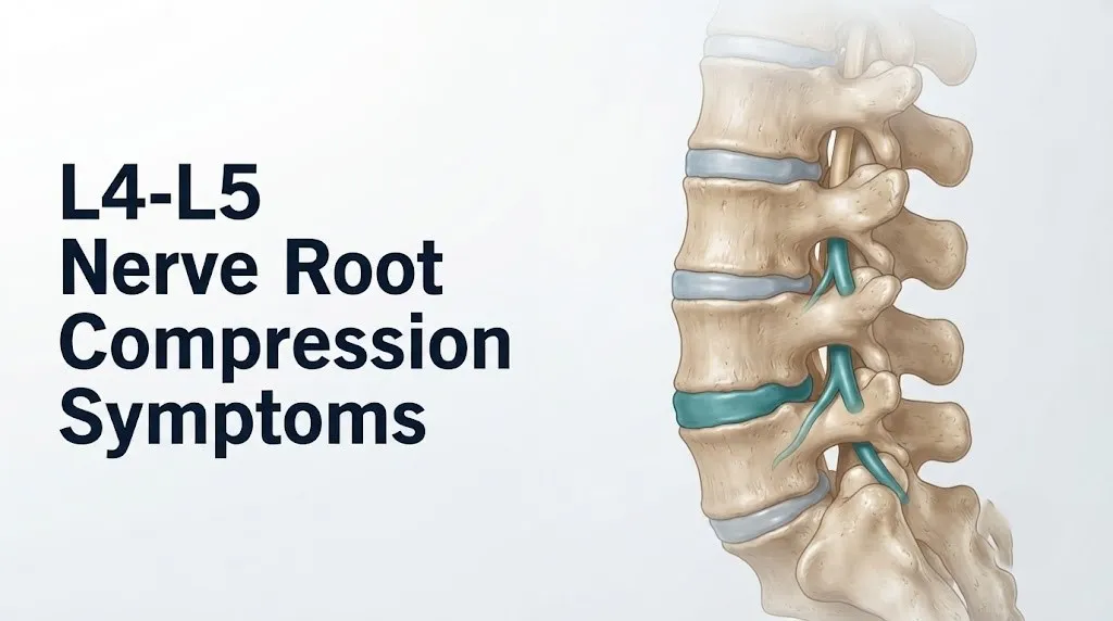
Two distinct nerve roots are at risk at this single level. The L4 nerve root exits the spinal canal through the L4-L5 neural foramen (the opening between the L4 and L5 vertebral bodies). The L5 nerve root has not yet exited at this level: it is traversing the spinal canal diagonally, crossing the L4-L5 disc in the lateral recess before descending to exit at the L5-S1 foramen below. A disc herniation that bulges posterolaterally compresses the traversing L5 root. A herniation that is more lateral or foraminal compresses the exiting L4 root instead. The same disc space. Two completely different clinical presentations.
Additionally, at L4-L5, the facet joints (zygapophyseal joints) are under the greatest degenerative stress in the lumbar spine. Facet arthrosis produces osteophytes that narrow the neural foramen from behind. Ligamentum flavum hypertrophy narrows the central canal and lateral recesses. These age-related changes produce a form of compression that develops gradually over years, quite differently from the acute herniation mechanism.
L4 Nerve Root Symptoms vs. L5 Nerve Root Symptoms at the Same Disc Level
At the L4-L5 spinal level, L4 nerve root compression produces pain and sensory changes along the medial lower leg and foot with quadriceps weakness, while L5 nerve root compression produces pain and sensory changes along the lateral lower leg, dorsal foot, and first web space with foot and great toe weakness.
This distinction is the most clinically important piece of information missing from every competing article on this topic, and it directly affects how you describe your symptoms to your physician and how a surgeon plans any intervention.
| Feature | L4 Nerve Root Compression | L5 Nerve Root Compression |
|---|---|---|
| Pain radiation | Front/medial thigh, medial lower leg, inner ankle | Lateral hip, posterolateral thigh, lateral lower leg, dorsal foot |
| Sensory loss location | Medial lower leg, inner aspect of foot | Dorsal foot, first web space (between big toe and second toe), lateral calf |
| Primary muscle weakness | Quadriceps (knee extension), hip adductors | Tibialis anterior (foot dorsiflexion), extensor hallucis longus (great toe extension), gluteus medius (hip abduction) |
| Reflex change | Diminished or absent patellar reflex (knee jerk) | No reflex change (L5 does not contribute to patellar or Achilles reflex) |
| Functional difficulty | Difficulty climbing stairs, knee buckling | Difficulty lifting foot off ground, tripping, heel walking |
| Provocative test | Femoral nerve stretch test (prone knee flexion) | Straight leg raise test (Lasegue sign) |
The straight leg raise test, performed by a clinician raising the patient’s leg with the knee straight while the patient lies on their back, stretches the L5 and S1 nerve roots. Reproduction of radiating leg pain below the knee (not just back pain) at angles below 60 degrees is considered a positive result. According to a systematic review published in Spine, the straight leg raise test has a sensitivity of approximately 80 percent for L4-L5 and L5-S1 disc herniation but a specificity of only around 40 percent, meaning a positive test strongly suggests nerve root involvement but does not confirm the specific level or cause.
For people over age 60, reflexes may be diminished bilaterally from age-related peripheral nerve changes unrelated to disc pathology. A single diminished patellar reflex in an older adult therefore requires correlation with other clinical findings before being attributed to L4 radiculopathy.
Key Takeaway: If your sensory loss is on the top of your foot and you can’t lift your toes well, your L5 root is the likely culprit. If your knee feels weak and your inner lower leg is numb, suspect L4. These are different clinical pictures requiring different clinical conversations.
Lower Back Pain From L4-L5 Nerve Root Compression
Lower back pain from L4-L5 nerve root compression originates from two simultaneous sources: nociceptive pain from irritated and degenerated disc and facet joint structures at the L4-L5 level, and referred pain from the inflamed nerve root itself radiating into the lower back from its attachment at the spinal cord.
The lower back pain associated with L4-L5 nerve root compression is typically described as a deep, aching pain centered in the lumbar region, usually to one side of midline (ipsilateral to the herniation), and often worse with sitting, bending forward, and coughing or sneezing. Forward bending and sitting increase intradiscal pressure at L4-L5, which can worsen disc herniation and increase compressive force on the nerve root. Coughing and sneezing produce a Valsalva maneuver (involuntary bearing-down action) that transiently raises intraspinal pressure, stretching the nerve root against the disc material and producing a sudden, sharp radicular jab.
The severity of lower back pain does not reliably predict the degree of nerve root compression. Research published in Spine has shown that patients with small disc herniations can have severe back pain, while patients with large herniations occasionally present with minimal local back pain but severe leg symptoms. This is partly because the disc itself (through its outer annular fibers and the adjacent posterior longitudinal ligament, which are richly innervated by the sinuvertebral nerve) generates its own pain signals independent of nerve root compression.
One underappreciated pattern: lower back pain that is worse when walking and relieved by sitting or leaning forward suggests lumbar spinal stenosis rather than acute disc herniation. In stenosis, the spinal canal narrows further in extension (upright walking position) and opens slightly in flexion (leaning forward or sitting). People with stenosis characteristically walk a short distance, develop lower back and leg pain, and find relief by sitting or leaning on a shopping cart. This is neurogenic claudication, and it is anatomically and mechanically distinct from the leg pain of acute disc herniation. People over age 65 presenting with L4-L5 symptoms are substantially more likely to have stenosis than herniation as the primary mechanism.
Leg Pain and Sciatica From L4-L5 Nerve Root Compression
Radiating leg pain from L4-L5 nerve root compression, often called sciatica, occurs when the compressed nerve root transmits abnormal pain signals along its entire length from the lumbar spine down to the foot, producing pain that the brain perceives as coming from the leg despite the actual source being in the spine.
The term sciatica is used colloquially to describe any radiating leg pain originating from the lumbar spine, but it is technically most accurate when applied to pain traveling along the distribution of the sciatic nerve (formed from the L4, L5, S1, S2, and S3 nerve roots). Compression specifically at L4-L5 most commonly affects the L5 component of the sciatic nerve, producing pain that travels from the lower back through the buttock, down the posterolateral thigh and lateral lower leg, and into the dorsal foot and toes.
The pain character is typically quite different from musculoskeletal pain. Nerve root pain is most often described as burning, sharp, electric, shooting, or lancinating. It follows a linear track down the leg rather than being diffuse or cramping. It may be continuous or may come in waves. Certain positions dramatically change its intensity: sitting typically worsens it (increases intradiscal pressure and nerve stretch), while lying flat often provides partial relief. Standing for extended periods also tends to worsen it in disc herniation cases.
According to the North American Spine Society clinical practice guidelines for lumbar disc herniation with radiculopathy, radiating leg pain that extends below the knee is a more specific indicator of true nerve root involvement than pain that stays above the knee or in the buttock alone. Below-knee radiation in the dermatomal pattern of L4 or L5 is a strong clinical signal that the compression is at the nerve root level.
Pregnant women in the third trimester may experience a similar radiating leg pain pattern from lumbar disc compression, driven by increased lumbar lordosis, disc loading from weight gain, and ligament laxity from relaxin. The clinical presentation can closely mimic non-pregnancy-related L4-L5 radiculopathy. MRI without contrast is safe after the first trimester and can be used to evaluate nerve root compression in pregnant women when clinical symptoms warrant imaging.
Numbness and Tingling From L4-L5 Nerve Compression
Numbness and tingling from L4-L5 nerve compression occur when the compressed nerve root’s sensory axons are sufficiently disrupted to produce abnormal electrical signals perceived by the brain as tingling (paresthesia) or the complete absence of sensation (numbness, technically termed hypoesthesia or anesthesia depending on severity).
The location of numbness and tingling directly maps to the dermatome of the compressed nerve root. A dermatome is the specific patch of skin whose sensory signals are carried by a single nerve root. The L4 dermatome covers the medial lower leg from just below the knee, the inner aspect of the ankle, and the medial arch of the foot. The L5 dermatome covers the lateral lower leg, the dorsal (top) surface of the foot, the first web space between the big toe and second toe, and extends to the big toe itself.
Tingling (paresthesia) typically occurs first, as partial compression produces irregular electrical discharge in the sensory axons. This feels like pins and needles, buzzing, or an electrical sensation. If compression worsens or persists, the sensory axons stop conducting entirely, and tingling is replaced by numbness. This progression from paresthesia to anesthesia indicates increasing severity of nerve root injury and warrants earlier clinical evaluation.
An important clinical distinction: numbness from nerve root compression is dermatomal (specific, map-like distribution). Numbness from peripheral neuropathy (as in diabetes) is typically stocking-like: symmetrical, affecting both feet, and involving the entire foot from the toes upward rather than a specific strip of skin. If your numbness is confined to a specific track on one leg and one foot corresponding to the L4 or L5 dermatome, this pattern is far more consistent with radiculopathy than peripheral neuropathy, though only clinical examination and EMG/NCV testing can reliably distinguish the two in people with diabetes.
Quick Tip:
- You can roughly self-assess your L5 dermatome by testing sensation on the top of your foot between your big and second toe. Reduced sensation there, compared to the same spot on the other foot, is a clinically relevant finding to report to your primary care physician.
- Numbness that is rapidly spreading, involving both legs or the groin and inner thighs, is not a dermatomal pattern. It requires emergency evaluation.
- People taking anticoagulant medications (blood thinners) who develop sudden severe back pain with leg weakness should go to the emergency room, as epidural hematoma can produce rapid nerve compression.
Key Takeaway: Tingling that becomes numbness in a specific dermatomal track means the nerve root compression is progressing, and that progression warrants medical evaluation within days, not weeks.
Muscle Weakness From L4-L5 Nerve Root Compression
Muscle weakness from L4-L5 nerve root compression occurs when the motor axons within the compressed nerve root are unable to transmit sufficient electrical signals to the muscles they supply, reducing the muscle’s ability to contract against resistance.
Each nerve root supplies a specific group of muscles called a myotome. The L4 myotome includes the quadriceps femoris (knee extension), the tibialis anterior (foot dorsiflexion, the motion of pulling the foot upward), and the hip adductors. L4 nerve root compression most often manifests as quadriceps weakness, noticed as difficulty fully extending the knee, a sense of knee instability on stairs, or occasional knee buckling when walking downhill. The L5 myotome includes the tibialis anterior, the extensor hallucis longus (great toe extension), the extensor digitorum longus (extension of the other toes), the peroneus longus and brevis (foot eversion), and the gluteus medius (hip abduction). L5 weakness is most often first noticed as difficulty lifting the front of the foot during walking, causing the toe to catch on flooring or steps.
Muscle weakness is graded clinically on the Manual Muscle Testing (MMT) scale from 0 (no contraction) to 5 (full strength against full resistance). Grades 4 and 4+ (movement possible against partial or moderate resistance) represent mild weakness that the patient may not notice during normal daily activities. Grade 3 (movement possible only against gravity, not added resistance) represents moderate weakness that affects function. Grades 2 and below represent severe weakness requiring immediate evaluation.
According to the American Academy of Orthopaedic Surgeons, any motor deficit at the time of initial presentation for lumbar disc herniation is an indication for MRI within one week, not watchful waiting. Motor deficits that are rapidly progressive (worsening over days to a week) indicate deteriorating nerve root function and require urgent evaluation by a board-certified orthopedic spine surgeon or board-certified neurosurgeon within 24 to 48 hours to determine whether surgical decompression is warranted.
Older adults with longstanding L4-L5 stenosis may have developed gradual, progressive weakness that they have normalized over months to years, attributing it to “aging” or “being out of shape.” Quadriceps weakness from L4 compression in particular can significantly increase fall risk in people over 70, a clinical consequence that primary care physicians specifically monitoring gait and balance in older patients should actively screen for.
Foot Drop as a Symptom of L4-L5 Nerve Compression
Foot drop is the inability to dorsiflex the foot (lift the front of the foot upward toward the shin) and is the most clinically recognizable functional deficit of L5 nerve root compression at the L4-L5 level, occurring when the motor fibers supplying the tibialis anterior muscle lose adequate nerve signal to sustain antigravity foot lifting during the swing phase of walking.
During normal walking, the tibialis anterior contracts immediately after the heel strikes the ground, keeping the toes lifted during the swing phase so they clear the floor. When L5 nerve root compression severely reduces tibialis anterior function, the foot hangs limp during the swing phase. The person compensates by raising the knee abnormally high (steppage gait) or swinging the leg out to the side, both visible to an observer watching the person walk from the front.
Foot drop from L4-L5 nerve root compression is a neurological motor deficit, not a musculoskeletal problem. This distinction is clinically important because foot drop can also be caused by peroneal (common fibular) nerve compression at the fibular head (from crossing the legs, prolonged squatting, or a cast), making EMG/NCV testing essential to confirm the lesion location is at the L5 nerve root rather than at the peripheral peroneal nerve.
The North American Spine Society clinical guidelines state that foot drop from lumbar disc herniation represents an absolute indication for surgical consultation, even if pain is manageable. The reason is time-sensitive: motor nerve axons that have been severely compressed for extended periods undergo Wallerian degeneration (axonal breakdown distal to the compression site). Recovery of foot drop is substantially better when decompression occurs within the first few weeks of onset than when it occurs months later. A person who develops foot drop should contact a board-certified orthopedic spine surgeon or board-certified neurosurgeon within one to three days, not at a routine appointment weeks away.
People with pre-existing type 2 diabetes are at elevated risk for poor motor recovery after nerve decompression because concurrent diabetic axonal damage impairs the nerve’s regenerative capacity. This population-specific factor should be discussed explicitly during surgical planning.
L4-L5 Symptoms From Disc Herniation vs. Spinal Stenosis vs. Spondylolisthesis
L4-L5 symptoms from disc herniation, spinal stenosis, and spondylolisthesis overlap substantially in their final common pathway (nerve root compression), but each has a distinct onset pattern, pain behavior, and clinical context that a clinician and an informed patient can use to guide diagnosis.
Understanding these distinctions matters because the natural history and treatment approach differ substantially between the three causes.
Disc herniation produces acute onset of radicular symptoms, often following a precipitating event (bending, lifting, twisting), in adults aged 30 to 50. Pain is typically severe at onset, made worse by sitting and forward bending (which increase intradiscal pressure), and relieved by lying flat. The straight leg raise test is frequently positive. The majority of disc herniations improve substantially without surgery: the SPORT trial showed that approximately 70 percent of patients treated nonoperatively achieved a good outcome at two years.
Lumbar spinal stenosis produces gradual onset of leg pain and weakness, often described as heaviness or cramping rather than sharp radicular pain, in adults over 60. Pain is worsened by walking and standing (extension positions that narrow the canal) and relieved by sitting or forward flexion (neurogenic claudication). The straight leg raise test is typically negative or equivocal. The Cleveland Clinic notes that spinal stenosis is the most common reason for lumbar spine surgery in adults over 65 in the United States.
Spondylolisthesis at L4-L5 occurs when the L4 vertebral body slips forward relative to L5, typically from degenerative facet joint disease in adults over 50 (degenerative spondylolisthesis) or from a pars interarticularis stress fracture in younger athletes (isthmic spondylolisthesis). The forward slip narrows the canal and foramen, compressing nerve roots. Symptoms combine features of both disc herniation and stenosis, and are often bilateral or alternating.
| Feature | Disc Herniation | Spinal Stenosis | Spondylolisthesis |
|---|---|---|---|
| Typical age at onset | 30 to 50 | Over 60 | Over 50 (degenerative); teens to 30s (isthmic) |
| Onset | Often acute | Gradual | Gradual |
| Pain worsened by | Sitting, flexion | Walking, extension | Walking, extension |
| Pain relieved by | Lying flat | Sitting, forward lean | Sitting, forward lean |
| Straight leg raise | Often positive | Usually negative | Variable |
| Bilateral symptoms | Uncommon at onset | Common | Common |
| Natural history | Most improve without surgery | Progressive without treatment | Variable |
Key Takeaway: If your symptoms started suddenly after lifting something and are worst when you sit, a disc herniation is the most likely cause. If your leg aches and cramps after walking two blocks and you feel better leaning on a shopping cart, stenosis is more likely, and your primary care physician should order lumbar spine MRI.
Atypical L4-L5 Symptoms: Nerve Pain Without Back Pain and Bilateral Presentations
A meaningful proportion of people with confirmed L4-L5 nerve root compression experience leg pain, numbness, or weakness without any lower back pain, a presentation that frequently delays diagnosis because both the patient and the initial clinician may not consider the spine as the source of isolated leg symptoms.
According to data from PubMed-indexed studies on lumbar radiculopathy, approximately 10 to 15 percent of patients with confirmed disc herniation and nerve root compression on MRI report no lower back pain at the time of initial presentation. The radicular symptoms (leg pain, numbness, weakness) can be the sole complaint. The absence of back pain does not exclude a spinal origin. The disc herniation may have occurred at a moment that was not perceived as painful, or the back pain may have been present briefly and resolved while the nerve root remained compressed. This pattern is more common in people who have experienced a gradual, progressive disc herniation rather than an acute event.
Bilateral L4-L5 symptoms (affecting both legs simultaneously) represent a more serious clinical picture than unilateral radiculopathy. Bilateral radicular symptoms suggest that the compressive process is centrally located (large central disc herniation or severe central canal stenosis) and is impacting the descending nerve roots of the cauda equina rather than a single unilateral nerve root. Bilateral symptoms are not automatically a cauda equina emergency, but they substantially narrow the safety margin: the cauda equina is already compromised bilaterally, and any further compression or swelling from an additional disc event could produce the full cauda equina syndrome.
People with bilateral L4-L5 symptoms should be evaluated by a primary care physician or physiatrist within 24 to 48 hours with MRI imaging ordered, and they should be explicitly counseled on the specific symptoms of cauda equina syndrome (detailed in a later section) so they know precisely when to go to the emergency room without waiting for a scheduled appointment.
Synovial cysts arising from degenerated L4-L5 facet joints represent another atypical cause worth noting. These cysts can compress the nerve root from a posterior direction rather than the anterior direction typical of disc herniation. On MRI, they appear as a fluid-filled structure adjacent to the facet joint and within the lateral recess. They tend to cause symptoms that fluctuate with position changes and activity level, because the cyst may change in size with joint fluid dynamics.
L4-L5 Nerve Compression Symptoms by Population: Older Adults, Pregnant Women, Athletes
L4-L5 nerve root compression symptoms present with meaningful differences across specific populations that affect both the likelihood of certain diagnoses and the urgency and type of evaluation needed.
Older adults (over 65): In this population, degenerative spinal stenosis and spondylolisthesis are far more likely causes of L4-L5 nerve compression than acute disc herniation. Older adults are also more likely to have multilevel disease (compression at L3-L4 and L4-L5 simultaneously) and superimposed peripheral neuropathy from diabetes, cardiovascular disease, or other systemic conditions. Symptoms may be less acutely painful and more insidious in onset. A gradual decline in walking tolerance, combined with relief on leaning forward, is the characteristic presentation that should prompt lumbar spine imaging. Older adults with significant stenosis are also at higher risk of epidural hematoma from falls, and any sudden onset of bilateral leg weakness or bowel/bladder symptoms in this group requires emergency evaluation even if a fall seemed minor.
Pregnant women: The third trimester is the highest-risk period for lumbar disc herniation due to peak lumbar lordosis, maximum disc loading from weight gain, and relaxin-mediated ligament laxity. MRI without gadolinium contrast is considered safe during pregnancy (after the first trimester) when clinical findings warrant imaging. Surgical intervention during pregnancy is reserved for severe motor deficits or cauda equina syndrome. Epidural steroid injections require careful risk-benefit discussion with the managing obstetrician-gynecologist before proceeding. Most pregnancy-related radiculopathy resolves after delivery as disc loading normalizes.
Athletes and active adults: Younger athletes, particularly those involved in heavy loading sports (powerlifting, football linemen, gymnasts, rowers), can sustain disc herniation from repetitive high-load spine compression. Isthmic spondylolisthesis from pars interarticularis fracture is also more prevalent in young athletes engaged in extension-loaded sports (gymnastics, wrestling, cricket bowling). Return-to-play decisions after lumbar disc herniation or spondylolisthesis require evaluation by a sports medicine physician alongside an orthopedic spine surgeon, and should not be made based on pain resolution alone without structural clearance.
People with diabetes: As noted previously, diabetic peripheral neuropathy can obscure the dermatomal pattern of radiculopathy and reduce the perceived severity of pain despite significant nerve root compression. EMG/NCV testing is particularly valuable in this population to distinguish and quantify both conditions simultaneously.
Cauda Equina Syndrome: The Emergency Presentation of Severe L4-L5 Compression
Cauda equina syndrome is a neurological emergency that occurs when massive compression of the cauda equina (the bundle of lumbar and sacral nerve roots descending through the central spinal canal below the conus medullaris) causes acute dysfunction of bowel, bladder, and lower extremity motor and sensory function simultaneously.
At the L4-L5 level, a large central disc herniation that suddenly expands into the central canal can compress the entire cauda equina rather than a single nerve root. This produces a bilateral, multi-level neurological deficit that cannot be explained by single nerve root compression. The surgical literature consistently identifies cauda equina syndrome as requiring decompressive surgery within 24 to 48 hours of onset to prevent permanent neurological deficits.
The defining symptoms of cauda equina syndrome are:
- Urinary retention (inability to urinate) or urinary incontinence (inability to hold urine): this is the most common and clinically specific finding, occurring in approximately 90 percent of confirmed cases per data reviewed by the North American Spine Society
- Bowel dysfunction: typically constipation or loss of voluntary bowel control
- Saddle anesthesia: numbness or reduced sensation in the perineum, inner thighs, buttocks, and genitals (the area that would contact a saddle), corresponding to the sacral nerve root territory
- Bilateral leg weakness: affecting both legs rather than one, often more severe than in typical unilateral radiculopathy
- Bilateral radiating leg pain: severe pain in both legs simultaneously
Cauda equina syndrome can be incomplete (some function preserved) or complete (total loss of function). Both presentations are emergencies. Incomplete cauda equina syndrome has better outcomes with early surgery, but waiting to see whether it becomes complete before seeking care is clinically indefensible.
The Mayo Clinic states that delay in surgical decompression for cauda equina syndrome beyond 24 to 48 hours significantly increases the risk of permanent urinary incontinence, bowel incontinence, sexual dysfunction, and bilateral lower extremity weakness. These are not theoretical risks: they are documented, permanent neurological sequelae of delayed treatment.
Emergency Symptoms: When to Call 911 or Go to the ER
Certain symptoms associated with L4-L5 nerve root compression require immediate emergency evaluation. Do not wait to see if these resolve on their own.
Call 911 or go to the nearest emergency room immediately if you experience:
- Any new loss of bladder control (urinary retention or urinary incontinence) combined with lower back or leg pain. This is the cardinal symptom of cauda equina syndrome and constitutes a surgical emergency. Every hour of delay increases the risk of permanent neurological deficit.
- Any new loss of bowel control (fecal incontinence or inability to defecate voluntarily) combined with lower back or leg symptoms. This represents cauda equina-level sacral nerve dysfunction.
- Numbness, tingling, or loss of sensation in the perineum, genitals, inner thighs, or buttocks (saddle distribution). This is the clinical hallmark of central cauda equina compression and requires emergency imaging within hours.
- Sudden bilateral leg weakness: both legs becoming weak or giving out simultaneously, indicating bilateral nerve root or cauda equina compression.
- Severe, sudden onset of lower back pain combined with rapidly progressing leg weakness in a person taking anticoagulant medications (blood thinners), as this pattern suggests epidural hematoma, which is a spinal surgical emergency.
- Rapid progression of foot drop from mild to complete within 24 to 48 hours, indicating severe and accelerating L5 nerve root compression that has surpassed the threshold for watchful waiting.
These presentations can indicate cauda equina syndrome, epidural hematoma, or severe progressive nerve root ischemia and require emergency medical assessment, not an urgent care appointment or a phone call to your primary care physician’s office.
How L4-L5 Nerve Root Compression Symptoms Progress Without Treatment
Without appropriate management, L4-L5 nerve root compression symptoms follow one of three natural history trajectories depending on the cause, the degree of compression, and individual biological factors.
The most favorable trajectory is spontaneous improvement. In disc herniation specifically, the herniated nucleus pulposus material undergoes resorption over weeks to months through a process involving macrophage infiltration and phagocytosis of disc material at the nerve root-disc interface. Research published in Spine has documented spontaneous herniation regression on serial MRI in 60 to 90 percent of patients who are followed conservatively, with the rate of regression correlating with the size of the herniation at baseline. Larger herniations show more complete regression, possibly because they provoke a larger immune response. During this resorption period, symptoms typically peak in the first two to four weeks and then gradually improve over three to six months.
The second trajectory is plateau: symptoms stabilize at a level of persistent pain, numbness, and mild weakness that neither worsens significantly nor improves substantially. This pattern is more common in people with foraminal stenosis from osteophytes (which do not resorb) or with chronic degenerative stenosis. Without structural intervention, the underlying cause remains and symptoms persist indefinitely, though they may fluctuate with activity level.
The third trajectory, occurring in a minority of patients, is deterioration. Progressive nerve root compression without decompression can lead to Wallerian degeneration of axons: the myelin sheath breaks down, the axon itself degrades, and the nerve’s ability to regenerate is increasingly compromised the longer compression persists. Motor deficits that have been present for more than six to twelve months have significantly lower recovery rates after surgical decompression than deficits of recent onset.
People with longstanding uncontrolled type 2 diabetes face accelerated axonal deterioration because compromised microvascular supply to the nerve (diabetic vasa nervorum disease) reduces the metabolic support available to the compressed nerve root. This population-specific factor makes early evaluation and intervention more, not less, important in diabetic patients with L4-L5 nerve root symptoms.
Diagnosing L4-L5 Nerve Root Compression: What Tests Reveal and What They Miss
Accurate diagnosis of L4-L5 nerve root compression requires combining the clinical examination findings with targeted imaging and, when necessary, electrodiagnostic testing, because no single test is sufficient to confirm the diagnosis in all cases.
The clinical examination comes first. A primary care physician or physiatrist performing a focused neurological examination looks for specific findings: the pattern of dermatomal sensory loss, myotomal weakness on manual muscle testing, reflex changes (diminished patellar reflex for L4, normal reflex for L5), and provocation tests (straight leg raise for L5, femoral nerve stretch test for L4). These findings together can identify which nerve root is compressed and how severely, before any imaging is obtained.
MRI of the lumbar spine is the standard imaging test for evaluating nerve root compression. It shows disc anatomy, disc herniation direction and size, canal diameter, foraminal diameter, and nerve root displacement with high sensitivity. One critical limitation: MRI shows structure, not function. A disc herniation visible on MRI may or may not be causing the patient’s symptoms. Studies published in the New England Journal of Medicine and in Spine have found that 20 to 30 percent of asymptomatic adults over age 40 have disc herniations visible on lumbar MRI. This means that finding a herniation on MRI does not automatically confirm it is the cause of symptoms without clinical correlation.
EMG and NCV testing, performed by a neurologist or physiatrist, measures the electrical activity of muscles and the conduction speed of nerve fibers. EMG detects denervation changes in specific muscles that correspond to the affected myotome, providing direct physiological evidence of nerve root dysfunction rather than structural appearance. This testing is particularly valuable when the clinical picture is uncertain, when the MRI shows multiple levels of pathology, or when diabetic neuropathy complicates symptom interpretation.
To prepare for your diagnostic appointment:
- Record the exact location of your pain, numbness, and weakness with as much anatomical specificity as possible.
- Note the timing pattern: whether symptoms are constant or intermittent, worse with sitting or walking, and whether they are improving or worsening over days.
- Rate your pain on a scale of 0 to 10 for both the back component and the leg component separately, as these may reflect different pathological processes.
- Note any bowel or bladder changes, even subtle ones, and report them explicitly; do not assume they are unrelated.
- Bring a list of all medications including blood thinners, as these affect both imaging and procedural options.
- If you have had previous lumbar spine imaging, bring those reports and images for comparison.
Key Takeaway: An MRI showing a herniated disc does not automatically mean surgery is needed or even that the disc is causing your symptoms. Clinical examination findings, specifically the dermatomal and myotomal pattern, must match the imaging finding to justify treatment decisions.
When to Seek Urgent Care or Emergency Evaluation for L4-L5 Nerve Compression
The appropriate level of care for L4-L5 nerve compression depends on the specific symptoms present, and matching the right urgency level to the right symptom pattern is one of the most practically important things this article can give you.
| Symptom Pattern | Urgency Level | Appropriate Action |
|---|---|---|
| Leg pain with no weakness, no numbness, no bowel/bladder change | Low | Primary care physician appointment within 1 to 2 weeks |
| Leg pain with mild numbness in dermatomal distribution | Moderate | Primary care physician within one week; request lumbar MRI |
| Any new motor weakness (foot, toes, knee) | High | Primary care physician or physiatrist within 2 to 3 days; urgent MRI |
| Progressive weakness worsening over days | Very High | Orthopedic spine surgeon or neurosurgeon within 24 to 48 hours |
| Foot drop of recent onset (days) | Very High | Orthopedic spine surgeon or neurosurgeon within 24 to 48 hours |
| Any bowel or bladder dysfunction combined with spine symptoms | Emergency | Go to the emergency room immediately |
| Saddle area numbness | Emergency | Go to the emergency room immediately |
| Bilateral leg weakness or bilateral radiating pain | Emergency | Go to the emergency room immediately |
Go to the emergency room immediately for any bowel or bladder change, saddle area numbness, or bilateral leg weakness. These are the cardinal signs of cauda equina syndrome, which is a surgical emergency where outcome is directly tied to the speed of decompression.
For all other symptom patterns, the referral chain typically begins with a primary care physician, who can order lumbar spine MRI and refer to a physiatrist, orthopedic spine surgeon, or neurosurgeon as findings warrant. People who already have a confirmed L4-L5 compression diagnosis from prior imaging and experience a sudden worsening of their neurological symptoms (new weakness, new bowel/bladder symptoms) should go directly to the emergency room rather than waiting for a clinic appointment, because acute-on-chronic compression can still produce cauda equina syndrome in people with longstanding disease.
How Long Do L4-L5 Nerve Root Compression Symptoms Last and What Affects Recovery
L4-L5 nerve root compression symptoms from acute disc herniation resolve in the majority of patients within six to twelve weeks with appropriate non-surgical management, but the timeline and degree of recovery depend on the cause of compression, the severity and duration of nerve root injury, and specific individual factors.
The SPORT trial, published in the New England Journal of Medicine by Weinstein and colleagues in 2006, followed 501 patients with lumbar disc herniation and radiculopathy over two years. In the nonoperatively treated group, approximately 65 to 70 percent of patients had meaningful improvement in pain and function by three months, with continued gradual improvement through twelve months. However, about 25 to 30 percent of the nonoperative group had persistent symptoms at two years that significantly limited function. Patients with larger herniations and more severe nerve root compression at baseline tended to have slower and less complete recovery.
Factors that support faster recovery:
- Shorter duration of symptoms before treatment begins (symptoms present for less than six weeks at the start of management)
- Younger age (regenerative capacity of nerve tissue is greater)
- Smaller herniation size on MRI
- Predominantly pain symptoms without significant motor deficit
- Normal body weight (reduced ongoing mechanical load on the disc and nerve root)
- Non-smoking status (smoking impairs disc and nerve microvasculature recovery)
Factors associated with slower or incomplete recovery:
- Motor deficit (foot drop, quadriceps weakness) present at onset
- Symptoms present for more than three months before intervention
- Diabetes mellitus with peripheral neuropathy (impairs axonal regeneration)
- Spinal stenosis or spondylolisthesis as the cause (structural narrowing does not resolve spontaneously)
- Advanced age (slower nerve regeneration, more comorbid degenerative changes)
- Heavy physical occupation requiring early return to spine-loading activities
Recovery of sensory symptoms (numbness and tingling) typically precedes recovery of motor strength, because sensory axons have a higher density and begin signaling recovery before muscle strength is measurable. Return of a previously absent reflex, such as the patellar reflex in L4 radiculopathy, is one of the most reliable clinical indicators that nerve root recovery is occurring.
Frequently Asked Questions About L4-L5 Nerve Root Compression Symptoms
What does L4-L5 nerve root compression feel like?
L4-L5 nerve root compression feels like sharp, burning, or electric pain that radiates from the lower back down into one leg, typically following a specific track toward the foot.
Associated sensations include numbness or tingling in the lateral lower leg, the top of the foot, or the inner lower leg depending on which nerve root is compressed.
Many people describe the leg pain as more distressing than the back pain, and it typically worsens with sitting or bending forward.
How do I know if I have L4 or L5 nerve root compression?
L4 nerve root compression produces pain and numbness along the inner lower leg and medial foot, weakness of the quadriceps (knee extension), and a diminished patellar (knee jerk) reflex.
L5 nerve root compression produces pain and numbness along the outer lower leg, the top of the foot, and the first web space between the big and second toe, with weakness of foot dorsiflexion and great toe extension.
A primary care physician or physiatrist can distinguish the two through a focused neurological examination and confirmation with lumbar spine MRI.
Can L4-L5 nerve compression cause foot drop?
L4-L5 nerve compression can cause foot drop when the L5 nerve root is sufficiently compressed to impair function of the tibialis anterior muscle, which is responsible for lifting the front of the foot during walking.
Foot drop is a motor deficit requiring urgent evaluation by a board-certified orthopedic spine surgeon or board-certified neurosurgeon within one to three days of onset, not a routine appointment.
Early surgical decompression produces substantially better motor recovery than decompression performed weeks or months after the onset of foot drop.
What are the warning signs of cauda equina syndrome from L4-L5 compression?
The warning signs of cauda equina syndrome are urinary retention or incontinence, loss of bowel control, numbness in the saddle area (perineum, inner thighs, and genitals), and bilateral leg weakness.
These symptoms indicate that a large disc herniation or central stenosis is compressing the entire cauda equina rather than a single nerve root.
Go to the emergency room immediately if any of these symptoms develop: cauda equina syndrome requires emergency surgical decompression within 24 to 48 hours to prevent permanent neurological damage.
Can L4-L5 nerve compression symptoms go away without surgery?
L4-L5 nerve compression symptoms from disc herniation improve without surgery in approximately 70 percent of patients within three to six months, according to the SPORT trial published in the New England Journal of Medicine.
Spontaneous improvement occurs as the herniated disc material undergoes macrophage-mediated resorption and the inflammatory chemical irritation of the nerve root subsides.
Symptoms from spinal stenosis, spondylolisthesis, or synovial cysts do not resolve spontaneously because the underlying structural cause does not resorb, and non-surgical management provides symptom control rather than structural correction.
When should I go to the emergency room for L4-L5 nerve compression?
Go to the emergency room immediately if you develop any change in bladder or bowel function, numbness in the saddle area, or bilateral leg weakness alongside your back or leg symptoms.
These are the defining features of cauda equina syndrome, and every hour of delay before surgical decompression increases the risk of permanent incontinence and bilateral lower extremity weakness.
For all other symptom presentations, including new motor weakness such as foot drop, contact a primary care physician or specialist within one to three days rather than waiting for a routine appointment.
Closing
The single most practically important thing to know about L4-L5 nerve root compression is the emergency threshold: bowel or bladder dysfunction, saddle area numbness, or bilateral leg weakness means go to the emergency room now, not tomorrow, not after calling your doctor’s office. That threshold is supported by clear surgical evidence, and missing it has permanent consequences.
For everything short of that emergency, the roadmap is straightforward. Identify your specific symptom pattern, note whether it matches the L4 or L5 dermatome, track whether weakness is present or progressing, and contact your primary care physician within one to seven days depending on severity. Request a lumbar MRI and bring your complete symptom description.
You now have the anatomical and clinical framework to describe your symptoms with precision. Use it when you talk to your physician, and be specific: which leg, which part of the leg, what the pain feels like, and whether anything has changed in your bladder or bowel function. That level of precision changes the quality of care you receive