What Causes Arthritis in Fingers? A Full 2026 Guide
What causes arthritis in fingers depends on which type is involved, but every form shares a common endpoint: structural damage to one or more of the small synovial joints of the hand, producing pain, swelling, stiffness, and eventually altered joint shape. The three most common causes are osteoarthritis from articular cartilage breakdown, rheumatoid arthritis from an autoimmune attack on the joint lining, and gout from monosodium urate crystal deposition within the joint space.
According to the National Institute of Arthritis and Musculoskeletal and Skin Diseases (NIAMS), arthritis is the leading cause of work disability in the United States, and hand and finger arthritis represents a disproportionate share of the functional burden, given how much daily life depends on grip strength, fine motor control, and pain-free finger movement. Radiographic evidence of hand osteoarthritis is present in more than 70% of adults over age 70.
This article covers the specific mechanisms behind each cause of finger arthritis, why certain joints in the finger are affected by certain types, why women are at higher risk, which causes affect younger adults, and the specific symptoms that mean you should see a board-certified rheumatologist rather than continuing to self-monitor.
What Causes Arthritis in Fingers: The Clinical Overview
What causes arthritis in fingers is not a single answer: six distinct pathological processes can damage the small joints of the hand, each through a different anatomical mechanism, affecting different finger joints, and requiring different clinical management.
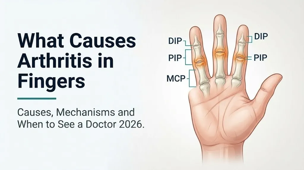
The finger joints most commonly affected by arthritis are the distal interphalangeal (DIP) joints (the outermost knuckle of each finger), the proximal interphalangeal (PIP) joints (the middle knuckle), and the metacarpophalangeal (MCP) joints (the large knuckles at the base of each finger where the finger meets the hand). The carpometacarpal (CMC) joint of the thumb is also a frequent site, particularly in osteoarthritis.
Each of these joints is a synovial joint: a fluid-filled cavity enclosed by a fibrous joint capsule, lined by the synovial membrane, and buffered by articular cartilage (hyaline cartilage) covering the bone ends within the joint. Arthritis develops when either the articular cartilage degrades, the synovial membrane becomes inflamed and destructive, or foreign material (crystals, bacteria, or debris) enters the joint space and triggers an inflammatory response.
The six principal causes of finger arthritis:
- Osteoarthritis: progressive articular cartilage loss from enzymatic degradation
- Rheumatoid arthritis: autoimmune synovial membrane attack producing pannus and bone erosion
- Psoriatic arthritis: IL-17 pathway-driven enthesitis and joint inflammation, often at DIP joints
- Gout: monosodium urate crystal deposition triggering NLRP3 inflammasome activation
- Post-traumatic arthritis: mechanical joint damage following fracture or ligament injury
- Reactive or septic arthritis: infection-triggered or direct bacterial invasion of the joint
Knowing which cause is most likely in your specific situation requires understanding which joints are affected, what the pain pattern is, what your personal risk factors are, and what diagnostic tests a physician will use to confirm the specific type.
How Finger Joint Anatomy Makes the Joints Vulnerable to Arthritis
The finger joints are vulnerable to arthritis because their small joint surface areas concentrate mechanical forces, their synovial membranes are highly reactive to inflammatory triggers, and their articular cartilage lacks its own blood supply, making it slow to repair once damaged.
Each finger joint relies on a thin coating of articular cartilage, typically 1 to 2 millimeters thick in the small joints of the hand, far thinner than the 4 to 6 millimeters found in larger weight-bearing joints like the knee. This cartilage is avascular: it receives all its nutrition through diffusion from the synovial fluid rather than through direct blood supply. When the synovial fluid composition changes from inflammation or when the cartilage matrix begins to degrade, the repair capacity of the tissue is extremely limited.
The articular chondrocytes (the only cells within cartilage) are responsible for maintaining the balance between anabolic processes (building new collagen and proteoglycans) and catabolic processes (breaking them down). Under normal conditions, this balance is maintained. When mechanical overload, inflammatory cytokines, or genetic predisposition tips this balance toward catabolism, the degradative enzymes matrix metalloproteinase-13 (MMP-13) and ADAMTS-5 begin outpacing repair, and cartilage loss becomes progressive.
Think of articular cartilage like a shock-absorbing gel pad on the end of each bone. The pad has no way to call for replacement parts. Once the enzymes start thinning it, the bones begin to contact each other during movement, producing the grinding, stiffness, and pain characteristic of osteoarthritis.
According to the American College of Rheumatology, the finger joints are particularly susceptible because repetitive pinch and grip forces concentrate pressure over very small articular surface areas, amplifying the per-unit stress on the cartilage compared to larger joints.
In people with diabetes mellitus, advanced glycation end-products (AGEs) accumulate in articular cartilage collagen, making it stiffer and more prone to mechanical failure. This explains why hand OA progresses faster in people with poorly controlled blood glucose.
Osteoarthritis in Fingers: Cartilage Breakdown Mechanism
Osteoarthritis (OA) of the fingers is the most frequent form of finger arthritis and occurs when the articular cartilage lining the DIP, PIP, and CMC joints degrades through a process driven by matrix metalloproteinase-13 (MMP-13) and ADAMTS-5, two cartilage-destroying enzymes upregulated by chondrocytes in response to mechanical stress and inflammatory cytokines.
The process begins when chondrocytes detect abnormal mechanical loading or low-grade synovial inflammation and shift from a cartilage-maintenance state to a catabolic state. MMP-13 cleaves type II collagen fibrils, the structural backbone of cartilage. ADAMTS-5 simultaneously degrades aggrecan, the large proteoglycan molecule responsible for cartilage hydration and compressive resilience. As both components are lost, the cartilage thins, softens, and eventually fibrillates (develops surface cracks).
The subchondral bone beneath the thinning cartilage responds by becoming denser and harder (subchondral sclerosis) and by developing bony outgrowths at the joint margins called osteophytes. These osteophytes are the bony enlargements that become visible and palpable at the DIP joints as Heberden nodes and at the PIP joints as Bouchard nodes.
The joint also produces excess synovial fluid as a secondary response to cartilage fragments and bone debris entering the joint space, contributing to visible joint swelling alongside the bony enlargement.
According to the NIAMS, OA of the hand affects an estimated 26.2% of women and 13.4% of men over age 71 in the United States, based on radiographic prevalence data. This nearly 2:1 sex ratio reflects the biological role of estrogen in maintaining articular cartilage health, which is discussed in a dedicated section below.
People who take long-term corticosteroid medications face an elevated risk of accelerated OA in finger joints because corticosteroids suppress the anabolic activity of chondrocytes and reduce synovial fluid quality, accelerating the cartilage thinning process.
Rheumatoid Arthritis in Fingers: The Autoimmune Mechanism
Rheumatoid arthritis (RA) causes finger joint destruction through a fundamentally different mechanism than OA: an autoimmune cascade in which the body’s immune system attacks the synovial membrane of the finger joints, producing a destructive tissue called pannus that erodes both cartilage and bone.
The process begins years before clinical symptoms appear. Peptidylarginine deiminase (PAD) enzymes, elevated in susceptible individuals, convert arginine residues in proteins to citrulline, creating citrullinated neoantigens that the immune system does not recognize as self. Autoreactive B cells and T cells mount a response against these neoantigens, producing anti-citrullinated protein antibodies (anti-CCP) and rheumatoid factor (RF). Both can be detected in blood years before joint symptoms develop.
When this autoimmune response targets the synovial membrane of the finger joints, the normally thin synovial lining proliferates abnormally into a hyperplastic, invasive tissue called pannus. Activated synovial fibroblasts and macrophages within the pannus release TNF-alpha, IL-1 beta, and IL-6, driving sustained inflammation. TNF-alpha additionally stimulates production of RANK-L (receptor activator of NF-kB ligand), which activates osteoclasts (bone-resorbing cells) at the pannus-bone interface, producing the characteristic bone erosions visible on plain radiograph at the MCP and PIP joint margins.
RA preferentially affects the MCP joints and PIP joints of the fingers while typically sparing the DIP joints, which is the opposite of osteoarthritis. This joint distribution pattern is so consistent that it is one of the clinical criteria in the 2010 ACR/EULAR Classification Criteria for Rheumatoid Arthritis.
Women develop RA at approximately 2 to 3 times the rate of men, with peak incidence in the fourth to sixth decades. Postpartum women face a particular risk window: the relative immune suppression of pregnancy temporarily reduces RA disease activity, but the postpartum period is associated with RA flares, a pattern attributed to the sharp postnatal drop in estrogen and progesterone levels.
Key Takeaway: Rheumatoid arthritis attacks the synovial lining of the MCP and PIP joints through TNF-alpha and pannus formation, while osteoarthritis destroys cartilage at the DIP and CMC joints through MMP-13 enzyme activity; the joints affected tell you which type you are likely dealing with.
Psoriatic Arthritis in Fingers: The IL-17 Pathway and Dactylitis
Psoriatic arthritis (PsA) causes finger joint inflammation through dysregulation of the IL-23/IL-17 axis, a specific immune signaling pathway that drives both the skin plaques of psoriasis and a distinctive pattern of joint and tendon inflammation in the fingers, including the phenomenon of dactylitis.
The mechanism begins when dendritic cells in the skin and joints produce excess IL-23, which stimulates Th17 lymphocytes to produce IL-17A. IL-17A drives inflammation at both the entheses (sites where tendons and ligaments attach to bone) and the synovial membrane, explaining why psoriatic arthritis produces a combined pattern of tendon and joint involvement that is not seen in rheumatoid arthritis.
Dactylitis, sometimes called “sausage digit,” is the clinical hallmark of psoriatic arthritis. It occurs when both the finger joints and the flexor tendon sheath of the same digit become simultaneously inflamed, producing diffuse swelling of the entire finger from the MCP joint to the fingertip. The whole finger swells rather than just the knuckle, giving it the characteristic sausage-like appearance. This is mechanistically impossible in pure osteoarthritis and relatively uncommon in RA.
PsA also preferentially affects the DIP joints, in contrast to RA’s predilection for MCP and PIP joints. This DIP joint involvement, combined with nail changes (pitting, onycholysis), and the presence of psoriatic skin plaques, forms the clinical picture that distinguishes PsA.
According to the American College of Rheumatology, approximately 30% of people with psoriasis will develop psoriatic arthritis during their lifetime. Importantly, joint symptoms can precede skin disease in up to 15% of PsA cases, meaning some people have psoriatic arthritis without obvious psoriasis at the time of diagnosis.
Men and women are affected by PsA at approximately equal rates, which distinguishes it from RA (where women predominate) and from OA (where postmenopausal women predominate in hand joints).
Gout in Fingers: Uric Acid Crystal Deposition
Gout causes arthritis in finger joints when monosodium urate crystals form within the joint space due to sustained hyperuricemia (elevated serum uric acid), triggering an acute and intensely painful inflammatory response mediated by NLRP3 inflammasome activation in neutrophils.
Uric acid is the end product of purine metabolism, produced by xanthine oxidase acting on hypoxanthine and xanthine derived from dietary purines (found in red meat, organ meats, shellfish, and alcohol, particularly beer) and from endogenous nucleotide turnover. When serum uric acid exceeds the physiological solubility threshold (typically above 6.8 milligrams per deciliter), monosodium urate begins crystallizing in cooler, peripheral tissues such as the toes, ankles, and occasionally the fingers.
Within the finger joints, monosodium urate crystals are recognized as foreign particles by resident macrophages and newly recruited neutrophils. Neutrophils attempt to phagocytose the needle-shaped crystals, which pierce the phagolysosome membrane and activate the NLRP3 inflammasome inside the neutrophil. The activated NLRP3 inflammasome cleaves pro-IL-1 beta into the highly potent proinflammatory cytokine IL-1 beta, producing the characteristically explosive, rapid-onset joint inflammation of an acute gout attack.
A gout attack in a finger joint presents as sudden, severe pain reaching maximum intensity within 12 to 24 hours, with visible redness, warmth, and swelling that is exquisitely tender to even light touch. Chronic gout deposits visible tophi (chalky urate crystal deposits) in the soft tissue around the DIP joints and in the olecranon bursae.
Gout affects men at substantially higher rates and younger ages than women because estrogen increases renal uric acid excretion via urate transporter ABCG2, giving premenopausal women a uric acid excretion advantage. After menopause, as estrogen levels fall, women’s gout incidence approaches that of men.
Post-Traumatic Arthritis in Fingers After Injury
Post-traumatic arthritis develops in finger joints after fractures, dislocations, or ligament injuries when the mechanical and biological consequences of the original injury initiate the same MMP-13 and ADAMTS-5 cartilage degradation pathway seen in primary osteoarthritis, but driven by trauma rather than age-related wear.
When a finger joint sustains a fracture involving the articular surface (called an intra-articular fracture), the impact directly damages the cartilage matrix and kills articular chondrocytes at the site of injury. Even well-healed intra-articular fractures with anatomical reduction carry long-term risk of post-traumatic OA because the initial chondrocyte death and matrix disruption cannot be fully repaired. The surviving chondrocytes adjacent to the injury site enter a pro-catabolic state, upregulating MMP-13 and ADAMTS-5 in response to mechanical instability and local inflammatory mediators released from damaged tissue.
Joint dislocations cause post-traumatic arthritis through a slightly different pathway: the dislocation stretches or tears the collateral ligaments and volar plate of the finger joint, introducing joint laxity. This laxity creates abnormal movement patterns and stress concentrations on the articular cartilage during subsequent use, accelerating cartilage wear over months to years after the original injury appears to have healed.
According to the Journal of Bone and Joint Surgery, post-traumatic OA accounts for approximately 12% of all lower extremity OA, with similar proportions expected in hand joints given the frequency of finger fractures and dislocations in sports and workplace injuries.
Athletes in contact sports, particularly ball sports where finger fractures and “jammed fingers” (collateral ligament injuries or PIP joint dorsal dislocations) are frequent, face elevated lifetime risk of finger joint OA that may appear one to two decades after the original injuries.
People who did not receive properly aligned fracture reduction at the time of the original injury face higher rates of post-traumatic arthritis because residual incongruity of the joint surface concentrates stress on a smaller contact area, accelerating focal cartilage degradation.
Key Takeaway: Post-traumatic arthritis after finger fractures or dislocations can take ten to twenty years to become symptomatic, which is why a “healed” finger injury from decades ago can be the explanation for new-onset DIP or PIP joint pain and stiffness in middle age.
Heberden Nodes and Bouchard Nodes: What They Are and Why They Form
Heberden nodes are bony enlargements at the DIP joints of the fingers, and Bouchard nodes are bony enlargements at the PIP joints, both representing the visible, palpable osteophytes formed in response to articular cartilage loss in osteoarthritis of the hand.
Osteophyte formation is a biologically active process, not simply passive bone accumulation. As cartilage thins and subchondral bone stress increases, bone morphogenetic proteins (BMPs) and transforming growth factor-beta (TGF-beta) released from stressed subchondral bone stimulate periosteal progenitor cells at the joint margins to form new bone. This new bone forms as osteophytes around the perimeter of the joint, which is why Heberden and Bouchard nodes are located at the sides and dorsal (back) surface of the affected knuckles rather than through the middle of the joint.
From the outside, Heberden nodes appear as firm, sometimes tender nodular swellings on either side of the DIP joint, often more prominent on the dorsal surface. They may initially be mildly tender during active osteophyte formation, then become painless and hard once the bone has fully mineralized. Gelatinous cysts called mucous cysts sometimes form adjacent to Heberden nodes, arising from outpouchings of the DIP joint capsule.
Heberden nodes are strongly associated with hand OA and are found in women approximately 10 times more often than in men, making them one of the most sex-specific clinical findings in musculoskeletal medicine. The Arthritis Foundation notes that Heberden nodes often appear in middle-aged women and may be accompanied by a brief period of tenderness and inflammation before the node hardens and becomes asymptomatic.
Importantly, the presence of osteophytes on radiograph does not always correlate with pain severity. Many people have prominent Heberden nodes with little pain, while others have early cartilage loss without visible osteophytes but more significant discomfort.
Genetic Risk Factors for Finger Arthritis
Genetic factors play a documented role in determining who develops finger arthritis, with different genetic markers associated with different arthritis types and the specific genes involved differing by disease mechanism.
In rheumatoid arthritis, the strongest genetic risk factor is the HLA-DRB1 shared epitope, a cluster of amino acid sequences in the HLA-DR antigen-binding groove that facilitates the presentation of citrullinated peptides to autoreactive T cells. Individuals carrying two copies of shared epitope alleles (particularly HLA-DRB1*0401 and *0404) have substantially higher risk of developing seropositive RA and tend to develop more aggressive joint erosion. The Annals of the Rheumatic Diseases has published multiple genome-wide association studies (GWAS) confirming that HLA-DRB1 accounts for approximately 30 to 40% of the total genetic heritability of RA.
In psoriatic arthritis, HLA-B27 is associated with axial disease and some patterns of finger involvement; additional associations with HLA-C*0602 (the primary psoriasis risk allele) are well-established, explaining the family clustering of psoriasis and psoriatic arthritis.
For osteoarthritis of the hand, the genetic contribution is polygenic and less clearly mapped, but family studies consistently show that having a first-degree relative with hand OA roughly doubles individual risk. Twin studies suggest that the heritability of hand OA is approximately 65%, one of the highest heritability estimates for any common musculoskeletal condition.
Gout has significant genetic components related to urate transporter genes. Variants in the ABCG2 and SLC22A12 genes, which encode renal urate transporters, reduce the kidney’s ability to excrete uric acid, predisposing individuals to hyperuricemia and gout regardless of dietary purine intake.
People with a strong family history of any of these arthritis types should inform their primary care physician or a board-certified rheumatologist early, as genetic risk may affect the interpretation of borderline laboratory or imaging findings.
What Causes Arthritis in Fingers in Women: The Estrogen Connection
Women develop finger arthritis at substantially higher rates than men across nearly every arthritis type, and for osteoarthritis of the hand, the biological mechanism involves estrogen receptor alpha (ERa) on articular chondrocytes and the loss of estrogen’s chondroprotective signaling at menopause.
Articular chondrocytes express functional estrogen receptor alpha (ERa), which, when bound by estrogen (specifically 17-beta estradiol), upregulates the anabolic activity of the cells: stimulating production of type II collagen, aggrecan, and other matrix components that maintain cartilage integrity. Estrogen also downregulates the expression of MMP-13 and suppresses chondrocyte apoptosis (programmed cell death). In premenopausal women, this ERa-mediated signaling provides measurable chondroprotection at the articular level.
At menopause, the cessation of ovarian estrogen production removes this protective signaling. Chondrocytes become more vulnerable to catabolic cytokines, MMP-13 expression increases, and cartilage turnover shifts toward net degradation. This explains why the incidence of hand and finger OA rises sharply in women in the decade following menopause.
According to research published in Arthritis and Rheumatology, postmenopausal women who undergo hormone replacement therapy (HRT) show modified patterns of OA progression compared to those who do not, though the clinical evidence for HRT as an OA-prevention strategy remains inconclusive and HRT carries independent risks that make it inappropriate as an arthritis-prevention measure.
For rheumatoid arthritis, women’s higher risk (approximately 2 to 3 times that of men) is attributed to sex-linked immune regulatory differences, including higher baseline B-cell and T-cell reactivity in women and the immunomodulatory effects of estrogen and progesterone cycling on autoimmune responses.
Women experiencing new-onset finger joint swelling or pain in the perimenopause or postmenopause transition period should request an evaluation that includes anti-CCP antibody and RF testing alongside hand radiographs, to distinguish inflammatory RA from the more common postmenopausal OA.
Key Takeaway: The sharp rise in finger arthritis in women after menopause is explained by the loss of estrogen receptor alpha signaling in articular chondrocytes, which normally suppresses MMP-13 cartilage degradation; this is a biological mechanism, not just an age effect, and it specifically increases OA risk at the DIP and CMC joints.
Finger Arthritis in Young Adults: Causes Under 40
Finger arthritis in adults under 40 is uncommon but not rare, and when it occurs, the cause is far more likely to be an inflammatory or autoimmune process than the age-related cartilage degradation of osteoarthritis.
Psoriatic arthritis is one of the most frequent causes of inflammatory finger arthritis in young adults, typically presenting between ages 20 and 40. A young adult with DIP joint swelling, sausage digit, or nail pitting with or without skin plaques who develops new-onset finger pain should be evaluated for PsA by a board-certified rheumatologist before other diagnoses are assumed.
Reactive arthritis (formerly called Reiter’s syndrome) affects young adults, particularly those with a history of recent urogenital infection (Chlamydia trachomatis) or gastrointestinal infection (Salmonella, Shigella, Campylobacter, or Yersinia species). The mechanism involves a cross-reactive immune response in which T cells primed against bacterial antigens recognize self-antigens at the synovial surface through molecular mimicry. HLA-B27 is found in approximately 50 to 80% of people with reactive arthritis and confers higher disease severity. Finger joint involvement in reactive arthritis can include dactylitis.
Juvenile idiopathic arthritis (JIA), by definition presenting before age 16, can persist into adulthood and produce ongoing finger joint damage in young adults who were not adequately treated in childhood. Several JIA subtypes (particularly polyarticular RF-positive JIA, which is essentially early-onset RA) produce MCP and PIP joint involvement that directly parallels adult RA.
Gout in young men under 40 occurs and is increasing in prevalence, driven by dietary factors (high purine intake from processed foods, fructose-sweetened beverages, and alcohol), obesity, and genetic urate transporter variants.
Any young adult under 40 with swollen, painful finger joints lasting more than 6 weeks should be evaluated by a board-certified rheumatologist to rule out inflammatory arthritis, because disease-modifying therapy initiated early produces substantially better long-term joint outcomes than delayed treatment.
Work-Related and Overuse Causes of Finger Arthritis
Repetitive occupational use of the hands is a documented risk factor for hand and finger osteoarthritis, with the mechanical mechanism involving sustained or repeated compressive loading of small articular surfaces that exceeds the repair capacity of articular chondrocytes.
Research published in Arthritis and Rheumatology has found associations between high-intensity hand use occupations and elevated rates of hand OA. Occupations with consistently elevated hand OA risk include farmers, miners, textile workers, meat packers, and assembly-line workers. The mechanism is not “wearing out” the joint in a simple mechanical sense; rather, sustained compression of articular cartilage reduces the diffusion of nutrients from synovial fluid into the avascular cartilage matrix, while simultaneously generating mechanical signals that upregulate MMP-13 and ADAMTS-5 in chondrocytes.
The distinction between physiological joint loading (which maintains cartilage health) and pathological overloading (which initiates cartilage degradation) lies in force magnitude, frequency, and recovery time. Brief rest periods between repetitive motions allow the cartilage to re-expand and rehydrate from synovial fluid. Sustained loading without adequate recovery eliminates this rehydration window.
Vibrating hand tools (pneumatic drills, jackhammers) produce a specific risk pattern: vibration accelerates subchondral bone changes and cartilage degradation at the MCP joints of the dominant hand, a distribution sometimes called “vibration white finger” at the vascular level, with concurrent OA at the hand joints.
Workers in high-risk occupations who develop finger joint symptoms should inform a primary care physician or a board-certified rheumatologist about their occupational history specifically, as it directly affects the interpretation of radiographic and clinical findings and may have implications for occupational medicine referral and workplace modification.
People who perform fine motor tasks as musicians (particularly string and keyboard instrumentalists) develop a different pattern: PIP joint overuse from sustained pinch and grip in specific finger positions, with risk concentrated in the dominant hand and in the most-used finger positions of their instrument.
Which Finger Joints Are Affected by Which Type of Arthritis
The specific distribution of arthritis across the finger joints is one of the most clinically useful pieces of information for distinguishing between arthritis types, because each type has a characteristic joint preference driven by its specific pathological mechanism.
| Arthritis Type | Joint Distribution | Why That Joint |
|---|---|---|
| Osteoarthritis | DIP joints, PIP joints, CMC joint of thumb | Cartilage thinnest here; highest repetitive load concentration |
| Rheumatoid arthritis | MCP joints, PIP joints (DIP typically spared) | Synovial membrane most abundant at MCP and PIP; DIP joint has less synovium |
| Psoriatic arthritis | DIP joints, any finger digit (dactylitis) | IL-17 affects entheses and DIP synovium; tenosynovitis of flexor tendons |
| Gout | Any joint; fingers less common than toes | Urate crystals form in cooler peripheral joints; fingers cooler than wrists |
| Post-traumatic arthritis | Site of original injury (any joint) | Cartilage damaged at fracture or dislocation site |
| Reactive arthritis | Asymmetric; lower extremity dominant but fingers possible | Molecular mimicry targets any synovial joint |
| Septic arthritis | Single joint; any finger joint | Direct bacterial seeding at any accessible joint |
The clinical rule of thumb is this: DIP joint swelling in an older adult with bony nodules most likely represents OA; MCP joint swelling with morning stiffness lasting more than 30 minutes in a middle-aged woman most likely represents RA; whole-digit sausage swelling in someone with psoriasis most likely represents PsA; acutely hot, red, exquisitely tender single finger joint most likely represents gout or septic arthritis.
A board-certified rheumatologist will use this joint distribution pattern alongside specific serological tests and imaging to confirm which type is present, because treatment strategies differ fundamentally across types.
How Finger Arthritis Is Diagnosed
Finger arthritis is diagnosed through a combination of clinical examination findings, specific blood tests that vary by suspected arthritis type, and imaging studies, with no single test sufficient to confirm the diagnosis or differentiate between types without clinical integration.
The diagnostic sequence a primary care physician or rheumatologist typically follows:
- Clinical history: duration of symptoms, morning stiffness duration, bilateral vs. unilateral involvement, joint distribution, family history of arthritis, personal history of psoriasis, recent infections (for reactive arthritis), dietary and alcohol history (for gout), occupational history, prior finger injuries
- Physical examination: joint-by-joint palpation of DIP, PIP, and MCP joints for tenderness, warmth, swelling, and bony versus soft tissue enlargement; assessment for Heberden or Bouchard nodes; assessment for dactylitis; grip strength testing
- Serological testing: anti-CCP antibody and rheumatoid factor (RF) for RA; serum uric acid for gout; CRP and ESR as nonspecific inflammatory markers; complete blood count (CBC) for baseline
- Plain radiograph (X-ray) of the hands: shows joint space narrowing (cartilage loss), osteophytes (OA), periarticular osteopenia and bone erosions (RA), soft tissue tophi (gout)
- Musculoskeletal ultrasound: particularly useful for detecting early synovitis, tenosynovitis, and enthesitis not yet visible on plain radiograph; also used to guide joint injections
- MRI of the hand: most sensitive for detecting early bone erosions and synovitis, particularly useful in early RA before radiographic changes appear
- Synovial fluid analysis (arthrocentesis): if gout or septic arthritis is suspected, joint fluid is aspirated and examined under polarized light microscopy for urate crystals (negatively birefringent needle-shaped crystals in gout) and sent for bacterial culture
The 2010 ACR/EULAR Classification Criteria for Rheumatoid Arthritis requires positive anti-CCP or RF, elevated CRP or ESR, affected joint count and distribution, and symptom duration of at least 6 weeks to classify a case as RA. A board-certified rheumatologist applies these criteria alongside clinical judgment.
Risk Factors That Make Finger Arthritis More Likely
Multiple independent and interacting risk factors determine who develops finger arthritis, and understanding which factors apply to a specific individual helps both in estimating likelihood and in knowing when to seek earlier evaluation.
Risk factors by type and mechanism:
| Risk Factor | Type of Arthritis | Mechanism |
|---|---|---|
| Age over 60 | OA predominantly | Cumulative cartilage wear; reduced chondrocyte repair capacity |
| Female sex after menopause | OA, RA | Loss of ERa chondroprotection; sex-linked immune dysregulation |
| HLA-DRB1 shared epitope | RA | Facilitates anti-CCP autoimmune response |
| HLA-B27 | PsA, reactive arthritis | Predisposes to enthesitis-driven arthritis |
| Personal or family history of psoriasis | PsA | 30% lifetime PsA risk with psoriasis |
| Obesity (BMI above 30) | OA, gout | Increased joint loading; elevated uric acid from insulin resistance |
| High purine diet, alcohol | Gout | Increased uric acid production and reduced excretion |
| Prior finger fracture or dislocation | Post-traumatic OA | Intra-articular damage initiates catabolic cascade |
| Repetitive occupational hand use | OA | Mechanical overload of articular cartilage |
| Smoking | RA | Increases PAD enzyme activity; generates citrullinated antigens |
| Diabetes mellitus | OA | AGE accumulation in cartilage reduces matrix resilience |
| Long-term corticosteroid use | OA | Suppresses anabolic chondrocyte activity |
Smoking deserves particular attention as an RA risk factor. Research published in Arthritis and Rheumatology has documented that smoking activates PAD enzymes in the airways, producing citrullinated proteins in the lungs that may initiate the anti-CCP autoimmune response years before joints are affected. The duration and pack-year intensity of smoking correlate with RA risk.
When to See a Doctor for Finger Arthritis: Symptoms That Need Evaluation
Certain patterns of finger joint symptoms require timely professional evaluation because they suggest inflammatory or infection-related causes that worsen without specific treatment, as opposed to the gradual mechanical changes of mild osteoarthritis that can be monitored briefly.
Symptoms that warrant scheduling an appointment with a primary care physician within one to two weeks to request appropriate blood tests and imaging:
- Morning stiffness in the finger joints lasting more than 30 minutes, which suggests inflammatory rather than mechanical arthritis
- Bilateral symmetric swelling of the MCP or PIP joints (both hands affected similarly), which suggests RA
- Any new finger joint swelling accompanied by skin psoriasis or nail pitting
- Sudden onset of severe acute finger joint pain reaching peak intensity within 12 to 24 hours, suggesting gout
Symptoms that warrant referral to a board-certified rheumatologist, typically arranged through a primary care physician:
- Anti-CCP antibody positive on any laboratory test regardless of symptoms
- Any confirmed inflammatory arthritis diagnosis
- Finger joint symptoms not explained by radiographic findings
- Suspected psoriatic arthritis or gout requiring specialist management
The following requires same-day emergency evaluation, not a scheduled appointment:
Call 911 or go to the nearest emergency room immediately if you experience:
- A single finger joint that becomes suddenly hot, intensely red, and exquisitely painful within hours, accompanied by fever (suggesting septic arthritis, which is a bacterial joint infection requiring emergency irrigation and intravenous antibiotics)
- A finger joint injury with sudden locked position, severe swelling, and deformity after trauma (suggesting intra-articular fracture or complete tendon rupture requiring emergency orthopedic evaluation)
These presentations can indicate septic arthritis or acute intra-articular fracture, both of which cause irreversible joint damage and systemic spread of infection if not managed immediately.
Key Takeaway: Morning stiffness lasting more than 30 minutes in multiple finger joints is the single most reliable early warning sign of inflammatory arthritis (RA or PsA) rather than mechanical OA, and it warrants blood testing for anti-CCP antibody and RF within one to two weeks, not indefinite self-monitoring.
Frequently Asked Questions About What Causes Arthritis in Fingers
What is the most common cause of arthritis in fingers?
Osteoarthritis is the most common cause of arthritis in fingers, affecting the DIP and PIP joints through progressive cartilage degradation driven by MMP-13 and ADAMTS-5 enzyme activity.
According to the National Institute of Arthritis and Musculoskeletal and Skin Diseases, radiographic hand OA is present in more than 70% of adults over age 70.
Rheumatoid arthritis is the second most common inflammatory cause, affecting the MCP and PIP joints through autoimmune synovial membrane destruction mediated by TNF-alpha and IL-6.
Can you get arthritis in your fingers from cracking your knuckles?
Cracking your knuckles does not cause arthritis in fingers, based on clinical research including a widely cited self-experimental study and larger observational studies that found no association between knuckle cracking and radiographic OA.
The popping sound from knuckle cracking is produced by rapid collapse of gas cavitation bubbles within the synovial fluid of the MCP joints, not by cartilage damage.
A primary care physician can confirm the absence of radiographic joint changes if there is ongoing concern.
Why do women get finger arthritis more than men?
Women develop finger arthritis more often than men primarily because estrogen, acting through estrogen receptor alpha on articular chondrocytes, suppresses MMP-13 cartilage-degrading enzyme activity in premenopausal women.
After menopause, the loss of estrogen removes this chondroprotective signaling, accelerating cartilage degradation at the DIP and CMC joints, which explains why hand OA incidence rises sharply in women in the decade following menopause.
Women also develop rheumatoid arthritis at 2 to 3 times the rate of men, a difference attributed to sex-linked immune regulatory differences that are an active area of rheumatology research.
What does arthritis in fingers feel like at first?
Early osteoarthritis in fingers most often begins as mild stiffness or achiness in the DIP or PIP joints, particularly after periods of inactivity, that eases with gentle movement within 15 to 30 minutes.
Early rheumatoid arthritis typically presents as morning stiffness lasting more than 30 minutes in the MCP or PIP joints of both hands, often accompanied by mild swelling that is softer and spongier than the bony firmness of OA nodules.
Any stiffness lasting more than 30 minutes in the morning in finger joints warrants blood testing for anti-CCP antibody and RF, as early RA treatment produces substantially better outcomes than delayed treatment.
Can arthritis in fingers be stopped or reversed?
Osteoarthritis cartilage loss cannot be reversed with currently available treatments, but the rate of progression can be slowed through joint protection, weight management, and appropriate physical therapy, according to clinical guidelines from the American College of Rheumatology.
Rheumatoid arthritis joint damage can be halted and sometimes partially reversed in early disease with disease-modifying antirheumatic drugs (DMARDs) and biologic therapies that target TNF-alpha, IL-6, or other inflammatory mediators.
The earlier inflammatory arthritis is diagnosed and treated, the greater the probability that a board-certified rheumatologist can prevent irreversible structural joint damage.
When should I see a doctor for finger arthritis?
See a primary care physician within one to two weeks if you have morning finger joint stiffness lasting more than 30 minutes, bilateral MCP or PIP joint swelling, or new-onset joint pain with a personal or family history of psoriasis.
Go to the nearest emergency room immediately if a single finger joint becomes suddenly hot, red, intensely tender, and swollen within hours, particularly with fever, as this may represent septic arthritis requiring emergency treatment.
A primary care physician can order anti-CCP antibody, rheumatoid factor, serum uric acid, CRP, ESR, and hand radiographs as a first-line workup before referring to a board-certified rheumatologist if inflammatory disease is suspected.
What to Do With This Information
The most important thing this article can tell you is that the joint pattern matters. DIP joint bony nodules in a 65-year-old woman are almost certainly osteoarthritis. MCP joint swelling with 45 minutes of morning stiffness in both hands in a 42-year-old is RA until proven otherwise. A sausage finger in a 35-year-old with nail changes is PsA until proven otherwise. Knowing that distinction helps you ask the right question at your next appointment.
If your finger joint symptoms have been present for more than six weeks, are interfering with daily activities, or include morning stiffness lasting more than 30 minutes, request anti-CCP antibody, rheumatoid factor, serum uric acid, CRP, and hand radiographs from your primary care physician at the same visit. Do not wait for a referral to order baseline bloodwork.
A board-certified rheumatologist is the specialist who should manage any confirmed or suspected inflammatory arthritis. The structural joint damage from untreated RA and psoriatic arthritis accumulates over months, not years, which means the earlier that referral happens, the more function and joint structure can be preserved.