What Causes Pincer Toenails: Reasons, Risks & Relief
What causes pincer toenails is a combination of factors including genetic predisposition, chronic mechanical compression from footwear, age-related changes in periungual connective tissue, underlying conditions such as psoriasis and osteoarthritis, and certain medications that alter nail matrix biology. Each cause operates through a specific anatomical pathway involving the nail matrix, the bony architecture of the distal phalanx, or the soft tissue scaffolding that normally holds the nail plate flat.
Pincer toenail deformity, also called transverse overcurvature or trumpet nail deformity, is not rare. Clinical observation published in Dermatologic Surgery identifies it as one of the more common nail deformities seen in podiatric and dermatological practice, particularly in adults over 50. Understanding which cause applies to your situation determines both the most appropriate intervention and the level of urgency involved.
This article covers every recognized cause of pincer toenail deformity with the specific anatomical and physiological mechanism behind each one, distinguishes modifiable from non-modifiable causes, addresses the populations at highest risk for serious complications, and gives you a clear framework for deciding when self-care is appropriate and when you need professional evaluation.
What Causes Pincer Toenails: An Overview
Pincer toenails are caused by abnormal inward curvature of both lateral edges of the nail plate, produced when the nail matrix generates nail plate tissue that converges at the free edge rather than growing flat and straight distally.
The causes fall into five broad categories: genetic (inherited matrix architecture), mechanical (footwear-driven lateral nail fold compression), structural (subungual bony changes from osteoarthritis), systemic disease (psoriasis, peripheral arterial disease, thyroid dysfunction), and pharmacological (medications altering nail matrix keratinocyte biology or periungual vascular supply). Most individuals with pincer toenails have more than one contributing factor operating simultaneously.
| Cause Category | Specific Cause | Modifiable | Most Affected Population |
|---|---|---|---|
| Genetic | Inherited lateral matrix horn architecture | No | Family history of pincer nail |
| Mechanical | Narrow toe box, high heels, prolonged standing | Yes | Working adults, women |
| Structural | Subungual osteophyte from DIP osteoarthritis | Partially | Adults over 50 |
| Systemic disease | Psoriasis, PAD, thyroid disease | Partially | Varies by disease |
| Pharmacological | Beta-blockers, retinoids, taxane chemotherapy | Yes (if drug changed) | Patients on named medications |
The great toenail is affected in the majority of cases, because it bears the greatest compressive load from footwear and supports the largest surface area of nail matrix vulnerable to lateral compression. However, pincer deformity can affect any toenail and, less commonly, fingernails.
Individual variation note: Adults over 65 are disproportionately affected because aging compounds multiple contributing mechanisms simultaneously: accumulated footwear damage to the lateral nail folds, DIP joint osteoarthritis producing subungual osteophytes, declining periungual vascular supply, and reduced skin and connective tissue resilience from age-related collagen cross-linking changes. A 70-year-old presenting with pincer toenail almost certainly has several simultaneous causes rather than one isolated driver.
The Nail Matrix: How Toenail Curvature Is Produced
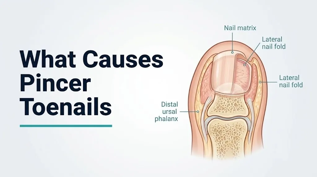
The nail matrix is the primary anatomical structure responsible for pincer nail deformity, because it is the site where nail plate keratinocytes are generated, and any process that alters the relative growth rate of the lateral matrix horns versus the central matrix directly determines nail plate curvature.
The nail matrix is a crescent-shaped region of specialized epithelium located beneath the proximal nail fold, extending distally to the visible pale lunula. The proximal portion generates the dorsal two-thirds of the nail plate by producing keratinocytes that undergo progressive keratinization (accumulation of hard nail keratins: keratin 81, keratin 86, and other hair-type keratins). The distal matrix, visible as the lunula, generates the ventral one-third of the nail plate.
Critically, the lateral extremities of the nail matrix, called the lateral nail matrix horns, generate the lateral edges of the nail plate. In a normally curved nail, these horns produce nail plate tissue that aligns with the nail bed and exits the nail fold in a relatively flat transverse arc. In a pincer nail, the lateral matrix horns are anatomically positioned to generate nail plate edges that curve progressively downward rather than growing flat.
Think of the nail matrix like a curved conveyor belt producing a continuous sheet of material. If the edges of the conveyor belt angle slightly downward, every sheet it produces will have edges that curl under. The sheet itself is not defective; it is the geometry of the production machinery that determines the final shape. In pincer nail deformity, the production geometry of the lateral matrix horns is the problem.
The American Academy of Dermatology recognizes nail matrix dysfunction and altered keratinocyte proliferation as the central anatomical basis of nail plate deformity across all nail dystrophies, including transverse overcurvature.
Individual variation note: Individuals who have had prior nail trauma, such as a crush injury to the distal phalanx or a nail avulsion, may develop secondary pincer nail deformity because the trauma disrupts the anatomical positioning of the nail matrix horns during healing, causing them to scar into an altered position that generates permanently curved nail plate edges.
Genetics as a Cause of Pincer Toenails
Genetics is a primary non-modifiable cause of pincer toenails, operating through inherited anatomical variation in the shape and positioning of the lateral nail matrix horns that determines the baseline transverse curvature angle of the nail plate from adolescence onward.
Pincer nail deformity runs in families. Clinical case series published in the Journal of the American Academy of Dermatology have documented familial clustering of pincer nail deformity across multiple generations, with affected family members sharing a characteristic overcurvature pattern that appears in adolescence or early adulthood, before significant footwear or age-related changes have accumulated. This pattern of early-onset symmetrical bilateral pincer nails with clear family history is the clinical presentation most consistent with a primarily genetic cause.
The specific genetic mechanism is not yet fully characterized at the molecular level. However, the available evidence points to inherited variation in the architecture of the distal phalanx and the lateral nail matrix horn positioning relative to the underlying bone. In individuals with a genetic predisposition, the nail matrix horns are anatomically oriented at a steeper angle relative to the nail bed, causing the lateral edges of every nail plate produced to curve inward from the moment of nail genesis.
What to look for in your family history:
- A parent, grandparent, or sibling with toenails that curl inward on both sides, particularly on the great toenail
- Family members who have had nail surgery or nail bracing for curved toenails
- A personal history of inward-curving nails dating to childhood or adolescence, before any significant footwear or health history factors were present
Individual variation note: Genetic pincer nail deformity tends to affect multiple toenails symmetrically, typically both great toenails and sometimes additional toes simultaneously. If only one toenail is affected and the others are flat, a non-genetic cause (subungual osteophyte, localized trauma, or footwear pressure concentrated on that specific toe) is more likely than a hereditary cause.
Key Takeaway: Genetic pincer toenails appear symmetrically and early in life; if your curved toenail appeared recently on one side or one toe only, the cause is more likely footwear, subungual osteophyte, or systemic disease than inherited nail matrix anatomy.
Footwear and Mechanical Pressure as Causes of Curved Toenails
Footwear causes pincer toenails by applying chronic compressive force to the lateral nail folds, which progressively pushes the lateral soft tissue inward against the nail plate edges and remodels the nail matrix position over years of cumulative loading.
The lateral nail folds are the soft tissue walls on either side of the nail plate. They normally sit parallel to the nail plate edge, providing structural support without compressing the nail. When footwear with a narrow toe box, pointed front, or rigid lateral walls is worn consistently, the shoe material compresses the lateral nail folds inward against the nail plate margins. This sustained lateral compression does two things simultaneously: it mechanically deforms the nail plate edges over time by redirecting their growth direction, and it gradually alters the anatomical positioning of the lateral nail matrix horns beneath the nail fold tissue.
The effect is cumulative. A single day in narrow shoes produces no lasting change. But years of daily compression gradually shift the nail fold tissue inward and alter the mechanics of nail plate generation at the lateral matrix horns.
High-heeled footwear compounds this problem by also shifting the body’s weight load forward onto the toes, increasing the compressive force on the nail folds at the same time as lateral compression from the shoe wall occurs.
Footwear factors most consistently associated with pincer nail deformity:
- Narrow toe box that compresses both lateral nail folds simultaneously
- Pointed-toe shoes that angle the lateral walls inward from the shoe’s front
- High heels that shift body weight toward the forefoot and toes
- Rigid work boots or dress shoes worn for prolonged standing occupations
- Footwear that is too short, causing the toenail to be chronically pushed against the front of the shoe
The American Podiatric Medical Association recommends that footwear provide at least a half-inch of space between the longest toe and the end of the shoe interior, measured while standing, as a minimum standard for avoiding nail and forefoot compression.
Individual variation note: Women who wear narrow-toe or high-heeled footwear professionally from their 20s through their 50s accumulate three to four decades of cumulative lateral nail fold compression before menopause also begins to reduce periungual connective tissue resilience. This compounded effect is one reason clinical observation consistently shows a higher prevalence of footwear-related pincer nail deformity in women than in men.
What Causes Toenails to Curl Inward With Age
Age causes toenails to curl inward through several simultaneous mechanisms: cumulative lateral nail fold compression from decades of footwear, progressive periungual connective tissue and skin elasticity decline, increasing prevalence of distal interphalangeal joint osteoarthritis producing subungual osteophytes, and reduced peripheral vascular perfusion of the nail matrix capillary bed.
The nail matrix requires a consistent blood supply from the subungual capillary plexus, which is supplied by branches of the digital arteries running along the sides of each toe. As peripheral vascular supply decreases with age, nail matrix keratinocyte metabolism slows, nail plate production rates decline, and the nail plate may become thicker and harder in response to altered keratinization dynamics. A thicker, harder nail plate is less flexible and more likely to maintain an abnormal curvature rather than adapting to normal nail bed contours.
Periungual connective tissue changes with age through reduced collagen type I and type III synthesis and increased collagen cross-linking, reducing the compliance of the lateral nail fold soft tissue. Less compliant nail folds transmit lateral pressure from footwear more directly to the nail matrix rather than absorbing it, accelerating the mechanical remodeling of nail matrix horn positioning.
The National Institute of Arthritis and Musculoskeletal and Skin Diseases identifies age-related changes in skin and connective tissue collagen architecture as a primary contributor to the increased prevalence of nail dystrophies in older adults, with pincer nail deformity among the most commonly reported nail shape changes in geriatric dermatological and podiatric practice.
Individual variation note: Postmenopausal women face an accelerated version of the age-related periungual connective tissue changes described above, because estrogen normally promotes collagen synthesis and periungual soft tissue hydration through estrogen receptor activity in fibroblasts. The estrogen decline of menopause reduces collagen synthesis rates in periungual skin, decreasing nail fold compliance and accelerating the progression of any pre-existing nail matrix maldirection into a clinically evident pincer deformity.
Subungual Osteophytes as a Cause of Pincer Nail Deformity
A subungual osteophyte is a bony spur that grows from the dorsal surface of the distal phalanx beneath the nail bed, and it causes pincer nail deformity by mechanically elevating and deforming the overlying nail bed and nail plate from below as the bony spur enlarges.
This mechanism is physically intuitive when you understand the anatomy. The nail plate lies over the nail bed, which lies over the periosteum of the distal phalanx. Normally, the dorsal surface of the distal phalanx is smooth and slightly convex, providing a smooth bony scaffold for the nail plate. In distal interphalangeal joint osteoarthritis, the bone remodeling process associated with cartilage degradation produces osteophytes (bony spurs) at the joint margins. When an osteophyte forms on the dorsal surface of the distal phalanx beneath the nail, it pushes upward against the nail bed from below. This upward pressure from the center of the nail bed forces the nail plate to bow at the lateral edges, producing the progressive transverse overcurvature of pincer nail deformity.
Think of pressing your thumb upward beneath the center of a flexible plastic card while holding both ends fixed: the center rises and the edges curl downward. The subungual osteophyte is the thumb; the nail plate is the card.
To identify whether a subungual osteophyte is contributing to a pincer toenail:
- Assess whether the nail is tender to firm palpation directly over the nail plate surface, particularly centrally
- Check whether the toenail appears elevated centrally (thicker in the center with edges that curve under rather than uniformly curled from the matrix)
- Ask a podiatrist or primary care physician to order a plain film X-ray of the affected toe; the osteophyte will appear as a bony protrusion on the dorsal aspect of the distal phalanx on the lateral view
- Note whether there is a visible Heberden’s node (bony enlargement of the DIP joint) on the same toe, which is a clinical marker of DIP osteoarthritis
- Report any history of osteoarthritis affecting the hands or other joints to the treating clinician
Individual variation note: Subungual osteophytes are more prevalent in the great toenail than in other toenails because the first DIP joint bears greater axial compressive loads during gait, accelerating the osteoarthritic remodeling that produces dorsal osteophyte formation.
Key Takeaway: If your pincer toenail appeared after age 50 and affects primarily the great toe, particularly if the toe shows a bony prominence at the DIP joint, a subungual osteophyte from osteoarthritis should be specifically evaluated with a plain film X-ray before any nail treatment is planned.
Systemic Diseases That Cause Curved Toenails
Several systemic diseases cause curved toenails by altering nail matrix keratinocyte function, nail bed vascular supply, or periungual connective tissue architecture through specific disease mechanisms that operate throughout the body but produce visible changes at the nail unit.
The systemic diseases most consistently associated with pincer or curved toenail deformity in clinical practice are:
- Psoriasis: inflammatory disease producing psoriatic nail disease in approximately 50 percent of patients with skin psoriasis and up to 80 percent of patients with psoriatic arthritis, according to research published in the Journal of the American Academy of Dermatology. The psoriatic nail shows multiple changes including onycholysis, subungual hyperkeratosis, and nail plate deformity from inflammatory disruption of the nail matrix.
- Hypothyroidism: reduced thyroid hormone levels slow the metabolic rate of nail matrix keratinocytes, producing thickened, brittle, and sometimes transversely curved nail plates. Thyroid-stimulating hormone (TSH) testing is appropriate when unexplained nail changes appear alongside other hypothyroid symptoms.
- Systemic lupus erythematosus (SLE): periungual vascular changes and connective tissue disease can alter nail matrix architecture and produce nail plate shape abnormalities.
- Renal failure: uremia affects nail keratinocyte function, producing characteristic nail changes including thickening and abnormal curvature in some patients with advanced chronic kidney disease.
- Arteriovenous fistula for hemodialysis: alters digital blood flow distribution and can produce secondary nail changes in the affected arm; distinct from the toenail deformity context but relevant in the systemic disease framework.
The National Institute of Arthritis and Musculoskeletal and Skin Diseases identifies psoriasis-associated nail disease and systemic connective tissue disorders as important non-mechanical causes of nail plate deformity that require systemic management rather than local nail treatment alone.
Individual variation note: Patients with psoriatic arthritis face a compounded mechanism: the psoriatic nail matrix inflammation alters nail plate geometry directly, and the DIP joint arthritis of psoriatic arthritis produces periarticular and possibly subungual bony changes that further deform nail architecture. Treating the nail alone without managing the underlying psoriatic disease will not produce durable nail improvement.
Psoriasis and Curved Toenail Causes
Psoriasis causes curved toenails through psoriatic nail disease, a specific pattern of nail matrix and nail bed inflammation driven by the same T-cell-mediated immune cascade responsible for the skin plaques of psoriasis, producing structural changes to the nail plate that include thickening, pitting, onycholysis, and in some cases transverse overcurvature.
The mechanism at the nail matrix is distinct from the mechanism in the skin. In the nail matrix, psoriatic inflammation driven by TNF-alpha, IL-17, and IL-22 cytokines disrupts the orderly keratinization of nail matrix keratinocytes. Instead of producing a uniformly keratinized nail plate, the psoriatic matrix generates nail plate tissue with variable thickness and abnormal keratin protein composition. Subungual hyperkeratosis (accumulation of keratinized debris beneath the nail plate) can elevate the nail plate from the nail bed and alter the mechanical support that normally keeps the nail plate flat.
The inflammatory involvement of the nail matrix in psoriatic disease also affects the lateral matrix horns, where disrupted keratinocyte proliferation can alter the curvature of the nail plate edges in ways that predispose to pincer-type transverse overcurvature in some patients.
Research published in the Journal of the American Academy of Dermatology has found that nail disease activity in psoriasis correlates strongly with psoriatic arthritis activity, particularly DIP joint involvement, making nail examination a clinically informative indicator of joint disease progression.
Individual variation note: Patients with psoriatic nail disease who are treated with biologic therapies targeting TNF-alpha (adalimumab, etanercept) or IL-17 (secukinumab, ixekizumab) often experience improvement in nail disease alongside skin and joint improvement. A board-certified dermatologist managing a patient’s psoriasis should document nail changes at each visit, as nail improvement is a recognized clinical endpoint in psoriasis treatment trials.
Medications That Cause Toenails to Curve Inward
Several medications cause toenails to curve inward by altering nail matrix keratinocyte biology, reducing periungual blood flow, or disrupting the keratinization process that determines nail plate structure and curvature.
The most clinically documented medication classes associated with pincer nail or nail curvature changes:
- Beta-adrenergic receptor blockers (beta-blockers): including propranolol, atenolol, and metoprolol. Beta-blockers reduce peripheral vascular resistance and can decrease the capillary perfusion pressure in the subungual capillary plexus supplying the nail matrix. Reduced vascular supply to the nail matrix alters the metabolic environment of keratinocyte production, potentially producing nail plate thickening and transverse curvature changes in some patients.
- Systemic retinoids: including isotretinoin (used for acne) and acitretin (used for psoriasis). Retinoids bind retinoic acid receptors (RAR-alpha and RAR-gamma) in nail matrix keratinocytes, altering their differentiation program and keratinization rate. This produces nail changes including brittleness, thinning, and sometimes abnormal curvature. Acitretin-associated nail changes are well-documented in dermatological literature.
- Taxane chemotherapy agents: including paclitaxel and docetaxel. Taxanes produce nail toxicity through disruption of microtubule dynamics in nail matrix keratinocytes, causing onycholysis, subungual hemorrhage, and nail plate deformity. Nail brace or tape-based preventive interventions during taxane therapy are being studied.
- Fluoroquinolone antibiotics: including ciprofloxacin and levofloxacin. Associated with onycholysis (nail plate separation from the nail bed) and in some reported cases, nail curvature changes, particularly in sun-exposed nails (photo-onycholysis mechanism).
- EGFR inhibitors: including erlotinib and cetuximab. These targeted cancer therapies inhibit the epidermal growth factor receptor that regulates nail matrix keratinocyte proliferation, producing a characteristic periungual inflammation (paronychia) and nail plate changes.
The American Academy of Dermatology recommends that patients beginning systemic retinoid therapy be counseled about expected nail changes and monitored for nail plate structural abnormalities throughout treatment.
Individual variation note: Women of reproductive age taking isotretinoin for acne are at particular risk for nail changes because their nail matrix keratinocytes express higher concentrations of retinoic acid receptors than adult male keratinocytes, potentially amplifying the drug’s effect on nail keratinization.
Key Takeaway: If your curved toenail appeared or worsened after starting a new medication, particularly a beta-blocker, retinoid, or chemotherapy agent, document the timeline and bring this to the attention of the prescribing physician or a dermatologist, because medication-induced nail changes may improve with dose adjustment or drug substitution.
What Causes Pincer Nails in Diabetic Patients
Pincer toenails in patients with diabetes mellitus develop through the same mechanical and structural mechanisms as in the general population, but the consequences are more serious because peripheral neuropathy reduces the pain warning that would normally prompt early care, and impaired wound healing converts the minor skin injury from a curling nail edge into a chronic ulceration.
In a person without diabetes, a pincer toenail that curves enough to press into the lateral nail fold soft tissue produces pain. That pain prompts the person to seek care before tissue breakdown occurs. In a patient with diabetic peripheral neuropathy, the sensory C-fibers and A-delta fibers of the digital nerves supplying the toe have undergone segmental demyelination and axonal loss from chronic hyperglycemia-mediated oxidative damage to Schwann cells. The warning pain is reduced or absent. The nail edge presses into the soft tissue for weeks or months before the patient notices.
Simultaneously, the hyperglycemic environment impairs neutrophil chemotaxis, phagocytosis, and oxidative burst capacity. Skin breaks from the pincer nail edge heal slowly, creating an entry point for bacterial colonization. What begins as a minor skin indentation from a curved nail can progress to:
- Chronic paronychia (periungual infection of the nail fold)
- Cellulitis of the toe or forefoot
- Osteomyelitis of the distal phalanx (bone infection from direct contiguous spread)
- In patients with concurrent peripheral arterial disease, non-healing ulceration requiring specialist wound care
What patients with diabetes should tell their podiatrist at every visit:
- Any toenail that appears curved or pressing into the adjacent skin, even without pain
- Any redness, warmth, or swelling around a toenail
- Any dark discoloration under a curved toenail (may indicate subungual hematoma or infection)
- Any non-healing skin break near a toenail
The American Podiatric Medical Association recommends annual comprehensive foot examinations for all patients with diabetes, including nail assessment, and more frequent visits for any patient with prior foot ulceration or peripheral neuropathy.
Individual variation note: Patients with diabetes who are also taking beta-blockers for cardiovascular disease face a dual mechanism for toenail changes: the metabolic impairment from hyperglycemia affecting nail matrix keratinocyte function, and the reduced periungual blood flow from beta-blocker-mediated vascular effects, potentially accelerating nail plate thickening and curvature beyond what either factor would produce alone.
Peripheral Arterial Disease and Curved Toenails
Peripheral arterial disease (PAD) causes curved and thickened toenails by reducing arterial blood flow to the digital arteries and the subungual capillary plexus that supplies the nail matrix, producing a hypoxic and nutrient-depleted environment for nail matrix keratinocytes that alters their keratinization pattern and nail plate production.
The nail matrix requires consistent arterial perfusion through the digital arteries that run bilaterally along each toe. In PAD, atherosclerotic narrowing of the femoral, popliteal, and tibial arteries reduces the driving pressure available to these terminal digital arteries. The subungual capillary plexus, which supplies the nail matrix, operates at the lowest pressure in the entire lower extremity vascular tree. It is, therefore, the first site to show perfusion changes when arterial pressure drops.
With reduced nail matrix perfusion, keratinocyte metabolism slows. Nail plate production rate decreases and nail plate thickness increases as the slower-moving nail plate undergoes more keratinization before reaching the free edge. The resulting nail plate is thicker, harder, and less flexible, properties that favor transverse overcurvature rather than the flat flexible growth of a normally perfused nail.
The ankle-brachial index (ABI) is the standard clinical test for screening peripheral arterial disease. An ABI below 0.9 indicates at least moderate arterial disease in the lower extremity. A value below 0.5 indicates severe disease associated with rest pain and tissue ischemia. Any patient with toenail changes alongside symptoms of claudication (calf pain with walking that resolves with rest), rest pain in the foot, or non-healing foot wounds should have an ABI measured by their primary care physician or vascular specialist.
Individual variation note: Patients over 65 who smoke or have a history of smoking carry substantially higher PAD risk than non-smoking peers of the same age, because nicotine and cigarette combustion products accelerate atherosclerosis in peripheral arteries. A 65-year-old smoker with curved, thickened toenails should have PAD specifically considered as a contributing cause, not attributed to aging or footwear alone.
Pincer Toenail vs Ingrown Toenail: Key Differences
A pincer toenail and an ingrown toenail are distinct anatomical problems, but they share overlapping symptoms and can be confused: the pincer toenail involves bilateral inward curvature of the entire nail plate from the matrix, while the ingrown toenail involves a single lateral nail edge growing into the adjacent soft tissue, typically from incorrect trimming or narrow footwear.
Understanding the difference matters because the treatment approach is different for each condition, and misidentifying one as the other can delay the correct management.
| Feature | Pincer Toenail | Ingrown Toenail |
|---|---|---|
| Nail curvature pattern | Both lateral edges curve inward along the full nail length | One lateral edge (usually medial) grows into the nail fold at the free edge |
| Primary cause | Nail matrix deformity, genetics, systemic disease, osteophyte | Incorrect trimming (curved rather than straight), narrow footwear, lateral fold hypertrophy |
| Pain location | Pressure pain along both nail edges, worse with footwear | Sharp pain and swelling at one corner of the nail, often with discharge |
| Visible nail shape | Nail plate narrows from base to free edge; may appear to pinch the soft tissue from both sides | Nail plate appears otherwise normal but one corner is buried in swollen red tissue |
| Associated findings | Possible subungual osteophyte, psoriatic nail changes | Possible granuloma formation (hypergranulation tissue) at the affected corner |
| Treatment approach | Nail bracing, chemical or surgical matrixectomy, footwear change, systemic disease management | Correct trimming technique, gutter splinting, partial nail avulsion of the offending edge |
Both conditions can coexist: a pincer toenail produces nail plate edges that press into both lateral folds, effectively creating bilateral ingrown nail complications. In this scenario, treating only the ingrown edges without addressing the underlying transverse overcurvature will result in recurrence.
Individual variation note: Adolescents and young adults are more likely to present with ingrown toenails from incorrect trimming or aggressive nail cutting, while middle-aged and older adults presenting with bilateral, symmetric nail edge tissue involvement are more likely to have a pincer deformity as the underlying cause.
Key Takeaway: If both sides of your toenail are pressing into the surrounding skin symmetrically and the nail plate appears narrow and pinched from base to tip, the underlying problem is pincer deformity of the nail matrix rather than a simple ingrown toenail edge, and treating only the skin edge without correcting the nail matrix curvature will not produce lasting relief.
What Causes Pincer Toenails on the Big Toe Specifically
The big toe is disproportionately affected by pincer toenail deformity because the first distal phalanx is wider and more exposed to compressive forces from footwear and gait mechanics than any other toe, making the nail matrix of the great toenail more vulnerable to every mechanical and structural cause of transverse overcurvature.
The first toenail has the largest surface area of nail matrix of any toenail. This means a larger total amount of nail plate tissue is being generated, providing a greater surface over which any curvature abnormality of the lateral matrix horns will be expressed. The greater the nail plate width, the more visible the transverse overcurvature becomes when the lateral matrix horn geometry is even slightly abnormal.
The great toe also bears the highest compressive load during the push-off phase of gait. As the foot rolls from heel strike through midstance to toe-off, the body’s full weight is transferred sequentially through the foot until the forefoot and great toe provide the final push. Narrow or rigid footwear compresses the first lateral nail folds at precisely the moment of maximum toe loading, producing the highest mechanical stress on the lateral nail matrix horns of any toe during every step.
The first DIP joint is also among the most commonly affected joints in both osteoarthritis and gout, two conditions that produce subungual and periarticular bony changes beneath the great toenail. Gout-associated tophi (uric acid crystal deposits) within the soft tissues surrounding the first DIP joint can produce direct mechanical deformation of the nail matrix from below and laterally.
The American Podiatric Medical Association identifies the hallux (great toe) as the site of the majority of nail pathology presentations in podiatric clinical practice, including both pincer nail deformity and ingrown toenail.
Individual variation note: Patients with hallux valgus (bunion deformity), where the great toe deviates medially and the first metatarsal head protrudes laterally, experience altered load distribution across the great toenail during gait. The medial deviation of the toe relative to the shoe can create asymmetric lateral nail fold compression, sometimes producing a predominantly medial-lateral asymmetry in the pincer deformity pattern rather than the symmetric bilateral overcurvature seen in genetic or aging-related cases.
How Pincer Toenails Are Diagnosed
Pincer toenails are diagnosed through clinical examination by a licensed podiatrist or board-certified dermatologist, with plain film X-ray of the affected toe ordered when a subungual osteophyte is suspected as a contributing cause.
The diagnostic process typically unfolds as follows:
- Clinical history: The clinician establishes symptom onset and duration, footwear habits, family history of curved nails, systemic health conditions (psoriasis, diabetes, osteoarthritis, PAD), and current medications (particularly beta-blockers, retinoids, chemotherapy agents)
- Visual inspection: The nail plate is assessed for the degree of transverse overcurvature, whether the curvature is symmetric (bilateral) or asymmetric (one side more affected), and whether the lateral nail edges are penetrating the adjacent soft tissue
- Palpation of the nail plate and nail fold: Tenderness on direct pressure over the nail plate centrally (suggesting subungual osteophyte) versus tenderness only at the lateral nail fold edges (suggesting soft tissue compression without bony involvement)
- Assessment of the surrounding toe: Presence of Heberden’s node at the DIP joint (suggesting osteoarthritis), periungual erythema and scaling (suggesting psoriatic nail disease), skin breakdown or discharge at the lateral nail folds (suggesting ingrown complication)
- Plain film X-ray of the distal phalanx: Ordered when clinical examination suggests a subungual osteophyte. The lateral view of the distal phalanx will show the osteophyte as a bony protrusion on the dorsal surface of the terminal phalanx, often with associated DIP joint space narrowing confirming osteoarthritis
- Vascular assessment: Ankle-brachial index (ABI) measurement if PAD is suspected based on symptom history, smoking history, or clinical findings of reduced toe temperature or absent pedal pulses
Individual variation note: Patients with psoriasis who present with nail curvature changes should have their nails assessed using the Nail Psoriasis Severity Index (NAPSI), a validated clinical scoring tool that grades psoriatic nail disease severity by examining the nail matrix and nail bed for eight specific features. This tool helps track treatment response and is used in clinical trials of psoriasis therapies.
When Curved Toenail Causes Require Medical Evaluation
Any pincer toenail that is causing pain with footwear, producing skin penetration or discharge at the lateral nail folds, is worsening progressively, or occurs in a person with diabetes or peripheral arterial disease requires evaluation by a licensed podiatrist rather than home management.
The urgency of evaluation depends on which population and which complication is present:
- For people without diabetes or PAD: A podiatrist or dermatologist should evaluate any pincer toenail that has produced skin penetration, any discharge (which may indicate secondary infection of the nail fold), pain that interferes with normal footwear use, or any nail that has been worsening progressively over 6 to 12 months
- For people with diabetes: Any pincer toenail that is curving toward the lateral nail fold skin should be evaluated by a podiatrist at the next available appointment, even without pain. Any skin break adjacent to the nail, any redness or warmth around the nail fold, or any non-healing wound near the toenail warrants same-day or next-day evaluation
- For people with peripheral arterial disease: Same urgency as for diabetes; reduced healing capacity makes any nail edge skin penetration a potential non-healing wound
Signs that indicate urgent same-day or next-day evaluation (not waiting for a scheduled appointment):
- Rapid spreading redness and warmth extending from the toe onto the foot (cellulitis)
- Purulent (pus-containing) discharge from the nail fold that is worsening
- Fever combined with local toe infection signs
- In diabetic patients: any wound near a pincer toenail that has been present for more than 2 weeks without measurable improvement
According to the American Podiatric Medical Association, cellulitis of the foot originating from a nail-related infection in a patient with diabetes or PAD represents a clinical urgency requiring same-day evaluation, and any delay increases the risk of osteomyelitis and limb-threatening infection.
Individual variation note: Patients who are on immunosuppressive medications for organ transplant, autoimmune disease, or cancer treatment should treat any nail fold skin break or infection sign with the same urgency as a diabetic patient, because their impaired immune function prevents adequate containment of even superficial infections.
Treatment Approaches for Pincer Toenail Causes
Treatment for pincer toenails addresses either the nail plate curvature itself, the underlying cause, or both, with the appropriate approach depending on which cause is driving the deformity and the severity of the clinical presentation.
The primary treatment approaches used by podiatrists and dermatologists:
| Treatment | Mechanism | Best Indicated For | Provider |
|---|---|---|---|
| Footwear modification | Removes lateral nail fold compression; allows nail matrix to gradually reorient | Mild-to-moderate footwear-related pincer nail | Self-directed, podiatrist guidance |
| Nail bracing (orthonyxia) | Mechanical orthotic attached to the nail plate that gradually flattens the transverse curvature over months | Mild-to-moderate pincer nail without osteophyte | Podiatrist or nail technician with training |
| Gutter splinting | A flexible tube placed alongside the nail edge to redirect the nail away from the nail fold | Mild ingrown complication of pincer nail | Podiatrist |
| Partial nail avulsion with phenol-alcohol ablation | Removes the lateral nail edge and chemically destroys the responsible lateral nail matrix horn to prevent regrowth | Moderate-to-severe pincer nail with nail fold penetration | Podiatrist, dermatologist |
| Surgical matrixectomy | Surgical removal of the lateral nail matrix horns under local anesthesia | Severe or recurrent pincer nail | Podiatrist, dermatologist |
| Subungual osteophyte removal | Surgical or minimally invasive removal of the bony spur beneath the nail plate | Pincer nail caused by or worsened by subungual osteophyte | Podiatrist, orthopedic surgeon |
| Systemic disease management | Treating underlying psoriasis, hypothyroidism, or other systemic cause | Pincer nail driven by systemic disease | Dermatologist, rheumatologist, endocrinologist |
Research published in Dermatologic Surgery has found that partial nail avulsion combined with phenol-alcohol lateral matrixectomy produces lower recurrence rates than conservative nail bracing alone for moderate-to-severe pincer nail deformity, particularly in cases where subungual osteophyte has been ruled out.
Individual variation note: For patients with suspected or confirmed subungual osteophyte as the primary driver of their pincer nail, nail bracing alone will not produce lasting improvement because the bony protrusion will continue to push the nail plate into overcurvature regardless of the external force applied by a brace. X-ray confirmation of an osteophyte should direct treatment toward addressing the bony spur rather than focusing exclusively on the nail plate geometry.
Frequently Asked Questions About What Causes Pincer Toenails
What causes pincer toenails to form?
Pincer toenails form when the lateral horns of the nail matrix generate nail plate edges that grow downward and inward rather than flat, caused by genetic nail matrix architecture, chronic footwear-mediated lateral nail fold compression, subungual bony spurs from osteoarthritis, systemic diseases, or medications that alter nail matrix keratinocyte function.
Most people with pincer toenails have more than one contributing factor operating simultaneously, particularly older adults who combine genetic predisposition with decades of footwear compression and age-related periungual connective tissue changes.
A licensed podiatrist can evaluate which causes are present and plan treatment that addresses the underlying mechanism rather than only the external nail shape.
Can wearing tight shoes cause pincer toenails?
Tight shoes with narrow toe boxes or pointed fronts can cause pincer toenails by applying chronic compressive force to the lateral nail folds, gradually redirecting the nail matrix horn positioning over years of sustained loading.
The effect is cumulative: single instances of narrow footwear cause no lasting change, but years of daily compression progressively remodel the lateral nail fold soft tissue and alter nail matrix mechanics.
Switching to wide toe box footwear in the early stages of pincer nail development may slow or reduce progression, but established nail matrix deformity typically requires professional podiatric treatment to address.
Are pincer toenails hereditary?
Pincer toenails have a hereditary component, with family-based case series published in the Journal of the American Academy of Dermatology documenting consistent familial clustering across multiple generations.
Inherited pincer nail deformity typically appears symmetrically on both great toenails in adolescence or early adulthood before significant footwear or age-related factors have accumulated.
A personal or family history of early-onset bilateral pincer nails is the strongest indicator that genetics is the primary cause rather than footwear or systemic disease.
What is the difference between a pincer toenail and an ingrown toenail?
A pincer toenail involves bilateral inward curvature of the entire nail plate from the matrix outward, while an ingrown toenail involves a single lateral nail edge at the free margin growing into the adjacent soft tissue, typically from incorrect curved trimming.
The pincer nail is a structural nail matrix problem; the ingrown toenail is a nail edge directional problem that may not involve any abnormality in the rest of the nail plate.
Both conditions can coexist when a pincer nail’s edges penetrate both lateral nail folds, but correcting only the ingrown edges without addressing the underlying transverse overcurvature will result in recurrence.
Can pincer toenails be dangerous for diabetics?
Pincer toenails are particularly dangerous for diabetic patients because peripheral neuropathy reduces the pain that would normally prompt early care, allowing the curling nail edges to penetrate soft tissue undetected until significant tissue breakdown has occurred.
The impaired wound healing of diabetes means that small skin breaks from a pincer nail edge can progress to chronic paronychia, cellulitis, and in severe cases, osteomyelitis of the distal phalanx.
The American Podiatric Medical Association recommends that all diabetic patients have their toenails professionally monitored at regular intervals, and that any pincer nail in a diabetic patient be managed by a licensed podiatrist rather than self-treated at home.
What kind of doctor treats pincer toenails?
A licensed podiatrist is the primary specialist who diagnoses and treats pincer toenails, providing services including clinical assessment, plain film X-ray for subungual osteophyte evaluation, nail bracing, and surgical procedures including partial nail avulsion and lateral matrixectomy.
A board-certified dermatologist is the appropriate specialist when the pincer nail is associated with psoriatic nail disease, medication-induced nail changes, or other systemic skin conditions affecting nail matrix function.
When a pincer nail complication such as cellulitis or wound infection is present in a diabetic or vascular-compromised patient, a wound care specialist or vascular surgeon may be involved alongside the podiatrist.
Closing
Pincer toenails have specific causes that operate through named anatomical and physiological pathways, and identifying which cause applies to your situation determines the most effective path forward. For most people, a combination of genetic nail matrix architecture, cumulative footwear pressure, and age-related connective tissue changes is responsible, all of which are addressable through the right professional guidance.
The next practical step: if your toenail has been curving progressively and is now pressing against the surrounding skin on either side, make an appointment with a licensed podiatrist. Bring a list of your current medications and your health history, specifically noting any psoriasis, diabetes, peripheral arterial disease, or osteoarthritis.
If you have diabetes or poor circulation in your feet and notice any skin redness, warmth, or non-healing area near a curved toenail, that warrants same-day or next-day podiatric evaluation, not watchful waiting. Catching it early is what prevents a nail problem from becoming a foot problem.