Bile Reflux Symptoms: Causes, Warning Signs, and Care
Bile reflux symptoms include persistent upper abdominal burning, nausea, regurgitation of bitter or greenish fluid, and heartburn that does not fully respond to standard acid-reducing medications. Unlike acid reflux, bile reflux involves a different injurious substance and a different anatomical failure, which is exactly why it produces a distinct symptom pattern and why treating it with proton pump inhibitors alone often provides incomplete relief.
The National Institute of Diabetes and Digestive and Kidney Diseases identifies bile reflux as a distinct clinical condition from acid reflux, though the two frequently co-occur. It is particularly common after gallbladder removal surgery (cholecystectomy) and after gastric bypass procedures, where the normal anatomical barriers to retrograde bile flow are altered or removed entirely. Understanding which symptoms arise specifically from bile rather than acid is the key to getting the right diagnosis and the right treatment.
This article covers every clinically recognized symptom of bile reflux with the physiological reason it occurs, how bile reflux progresses from early to chronic stages, which populations carry the highest risk, which symptoms require emergency evaluation, and how the diagnosis is confirmed. The information here is built on the clinical evidence in gastroenterology literature rather than generic symptom lists.
Bile Reflux Symptoms: Overview and Clinical Picture
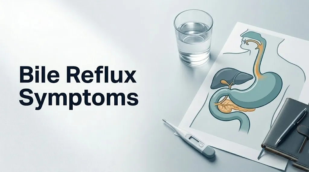
Bile reflux symptoms arise when bile, a digestive fluid produced by the liver and normally stored in the gallbladder, flows backward from the duodenum into the stomach and sometimes into the esophagus, producing a characteristic pattern of upper gastrointestinal distress.
The symptom picture differs from pure acid reflux in several clinically important ways. The burning is often described as more diffuse and persistent than acid heartburn, the regurgitated fluid is bitter or greenish rather than acidic, nausea and bile vomiting are more prominent, and the condition responds poorly to proton pump inhibitors (PPIs) because the injurious agent is not acid but bile acids and pancreatic enzymes.
According to the Mayo Clinic, the five most recognized symptoms of bile reflux are upper abdominal pain, nausea, vomiting of bile, heartburn, and unintended weight loss. These symptoms frequently overlap with those of gastroesophageal reflux disease (GERD), peptic ulcer disease, and gastroparesis, making accurate diagnosis dependent on clinical testing rather than symptom assessment alone.
| Symptom | Frequency Pattern | Key Distinguishing Feature |
|---|---|---|
| Upper abdominal burning or pain | Persistent, often postprandial | Not fully relieved by antacids or PPIs |
| Nausea | Frequent, often morning or nocturnal | May accompany bile vomiting |
| Vomiting of bile (greenish or yellow fluid) | Episodic | Bitter taste; distinctly different from acid regurgitation |
| Heartburn | Present but variable | Burning may be felt throughout the chest and throat |
| Unintended weight loss | Later-stage symptom | Signals chronic disease affecting oral intake |
| Cough or hoarseness | Present in some cases | From laryngopharyngeal bile reflux affecting the vocal cords |
People who have undergone gastric or biliary surgery should be especially alert to this symptom pattern. Symptom onset or worsening in the months following a cholecystectomy or gastric procedure warrants evaluation by a primary care physician or board-certified gastroenterologist rather than self-management with OTC antacids.
What Is Bile Reflux and How Does It Differ from Acid Reflux
Bile reflux is the retrograde flow of bile from the duodenum into the stomach (duodenogastric reflux) or further into the esophagus (duodenogastroesophageal reflux), caused by failure of the pyloric sphincter to prevent backward movement of intestinal contents.
Acid reflux, by contrast, involves stomach acid moving backward through the lower esophageal sphincter (LES) into the esophagus. The two conditions involve different valve failures in different anatomical locations. Bile reflux originates one level below acid reflux: it starts in the duodenum, passes through a failing pyloric sphincter, and may then also breach the LES if pyloric dysfunction is combined with LES incompetence.
Think of the gastrointestinal tract as a one-way highway with two tollgates. The pyloric sphincter is the lower tollgate, controlling traffic from the duodenum back into the stomach. The lower esophageal sphincter is the upper tollgate, controlling traffic from the stomach back into the esophagus. Bile reflux is what happens when the lower tollgate fails. Acid reflux is what happens when the upper tollgate fails. When both fail simultaneously, you get a combined reflux syndrome that is substantially harder to manage.
| Feature | Bile Reflux | Acid Reflux (GERD) |
|---|---|---|
| Primary injurious agent | Bile acids, pancreatic enzymes | Hydrochloric acid (gastric acid) |
| Anatomical failure site | Pyloric sphincter (lower) | Lower esophageal sphincter (upper) |
| Fluid character | Greenish-yellow, bitter | Acidic, sour |
| Response to PPIs | Partial or absent | Usually good |
| Associated with surgery | Strongly (cholecystectomy, gastric bypass) | Weakly |
| Risk of Barrett’s esophagus | Yes, especially in combination | Yes |
The American College of Gastroenterology notes that bile reflux and acid reflux co-exist in many patients, and that the combined reflux pattern is associated with a higher risk of esophageal mucosal injury than either alone. Anyone whose heartburn symptoms do not improve meaningfully after 4 to 8 weeks of PPI therapy should ask their primary care physician whether bile reflux may be contributing to their symptoms.
What Causes Bile Reflux
Bile reflux is caused by dysfunction or structural alteration of the pyloric sphincter, the muscular valve at the junction of the stomach and duodenum that normally prevents duodenal contents from moving backward into the stomach.
Under normal physiology, the pyloric sphincter maintains coordinated closure timed by the enteric nervous system and regulated partly by cholecystokinin (CCK), a hormone released by I-cells in the duodenal wall when fat or protein enters the duodenum. CCK signals the gallbladder to contract, the sphincter of Oddi to relax, and the pyloric sphincter to maintain tone. When this system is disrupted, retrograde bile flow becomes possible.
The most clinically common causes of bile reflux include:
- Cholecystectomy (gallbladder removal): After the gallbladder is removed, bile drains continuously into the duodenum rather than being stored and released in coordinated boluses. This constant bile flow increases duodenal bile pressure and the likelihood of retrograde movement through a transiently relaxed pyloric sphincter.
- Pyloric sphincter dysfunction from peptic ulcer disease: Chronic peptic ulceration near the pyloric channel can cause scarring, fibrosis, and disruption of normal pyloric smooth muscle coordination, impairing valve competence.
- Gastric surgery (Billroth II gastrectomy, Roux-en-Y procedures): These procedures surgically alter or bypass the pyloric sphincter entirely, creating a direct pathway for bile entry into the gastric remnant or esophagus.
- Gastroparesis: Delayed gastric emptying from diabetic autonomic neuropathy, post-viral injury, or idiopathic causes allows bile that has refluxed into the stomach to remain in prolonged contact with the gastric and esophageal mucosa.
- Enteric nervous system dysmotility: Conditions including scleroderma and other connective tissue disorders disrupt the coordinated peristalsis that keeps gastrointestinal contents moving in the correct direction.
People with type 2 diabetes are at elevated risk for bile reflux due to the high prevalence of gastroparesis in this population. Autonomic neuropathy from chronic hyperglycemia impairs the vagal nerve signals that coordinate pyloric sphincter tone and gastric emptying, creating a permissive environment for retrograde bile flow.
Key Takeaway: Bile reflux is not just acid reflux by another name. It involves a different valve failure, a different injurious substance, and different causes, which is why standard acid-reflux treatments often provide only partial relief.
Core Symptoms of Bile Reflux
The core symptoms of bile reflux are upper abdominal burning pain, nausea, vomiting of bile, heartburn, unexplained weight loss, and sometimes a chronic cough or hoarse voice from laryngopharyngeal involvement.
What makes bile reflux symptoms clinically distinct from pure acid reflux is the character of regurgitated fluid and the inadequate response to acid suppression therapy. Patients who have been taking PPIs for weeks without meaningful symptom improvement often have bile reflux contributing to their clinical picture, even if acid reflux is also present.
The symptom of vomiting bile deserves specific attention. Bile has a distinctive bitter taste and a greenish or yellow-green color. Regurgitating this fluid, particularly in the morning on an empty stomach, is a pattern seen in bile reflux that does not typically occur in pure acid GERD. The American Journal of Gastroenterology has published observational data indicating that morning bile regurgitation is one of the more specific indicators of duodenogastric reflux rather than pure gastric acid reflux.
Key clinical symptoms in order of clinical significance:
- Upper abdominal burning that is persistent and not fully relieved by antacids, PPIs, or H2 blockers
- Nausea, particularly in the morning or after fatty meals, often without the classic heartburn of acid reflux
- Vomiting of greenish or yellow-green fluid with a bitter taste (bile)
- Heartburn or chest discomfort, often burning in quality, spreading from the upper abdomen through the chest
- Unintended weight loss from pain-avoidance of eating and nausea affecting caloric intake
- Chronic nonproductive cough or voice hoarseness from bile reaching the laryngopharynx
- Abdominal bloating and early satiety from impaired gastric motility associated with the underlying cause
Older adults over 65 may present with atypical symptoms: anorexia, fatigue, and weight loss without prominent burning or nausea, because the pain perception threshold changes with age and because enteric nervous system sensitivity declines. A gastroenterologist should evaluate unexplained weight loss combined with any upper GI symptom in this age group.
Upper Abdominal Pain and Nausea from Bile Reflux
The upper abdominal pain of bile reflux is a persistent burning or gnawing discomfort located in the epigastric region, the area between the navel and the lower end of the breastbone, caused by bile acid contact with the gastric mucosa.
This pain differs from peptic ulcer pain in its relationship to meals. Peptic ulcer pain often improves briefly after eating because food buffers gastric acid. Bile reflux pain may worsen after meals, particularly after fatty or high-protein meals, because eating stimulates CCK release, gallbladder contraction (if the gallbladder is intact), and increased bile flow into the duodenum, raising the probability of retrograde movement across a poorly functioning pyloric sphincter.
The nausea of bile reflux has a specific physiological basis. When bile acids contact the gastric antrum and duodenal-gastric junction, they stimulate serotonin (5-HT3) receptors on enterochromaffin cells in the gastric wall, activating afferent vagal signals to the brainstem’s chemoreceptor trigger zone (CTZ) in the area postrema. This is the same pathway that produces chemotherapy-induced nausea, which explains why bile reflux nausea can be so persistent and distressing.
To describe upper abdominal pain accurately to your gastroenterologist:
- Note the exact location: point to where the pain is centered, whether above the navel, below the breastbone, or spreading to the left or right.
- Describe the character: burning, gnawing, aching, cramping, or sharp.
- Note the relationship to meals: does it begin immediately after eating, 30 to 60 minutes after eating, or independently of meals.
- Note the relationship to body position: does lying flat or bending forward worsen the pain, which suggests esophageal involvement.
- Note what relieves it: if antacids or PPIs provide no relief, tell the doctor specifically, as this is a key diagnostic indicator pointing away from pure acid reflux.
Women of reproductive age should note that progesterone-mediated relaxation of the lower esophageal sphincter and pyloric sphincter during pregnancy can worsen bile reflux symptoms, particularly in the third trimester when intra-abdominal pressure is elevated.
How Bile Reflux Damages the Esophagus and Stomach Lining
Bile reflux damages the esophageal and gastric mucosa through a mechanism entirely different from acid reflux: bile acids, particularly deoxycholic acid and chenodeoxycholic acid, act as biological detergents that dissolve the phospholipid bilayer of epithelial cell membranes, triggering apoptosis and inflammatory cascades.
The injury depends on pH. In the acidic stomach environment below pH 4, most bile salts remain conjugated and relatively water-soluble, limiting their ability to penetrate cell membranes. As bile enters the esophagus at a higher pH (above 5 to 6), gastric acid deconjugates bile salts into free bile acids. These free bile acids are highly lipid-soluble and penetrate cell membranes far more readily. This is why combined acid and bile reflux produces greater mucosal injury than either alone.
Research published in Gastroenterology has established that bile acid exposure activates mitochondrial apoptotic pathways in esophageal squamous epithelial cells, generates reactive oxygen species (ROS), and stimulates the release of interleukin-8 and TNF-alpha from injured cells. This inflammatory signaling recruits neutrophils to the mucosa, producing the histological pattern of esophagitis and, with chronic exposure, promoting the genetic changes that lead to Barrett’s esophagus.
Pancreatic enzymes in the duodenal refluxate, particularly phospholipase A2, add to the gastric mucosal injury by degrading the phospholipid component of the protective mucus layer coating the stomach lining. This protective layer normally keeps gastric acid from contacting the gastric epithelium directly. When phospholipase A2 removes it, both bile acids and gastric acid gain direct access to the gastric cells, producing the biliary reflux gastritis pattern seen on endoscopic biopsy.
People taking nonsteroidal anti-inflammatory drugs (NSAIDs) or aspirin regularly are at compounded risk because NSAIDs also suppress prostaglandin E2 production in the gastric mucosa, further weakening the mucosal barrier at the same time that bile acids are attacking it. Any patient with bile reflux symptoms who regularly uses NSAIDs should discuss this with their primary care physician, as this combination accelerates mucosal injury.
Key Takeaway: Bile acids damage the esophageal lining by dissolving cell membranes directly, not by burning with acid. This is why PPIs alone cannot stop the injury and why bile reflux requires specific diagnostic testing to identify and manage properly.
Bile Reflux Symptoms at Night and in the Morning
Bile reflux symptoms frequently worsen at night and on waking because lying flat removes gravity as a barrier to retrograde bile movement, and because fasting conditions alter the pattern of bile flow and gastric motility.
During sleep, the swallowing rate drops from approximately one swallow per minute while awake to one swallow every five minutes or less. Swallowing is the primary mechanism by which the esophagus clears refluxed material back into the stomach. With swallowing nearly absent and the body horizontal, bile that has refluxed into the esophagus remains in prolonged contact with the esophageal mucosa, extending the duration of mucosal injury per reflux episode significantly.
The morning symptom pattern of bile reflux has a distinct character. Waking with a bitter taste in the mouth, bile-tinged saliva, or an episode of vomiting greenish fluid on an empty stomach is a recognized clinical presentation of nocturnal bile reflux. This morning bile vomiting occurs because fasting overnight increases the relative concentration of bile in the stomach and proximal esophagus, with no food present to dilute or buffer it.
Quick Tip:
- Elevating the head of the bed by 6 to 8 inches using bed risers (not just extra pillows, which flex the spine rather than tilting the full body) reduces nocturnal reflux episodes by using gravity to assist esophageal clearance.
- Avoid eating within 3 hours of lying down; a full stomach dramatically increases the probability of bile moving retrograde across the pyloric sphincter when you transition from upright to horizontal.
- Left-lateral sleep position is associated with fewer nocturnal reflux events than right-lateral or supine positioning, based on observational data published in the American Journal of Gastroenterology, because the left-lateral position places the gastroesophageal junction above the level of gastric fluid.
Older adults with reduced esophageal motility due to age-related changes in the enteric nervous system are at particular risk for prolonged nocturnal bile exposure, as esophageal peristaltic clearance slows with age independent of the presence of reflux disease.
Chronic Bile Reflux Symptoms and Long-Term Complications
Chronic bile reflux produces progressive mucosal injury that extends beyond discomfort into structural changes in the esophageal and gastric tissue, including intestinal metaplasia, Barrett’s esophagus, and an elevated risk of esophageal adenocarcinoma.
The chronic symptom picture often involves a paradox: some patients with the most extensive mucosal damage report fewer or milder symptoms than those with early or intermittent reflux. This occurs because chronic bile acid exposure causes desensitization of esophageal nociceptors over time, producing what clinicians call “silent” bile reflux disease. The mucosal injury continues silently while symptoms appear to improve or stabilize.
Chronic bile reflux gastritis, clinically termed alkaline reflux gastritis or biliary reflux gastritis, produces a pattern of gastric inflammation characterized by foveolar hyperplasia (proliferation of surface gastric gland cells), edema of the gastric lamina propria, and vascular congestion visible on endoscopy. According to a study published in Gut, this pattern is histologically distinct from Helicobacter pylori gastritis and requires bile acid measurement in refluxate for definitive characterization.
Long-term complications of untreated bile reflux include:
- Erosive esophagitis from cumulative bile acid mucosal injury
- Barrett’s esophagus: replacement of esophageal squamous epithelium with intestinal-type columnar epithelium
- Esophageal adenocarcinoma: the most serious complication, arising from dysplastic changes in Barrett’s epithelium
- Iron deficiency anemia from chronic occult mucosal bleeding in the esophagus or stomach
- Significant unintended weight loss from pain-driven reduction in food intake
- Peptic stricture: fibrotic narrowing of the esophagus from repeated injury and scarring, producing progressive dysphagia
People with obesity carrying a body mass index above 30 are at compounded risk for both the progression and complications of bile reflux, as elevated intra-abdominal pressure adds a mechanical pressure gradient that worsens retrograde bile movement through both the pyloric sphincter and the lower esophageal sphincter simultaneously.
Bile Reflux and Barrett’s Esophagus
Barrett’s esophagus is a condition in which the normal squamous epithelium lining the esophagus is replaced by columnar epithelium with intestinal metaplasia, and bile reflux is now recognized as an independent promoter of this transformation alongside gastric acid reflux.
The mechanism by which bile drives Barrett’s progression involves epigenetic changes in esophageal stem cells. Research published in Gastroenterology has established that bile acids, particularly deoxycholic acid, activate the nuclear factor kappa-B (NF-kB) signaling pathway in esophageal cells, upregulating genes that promote cell survival, proliferation, and the intestinal cell identity associated with Barrett’s metaplasia. This is a distinct molecular pathway from acid-driven Barrett’s development.
The American College of Gastroenterology clinical practice guideline on Barrett’s esophagus recognizes that the combination of acid and bile reflux is associated with a higher rate of progression from Barrett’s metaplasia to dysplasia and adenocarcinoma than acid reflux alone. Patients with confirmed Barrett’s esophagus who also have documented bile reflux on pH-impedance monitoring require more frequent endoscopic surveillance than those with Barrett’s from acid reflux alone.
People who have had bile reflux symptoms for five or more years, those who have had gastric surgery that increases bile reflux risk, and those who are male, over 50, and overweight fall into the higher-risk category for Barrett’s esophagus per ACG criteria. These individuals should discuss upper endoscopy screening with a board-certified gastroenterologist rather than waiting for symptom escalation to prompt the conversation.
Quick Tip:
- Barrett’s esophagus produces no symptoms of its own. You cannot tell from how you feel whether Barrett’s changes are present.
- The only way to diagnose Barrett’s esophagus is through upper endoscopy with biopsy of the esophageal lining.
- Patients already diagnosed with Barrett’s esophagus should ask their gastroenterologist specifically about the contribution of bile reflux to their condition, as this affects both surveillance frequency and treatment approach.
Key Takeaway: Bile acids independently drive the molecular changes that produce Barrett’s esophagus, which means that controlling acid alone with PPIs does not fully protect against this complication in patients with combined acid and bile reflux.
Bile Reflux Symptoms After Gallbladder Removal
Bile reflux symptoms after gallbladder removal (cholecystectomy) represent one of the most common and underrecognized post-surgical upper GI syndromes, affecting a clinically meaningful proportion of cholecystectomy patients in the months to years following surgery.
Before cholecystectomy, the gallbladder stores bile between meals and releases it in concentrated boluses when CCK signals a fat-containing meal. After gallbladder removal, bile drains continuously from the liver through the common bile duct directly into the duodenum, regardless of meal timing. This produces a constant stream of bile in the duodenum rather than the coordinated, meal-synchronized release that the pyloric sphincter was anatomically designed to accommodate.
According to clinical observational data published in the American Journal of Gastroenterology, post-cholecystectomy patients show measurably higher duodenal bile acid concentrations between meals than intact-gallbladder controls. This increased inter-meal biliary pressure elevates the probability of bile crossing a transiently relaxed pyloric sphincter into the stomach, particularly in the overnight fasting period.
The symptom pattern in post-cholecystectomy bile reflux often includes:
- Upper abdominal burning and bloating, particularly in the epigastric area
- Nausea in the morning, worsened by fatty food intake
- Bitter regurgitation of greenish fluid, especially on an empty stomach
- Heartburn that predates cholecystectomy but worsens substantially after the procedure
- Loose stools or diarrhea from increased bile acid concentration in the colon (bile salt malabsorption diarrhea, a separate but related phenomenon)
Anyone who develops new or worsening upper GI symptoms in the 3 to 12 months following cholecystectomy should be evaluated by a board-certified gastroenterologist. The clinical evaluation should include 24-hour pH-impedance monitoring and possibly Bilitec monitoring to document bile in the esophagus, since standard acid pH monitoring alone may miss bile reflux episodes occurring at a non-acidic pH.
Bile Reflux After Gastric Bypass and Stomach Surgery
Bile reflux after gastric bypass surgery and other gastric procedures produces some of the most treatment-resistant and anatomically complex bile reflux cases seen in clinical gastroenterology practice.
The Roux-en-Y gastric bypass (RYGB), the most common bariatric procedure performed in the United States, creates a small gastric pouch connected directly to a loop of small intestine (the Roux limb). A separate limb carries bile and pancreatic secretions from the bypassed stomach and duodenum. These biliary secretions join the Roux limb at the jejunojejunal anastomosis, typically 75 to 150 cm downstream from the gastric pouch. When motility is abnormal or the anatomy creates a short efferent limb, bile can reflux retrograde up the Roux limb into the gastric pouch and even into the esophagus.
In the Billroth II partial gastrectomy, used historically for peptic ulcer disease and still performed for gastric cancer, the remaining gastric tissue is connected directly to the jejunum after removal of the pyloric valve. This eliminates the pyloric sphincter entirely, creating an unimpeded pathway for bile reflux into the gastric remnant and esophagus. The Gut journal has published data showing that alkaline reflux gastritis is a recognized and common long-term complication of Billroth II surgery, with symptom prevalence ranging from 15 to 30 percent of operated patients depending on follow-up duration.
Post-surgical bile reflux is harder to manage medically than de novo bile reflux because the anatomy that normally contains bile flow has been surgically altered. Dietary modifications and positional strategies remain useful, but refractory cases may require surgical revision, specifically conversion to a Roux-en-Y biliary diversion to physically redirect bile flow away from the gastric remnant and esophagus. This decision requires evaluation and management by both a board-certified gastroenterologist and a board-certified upper GI or bariatric surgeon.
The symptom picture in post-surgical bile reflux is often more severe than in non-surgical cases. Patients frequently report incapacitating nausea, recurrent bile vomiting, severe epigastric pain, and weight loss that significantly affects quality of life. These presentations require specialist evaluation rather than primary care management alone.
Key Takeaway: Post-surgical bile reflux, particularly after Billroth II gastrectomy and Roux-en-Y gastric bypass, produces a structurally unique reflux pattern that often requires both specialized diagnostic testing and surgical expertise to manage effectively.
Bile Reflux Symptoms in Specific Populations
Bile reflux symptoms present differently across specific population groups, and standard symptom descriptions may not accurately reflect how the condition manifests in older adults, women, people with diabetes, or those with connective tissue disorders.
Older adults experience bile reflux at higher rates than younger populations for several physiologically grounded reasons. Lower esophageal sphincter pressure declines with age, gastric emptying slows, and enteric nervous system coordination becomes less precise. The pain perception threshold also rises with age, meaning older adults may have extensive mucosal injury visible on endoscopy while reporting only mild or vague symptoms. According to the Mayo Clinic, older adults with symptoms of anorexia, unexplained weight loss, or new-onset anemia should be evaluated for upper GI pathology including bile reflux gastritis regardless of symptom severity.
Women with intact uteri experience progesterone-mediated smooth muscle relaxation that reduces both LES tone and pyloric sphincter contractility, increasing bile reflux risk during the luteal phase of the menstrual cycle and substantially during pregnancy. Women who had upper GI symptoms during pregnancy that resolved postpartum but then recurred later in life often have underlying pyloric or LES dysmotility unmasked by hormonal fluctuation.
People with type 1 or type 2 diabetes who have developed gastroparesis face compounded bile reflux risk. Delayed gastric emptying extends the contact time between any bile that has refluxed into the stomach and the gastric mucosa, increasing the severity of biliary reflux gastritis per reflux episode.
People with scleroderma (systemic sclerosis) have severe esophageal dysmotility from fibrosis of the esophageal smooth muscle, producing one of the most severe combined reflux syndromes in internal medicine. Both the LES and the esophageal peristaltic apparatus fail, allowing prolonged contact between bile and the esophageal mucosa without the normal peristaltic clearance mechanism. These patients require evaluation by both a gastroenterologist and a rheumatologist.
Helicobacter pylori infection complicates the bile reflux clinical picture by independently producing gastritis and disrupting the gastric mucosal defense layer. The American College of Gastroenterology recommends testing for H. pylori in all patients with upper GI symptoms before assuming a diagnosis of pure bile or acid reflux, as untreated H. pylori infection alters gastric mucosal biology and affects both symptom presentation and treatment response.
How Bile Reflux Is Diagnosed
Bile reflux is diagnosed through a combination of upper endoscopy, ambulatory esophageal monitoring, and in selected cases, bile-specific spectrophotometric testing, because standard acid pH monitoring alone cannot detect the bile component of reflux.
Upper endoscopy, formally called esophagogastroduodenoscopy (EGD), allows direct visualization of the esophageal and gastric mucosa. Endoscopic findings consistent with bile reflux include erythema and friability of the gastric antrum with bile staining of the mucosa, the presence of bile-tinged fluid in the stomach on an empty stomach examination, and characteristic histological changes on mucosal biopsy showing foveolar hyperplasia and minimal or absent H. pylori infection.
The gold standard for documenting the combined acid and bile reflux pattern is 24-hour ambulatory pH-impedance monitoring combined with Bilitec monitoring (bilirubin spectrophotometry). The Bilitec device uses a fiber-optic probe positioned in the esophagus to detect the spectroscopic signature of bilirubin in esophageal refluxate, directly confirming bile rather than acid as the refluxed substance. This testing is available at major academic medical centers and specialized esophageal motility laboratories.
To prepare for a diagnostic evaluation for bile reflux:
- Stop PPI medications for 7 to 14 days before pH-impedance monitoring if your gastroenterologist advises this, as PPIs can suppress acid enough to mask acid reflux events while bile events remain detectable.
- Keep a symptom diary for 7 days before your appointment: record meal timing, symptom onset timing, body position when symptoms occur, and what if anything relieves them.
- Bring a complete list of all medications including NSAIDs, aspirin, oral contraceptives, and any supplements, as these affect gastric mucosal physiology and may need to be disclosed before testing.
- Tell your gastroenterologist specifically about your surgical history, including any prior abdominal surgery, cholecystectomy, bariatric procedures, or peptic ulcer surgeries.
- Ask your gastroenterologist whether Bilitec monitoring or bile acid assay testing is appropriate for your specific clinical picture, particularly if your symptoms do not improve with acid suppression.
Gastric emptying scintigraphy (a nuclear medicine gastric emptying study) is ordered when gastroparesis is suspected as a contributing cause. It involves ingesting a radioactively labeled meal and imaging the stomach at intervals to measure the rate of gastric emptying, identifying delayed emptying that increases bile reflux risk.
Bile Reflux Treatment Overview
Bile reflux treatment targets three distinct goals: reducing the volume of bile that reaches the stomach and esophagus, protecting the gastric and esophageal mucosa from bile acid injury, and addressing any underlying motility disorder contributing to the reflux pattern.
Lifestyle modifications form the first tier of management. Eating smaller, more frequent meals reduces gastric volume and lowers the pressure gradient that drives retrograde bile movement. Avoiding high-fat meals reduces CCK-stimulated bile secretion. Avoiding alcohol reduces pyloric sphincter tone. Avoiding NSAIDs preserves prostaglandin-mediated gastric mucosal protection. These measures do not eliminate bile reflux in most patients but reduce symptom severity and slow mucosal injury.
Pharmacological approaches used in bile reflux management include:
- Ursodeoxycholic acid (UDCA): A hydrophilic bile acid that, when taken orally, displaces hydrophobic toxic bile acids (deoxycholic acid, chenodeoxycholic acid) in the bile acid pool, reducing the injurious potential of refluxed bile. Research published in Alimentary Pharmacology and Therapeutics has found that UDCA reduces bile-induced esophageal mucosal injury in patients with documented bile reflux.
- Bile acid sequestrants (cholestyramine): Bind bile acids in the duodenum, reducing the concentration of free bile acids available for reflux. Used primarily in post-cholecystectomy bile salt diarrhea but sometimes applied to gastric bile reflux.
- Prokinetic agents (metoclopramide, domperidone): Improve pyloric sphincter tone and gastric emptying, reducing the opportunity for bile to accumulate in the stomach. Their use is limited by side effect profiles and should only be managed by a supervising physician.
- Sucralfate: Forms a protective coating over the gastric and esophageal mucosa, providing some barrier protection against bile acid contact.
Surgical treatment, specifically Roux-en-Y biliary diversion, is reserved for patients with severe, refractory bile reflux who have failed medical management. This procedure redirects bile flow away from the stomach by routing biliary drainage through a jejunal loop that joins the gastrointestinal tract well below the gastric remnant or esophagus. The decision to pursue surgical management requires evaluation by a board-certified gastroenterologist and a board-certified upper GI or bariatric surgeon who can assess the individual anatomy and expected outcome.
Emergency Symptoms: When to Call 911 or Go to the ER
Certain symptoms associated with bile reflux and its complications require immediate emergency evaluation. Do not wait to see if these resolve on their own.
Call 911 or go to the nearest emergency room immediately if you experience:
- Vomiting blood or material that looks like coffee grounds (hematemesis), which signals active upper gastrointestinal bleeding from esophageal or gastric mucosal erosion
- Black, tarry, foul-smelling stools (melena), which indicate blood that has transited the upper GI tract and is a sign of significant upper GI bleeding
- Progressive difficulty swallowing (dysphagia) that worsens over days to weeks, signaling possible esophageal stricture or malignancy requiring urgent endoscopic evaluation
- Sudden severe upper abdominal or chest pain that is constant, does not change with position, and is accompanied by sweating, nausea, or radiation to the back or jaw
- Dizziness, fainting, rapid heart rate, or pallor accompanying upper GI symptoms, suggesting hemodynamic instability from blood loss
- Inability to keep any food or fluid down for more than 24 hours, causing signs of dehydration including no urination, extreme thirst, and rapid heart rate
- Sudden unintended weight loss of 10 percent or more of body weight over 6 months combined with any upper GI symptoms, which requires urgent endoscopic evaluation to rule out esophageal or gastric malignancy
These presentations can indicate upper gastrointestinal hemorrhage, esophageal obstruction, or malignant transformation of Barrett’s esophagus, all of which require emergency medical assessment and cannot be appropriately managed with an urgent care visit or telephone triage alone.
Frequently Asked Questions About Bile Reflux Symptoms
What does bile reflux feel like?
Bile reflux typically feels like a persistent burning sensation in the upper abdomen and lower chest that does not fully respond to antacids or proton pump inhibitors.
Many people also experience nausea, a bitter taste in the mouth, and sometimes the regurgitation of greenish or yellow-green fluid, particularly in the morning on an empty stomach.
The burning tends to be more diffuse and constant than acid heartburn and is often accompanied by bloating, early satiety, and nausea rather than the sharp, meal-triggered burning typical of pure acid reflux.
How do I know if I have bile reflux or acid reflux?
The most clinically telling sign that bile reflux is contributing to your symptoms is that standard acid-reducing medications (proton pump inhibitors or H2 blockers) provide only partial or no relief after 4 to 8 weeks of consistent use.
Regurgitating bitter, greenish fluid (bile), nausea rather than sour regurgitation, and worsening symptoms after gallbladder surgery or gastric surgery are additional indicators that point toward bile rather than pure acid reflux.
Accurate differentiation requires 24-hour ambulatory pH-impedance monitoring and possibly Bilitec monitoring performed by a board-certified gastroenterologist, as symptoms alone cannot reliably separate the two conditions.
Can bile reflux go away on its own?
Mild, intermittent bile reflux caused by transient pyloric sphincter relaxation may reduce with dietary changes and weight management, but it rarely resolves entirely without addressing the underlying anatomical or motility cause.
In post-surgical patients whose pyloric valve has been removed or bypassed, bile reflux cannot resolve spontaneously because the anatomical mechanism preventing it no longer exists.
Persistent bile reflux symptoms lasting more than 2 weeks warrant evaluation by a primary care physician to prevent progressive mucosal injury from continuing untreated.
What are the warning signs that bile reflux has become serious?
The warning signs that bile reflux has caused serious mucosal damage include difficulty swallowing (dysphagia), pain with swallowing (odynophagia), vomiting blood, black tarry stools, or unintended weight loss exceeding 10 percent of body weight over 6 months.
These symptoms indicate possible Barrett’s esophagus, esophageal stricture, upper GI bleeding, or malignant transformation and require urgent evaluation by a gastroenterologist with upper endoscopy rather than a routine appointment.
Any of these symptoms combined with a history of chronic reflux disease warrants same-day contact with a primary care physician or emergency department evaluation, depending on severity.
Can bile reflux cause cancer?
Bile reflux is an independent risk factor for the development of Barrett’s esophagus and, through Barrett’s esophagus, for esophageal adenocarcinoma.
Research published in Gastroenterology has established that bile acids activate the NF-kB signaling pathway in esophageal cells, driving the molecular changes that produce intestinal metaplasia and, with continued exposure, dysplasia and adenocarcinoma.
The American College of Gastroenterology recommends upper endoscopy surveillance for patients with chronic reflux symptoms and Barrett’s esophagus risk factors; the surveillance interval depends on the grade of dysplasia found on biopsy.
What should I tell my doctor if I think I have bile reflux?
Tell your primary care physician specifically that your reflux symptoms are not adequately controlled by proton pump inhibitors, describe the character of any regurgitated fluid (bitter taste, greenish color), and mention any prior abdominal surgeries including cholecystectomy, gastric bypass, or peptic ulcer operations.
Bring a 7-day symptom diary that records the timing of symptoms relative to meals and body position, a complete medication list including all NSAIDs and aspirin, and your full surgical history.
Ask your physician specifically for a referral to a board-certified gastroenterologist for 24-hour pH-impedance monitoring if your symptoms have not responded to 4 to 8 weeks of PPI therapy, as this test is the most accurate way to document bile in the esophagus.
Bile reflux is one of the most underdiagnosed and undertreated conditions in upper gastrointestinal medicine, largely because its symptoms overlap with acid reflux and because standard acid-suppression therapy does not address the actual injurious agent. The single most practically useful thing you can take from this article is this: if your reflux symptoms persist despite consistent PPI therapy, the problem may be bile rather than acid.
The next appropriate step is a conversation with your primary care physician about referral to a board-certified gastroenterologist for esophageal function testing. Ask specifically about 24-hour pH-impedance monitoring and whether Bilitec monitoring is available or indicated. If you have had gallbladder surgery or gastric surgery, make that part of the conversation from the start.
Your symptom pattern, surgical history, and response to prior treatment are the three most informative data points your gastroenterologist needs. You now know exactly what to bring to that appointment.