Neck Hump Causes in 2026: Buffalo Hump and Cushing Syndrome
A neck hump, medically termed a dorsocervical fat pad and commonly called a buffalo hump, has multiple distinct causes ranging from elevated cortisol levels and corticosteroid medications to obesity and postural changes. The underlying mechanism behind most medically significant neck humps is the effect of glucocorticoid hormones on adipose tissue distribution, which preferentially deposits fat between the shoulder blades while simultaneously breaking down muscle and fat in the arms and legs. This article explains the specific anatomical and hormonal mechanisms behind each cause, ranks them by how frequently they occur in clinical practice, and provides the exact diagnostic pathway an endocrinologist uses to determine whether a neck hump signals Cushing syndrome, medication side effects, or a structural spine condition.
According to the National Institutes of Health, endogenous Cushing syndrome affects approximately 10 to 15 people per million annually, and a dorsocervical fat pad is one of its most visually recognizable features. Yet the condition remains underdiagnosed because the hump develops gradually and many people attribute it to weight gain or aging. The distinction matters clinically because untreated hypercortisolism carries serious health consequences including osteoporosis, type 2 diabetes, hypertension, and increased infection risk.
A neck hump is not a single condition with a single cause. It is a physical finding that sits at the intersection of endocrinology, rheumatology, and metabolic health. Understanding which type of neck hump you are dealing with determines which specialist you need to see, which tests are appropriate, and whether the underlying condition requires medical treatment or can improve with targeted lifestyle changes. Here is exactly what causes a neck hump, broken down by mechanism, frequency, and clinical urgency.
What Is a Buffalo Hump and Dorsocervical Fat Pad
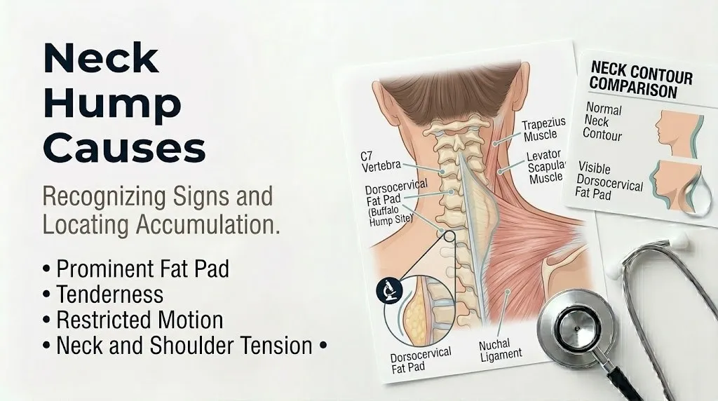
A buffalo hump is a visible accumulation of adipose tissue located at the posterior base of the neck, overlying the cervicothoracic junction at the C7 through T1 vertebrae. The medical term dorsocervical fat pad describes this anatomically specific fat deposit that creates a rounded protrusion visible when viewing a person from the side.
The dorsocervical region has a unique biological responsiveness to glucocorticoid hormones that distinguishes it from fat deposits elsewhere on the body. Adipose tissue in this area expresses a higher density of glucocorticoid receptors compared to fat in the arms, legs, or hips. When cortisol levels rise, whether from endogenous overproduction or exogenous corticosteroid medications, these receptors activate lipoprotein lipase, an enzyme that breaks down circulating triglycerides and facilitates their uptake into adipocytes for storage. The dorsocervical fat pad expands while peripheral fat stores shrink, a pattern that explains why someone with Cushing syndrome can develop a buffalo hump and central obesity while their arms and legs become thinner.
The hump itself is composed of normal white adipose tissue. It is not a tumor, a cyst, or a fluid collection. In hypercortisolism, the adipocytes in this region undergo hyperplasia, increasing in both size and number. The fat pad sits superficial to the trapezius muscle and deep to the subcutaneous tissue of the upper back. It can range in size from a subtle thickening noticeable only on close examination to a prominent protrusion that alters the contour of the neck and upper back profile.
| Feature | Buffalo Hump (Dorsocervical Fat Pad) | Dowager’s Hump (Kyphosis) |
|---|---|---|
| Primary tissue | Adipose (fat) tissue | Bone (vertebral bodies) |
| Texture on palpation | Soft, compressible, mobile | Firm, rigid, non-compressible |
| Underlying cause | Hypercortisolism, medications, obesity | Osteoporosis, vertebral compression fractures |
| Progression | Typically gradual, can be rapid with ectopic ACTH | Gradual over years |
| Reversibility | Can regress with treatment of underlying cause | Generally permanent structural change |
The Mayo Clinic notes that a dorsocervical fat pad itself is not harmful or painful. Its clinical significance lies entirely in what it reveals about underlying hormonal, metabolic, or medication-related processes. The hump is a visible sign of a systemic condition rather than a localized problem of the upper back.
Key Takeaway: A buffalo hump is not just upper back fat. It is a glucocorticoid-responsive fat pad at the C7-T1 junction whose presence should trigger consideration of cortisol excess, medication effects, or metabolic evaluation.
Buffalo Hump Cushing Syndrome Causes
Endogenous Cushing syndrome is the most clinically significant cause of a buffalo hump because it reflects a state of chronic cortisol excess produced internally by the body rather than from external medication exposure. In Cushing syndrome, a dorsocervical fat pad develops as part of a characteristic pattern of fat redistribution that also includes central obesity, supraclavicular fat pad fullness, and rounded facial appearance.
Cushing syndrome results from prolonged exposure to elevated circulating glucocorticoids. The source of excess cortisol determines the specific subtype. Cushing disease, responsible for approximately 70 percent of endogenous cases according to the Endocrine Society, occurs when a benign pituitary adenoma secretes excess adrenocorticotropic hormone (ACTH). ACTH stimulates the adrenal glands to produce cortisol far above normal physiological levels. The adrenal zona fasciculata cells respond to chronic ACTH stimulation with hyperplasia and unregulated cortisol output that loses the normal diurnal rhythm.
Adrenal Cushing syndrome accounts for roughly 15 to 20 percent of endogenous cases. An adrenal adenoma or, more rarely, an adrenocortical carcinoma, autonomously secretes cortisol independent of pituitary ACTH control. In these cases, ACTH levels are actually suppressed because the pituitary gland detects the high cortisol and reduces its own ACTH output. The adrenal tumor produces cortisol without needing the upstream signal.
Ectopic ACTH syndrome represents approximately 10 to 15 percent of Cushing syndrome cases and carries the most urgent clinical implications. Certain malignancies, most notably small cell lung cancer, bronchial carcinoid tumors, thymic carcinoids, and pancreatic neuroendocrine tumors, can produce ACTH ectopically. The ACTH output from these tumors is often extremely high and uncontrolled, producing rapid-onset, severe hypercortisolism. A buffalo hump that develops over weeks rather than months or years, especially when accompanied by facial rounding, severe muscle weakness, and unexplained weight loss, requires urgent evaluation to rule out an underlying malignancy.
- Cushing disease (pituitary ACTH-secreting adenoma): 70% of endogenous cases
- Adrenal Cushing syndrome (adrenal adenoma or carcinoma): 15-20% of cases
- Ectopic ACTH syndrome (malignancy secreting ACTH): 10-15% of cases
- Ectopic CRH syndrome: rare, less than 1% of cases
The National Institute of Diabetes and Digestive and Kidney Diseases (NIDDK) reports that Cushing syndrome affects women three times more frequently than men and is most commonly diagnosed between ages 20 and 50. The dorsocervical fat pad in Cushing syndrome does not develop in isolation. It appears alongside other Cushingoid features including moon face, violaceous abdominal striae wider than 1 centimeter, easy bruising from skin thinning, proximal muscle weakness making it difficult to stand from a chair, and menstrual irregularities in premenopausal women.
Key Takeaway: A buffalo hump occurring with facial rounding, wide purple abdominal stretch marks, and easy bruising strongly suggests endogenous Cushing syndrome and warrants endocrinology evaluation, not watchful waiting.
High Cortisol and Neck Hump Formation
The neck hump in hypercortisolism forms because cortisol directly stimulates fat storage in glucocorticoid-receptor-rich adipose tissue while simultaneously promoting the breakdown of protein in skeletal muscle and peripheral fat stores. This creates the characteristic body shape of central fat accumulation with thin extremities that clinicians recognize as a Cushingoid body habitus.
Cortisol is a steroid hormone produced by the adrenal cortex under the regulation of the hypothalamic-pituitary-adrenal (HPA) axis. Under normal conditions, the hypothalamus secretes corticotropin-releasing hormone (CRH), which signals the anterior pituitary to release ACTH, which in turn stimulates adrenal cortisol production. Cortisol follows a strict diurnal rhythm, peaking in the early morning and declining throughout the day to reach a nadir around midnight. This rhythm is essential for normal metabolic function, immune regulation, and tissue maintenance.
When cortisol is chronically elevated, whether from endogenous overproduction or exogenous medication, the normal diurnal pattern flattens. Adipose tissue is exposed to high glucocorticoid levels continuously rather than cyclically. The enzyme 11-beta-hydroxysteroid dehydrogenase type 1 (11-beta-HSD1), which is highly expressed in dorsocervical and visceral adipose tissue, converts inactive cortisone to active cortisol locally within fat cells. This tissue-level amplification means that even moderately elevated circulating cortisol can produce disproportionately high cortisol activity within the dorsocervical fat pad.
Glucocorticoid receptors within dorsocervical adipocytes, once activated by cortisol binding, translocate to the cell nucleus and alter gene transcription. Lipoprotein lipase expression increases, pulling more circulating triglycerides into the fat cell. Simultaneously, hormone-sensitive lipase activity decreases, reducing the breakdown of stored triglycerides. The fat cell grows larger and, over time, preadipocytes differentiate into mature adipocytes, increasing the total number of fat cells in the region. This dual mechanism of hypertrophy plus hyperplasia makes the dorsocervical fat pad progressively more prominent as hypercortisolism continues.
Peripheral tissues respond to cortisol very differently. In skeletal muscle, cortisol activates the ubiquitin-proteasome pathway, breaking down myofibrillar proteins into amino acids released into circulation. In peripheral subcutaneous fat, lipolysis increases, releasing free fatty acids. The amino acids and fatty acids are taken up by the liver for gluconeogenesis and by the dorsocervical and visceral adipocytes for triglyceride storage. What is lost from the arms and legs is effectively redistributed to the trunk and upper back.
The Journal of Clinical Endocrinology & Metabolism published research demonstrating that 11-beta-HSD1 expression is significantly higher in omental and dorsocervical adipose tissue compared to subcutaneous abdominal or gluteal fat. This enzyme distribution explains anatomically why cortisol excess deposits fat between the shoulder blades and within the abdomen rather than diffusely across the body.
Key Takeaway: Cortisol does not simply cause weight gain. It redistributes fat from peripheral stores to glucocorticoid-sensitive central depots, including the dorsocervical region, through tissue-specific differences in 11-beta-HSD1 enzyme expression and glucocorticoid receptor density.
Neck Hump From Steroids and Corticosteroid Medications
Exogenous corticosteroid medications, including prednisone, dexamethasone, hydrocortisone, and methylprednisolone, are the most common cause of buffalo hump in clinical practice because these drugs are widely prescribed for asthma, autoimmune diseases, inflammatory conditions, and organ transplant immunosuppression. The resulting condition is termed iatrogenic Cushing syndrome or exogenous hypercortisolism.
Corticosteroids bind to the same glucocorticoid receptors as endogenous cortisol and produce identical metabolic effects on adipose tissue distribution. The dorsocervical fat pad develops through the same mechanism of lipoprotein lipase activation and adipocyte differentiation described for endogenous Cushing syndrome. The difference is that the source of glucocorticoid excess is a prescription medication rather than an internal tumor or glandular disorder.
The dose, duration, and route of corticosteroid administration all influence whether a buffalo hump develops. Oral corticosteroids at doses equivalent to prednisone 7.5 milligrams daily or higher, taken for more than three to four weeks, carry the highest risk of producing visible Cushingoid features. However, potent topical corticosteroids applied to large body surface areas, high-dose inhaled corticosteroids in asthma management, and repeated intra-articular or epidural corticosteroid injections can all produce systemic absorption sufficient to cause fat redistribution over months to years of use.
The American College of Rheumatology guidelines on glucocorticoid therapy recognize that Cushingoid features, including dorsocervical fat pad accumulation, are among the most predictable adverse effects of long-term corticosteroid use. The risk correlates more strongly with cumulative dose over time than with any single dose level. A person taking prednisone 5 milligrams daily for two years may develop a more prominent buffalo hump than someone who took 40 milligrams daily for two weeks and then stopped.
- Prednisone (oral): most commonly prescribed oral corticosteroid, high risk with doses above 7.5 mg daily for more than 3 to 4 weeks
- Dexamethasone (oral, injectable): potent, long-acting corticosteroid, higher relative glucocorticoid receptor affinity
- High-potency topical corticosteroids: clobetasol, betamethasone dipropionate applied to large areas can cause systemic absorption
- Inhaled corticosteroids: fluticasone, budesonide at high doses can produce measurable systemic effects
- Injectable corticosteroids: repeated triamcinolone acetonide joint injections or epidural injections
Iatrogenic Cushing syndrome differs from endogenous Cushing syndrome in one critical diagnostic feature. A person taking exogenous corticosteroids will have suppressed endogenous cortisol production because the HPA axis detects the medication as cortisol and shuts down its own ACTH secretion. The adrenal glands atrophy over time from lack of stimulation. A morning cortisol level in someone on chronic corticosteroids will be low, not high, distinguishing iatrogenic from endogenous hypercortisolism. The person has Cushingoid features from the medication despite their own cortisol production being suppressed.
Key Takeaway: Corticosteroid medications are the single most common cause of buffalo hump. Any person taking prednisone, dexamethasone, or high-dose inhaled or topical steroids who notices upper back fat accumulation should discuss this with their prescribing physician.
What Causes Neck Fat and Obesity-Related Neck Hump
Generalized obesity can produce a dorsocervical fat pad through caloric excess and overall adipose tissue expansion that includes the upper back region, but the distribution pattern and associated features distinguish simple obesity from cortisol-mediated fat redistribution. Obesity-related neck fat develops through a different hormonal mechanism than glucocorticoid-driven dorsocervical fat pads.
In obesity, insulin resistance and compensatory hyperinsulinemia are the primary drivers of adipose tissue expansion. Insulin promotes glucose uptake and lipogenesis in adipocytes through activation of the insulin receptor and downstream signaling pathways including PI3K/Akt. Unlike cortisol, insulin promotes fat storage relatively uniformly across all subcutaneous and visceral depots, though with some individual genetic variation in distribution pattern. An obesity-related neck hump typically develops in proportion to overall body fat accumulation rather than as an isolated finding.
The clinical distinction between obesity-related neck fat and a cortisol-driven buffalo hump is essential. In simple obesity, fat distribution is generalized. The person gains weight proportionally in the face, trunk, arms, legs, and upper back. The dorsocervical fat pad, if present, is proportionate to overall adiposity. In Cushing syndrome, fat distribution is strikingly disproportionate. The arms and legs may be thin with visible muscle wasting while fat accumulates centrally in the face, abdomen, supraclavicular region, and dorsocervical area. This pattern, called truncal obesity with peripheral wasting, is highly characteristic of glucocorticoid excess.
A waist-to-hip ratio measurement can help distinguish generalized obesity from Cushingoid fat distribution, though it is not diagnostic on its own. Obesity typically produces a waist-to-hip ratio below 1.0 in women and near or above 1.0 in men. Cushing syndrome often produces a ratio significantly above 1.0 in both sexes because abdominal fat accumulation is disproportionate to hip and thigh fat, which may actually decrease.
| Feature | Obesity-Related Neck Fat | Cortisol-Driven Buffalo Hump |
|---|---|---|
| Fat distribution | Generalized, proportionate | Central with peripheral wasting |
| Arms and legs | Proportionate to trunk | Often thin with muscle wasting |
| Supraclavicular fat pads | Absent or proportionate | Prominent, disproportionate |
| Abdominal striae | Pale or silver, narrow | Purple (violaceous), wide (over 1 cm) |
| Muscle strength | Preserved relative to size | Proximal muscle weakness common |
| Rate of development | Gradual with weight gain | Can be rapid or gradual |
The Centers for Disease Control and Prevention estimates that over 40 percent of U.S. adults meet criteria for obesity. Given this prevalence, many people with a dorsocervical fat pad will have obesity as the primary cause. However, the presence of a neck hump should never be dismissed as simply weight-related without considering the possibility of concurrent hypercortisolism, particularly if the fat pad developed disproportionately to overall weight or appeared alongside other Cushingoid features.
Key Takeaway: Obesity-related neck fat develops proportionally with overall body adiposity. A dorsocervical fat pad that is disproportionate to arm and leg size, or that appears alongside facial rounding and purple stretch marks, should raise suspicion for hypercortisolism.
Dowager’s Hump vs Buffalo Hump Differences
A dowager’s hump and a buffalo hump are anatomically and clinically distinct conditions that are frequently confused because both produce a visible protrusion at the upper back and neck. The dowager’s hump is a structural deformity of the thoracic spine caused by vertebral bone changes, most commonly osteoporotic compression fractures. The buffalo hump is a soft tissue fat pad caused by glucocorticoid-mediated adipose accumulation.
The dowager’s hump represents thoracic hyperkyphosis, an exaggerated forward curvature of the thoracic spine. The normal thoracic kyphosis measures between 20 and 40 degrees. In hyperkyphosis, this angle increases, producing a visible rounded protrusion of the upper back. The hump consists of the posteriorly prominent spinous processes of the thoracic vertebrae, which become more visible as the vertebral bodies anteriorly compress and the spine angles forward. The tissue overlying the hump is skin and subcutaneous fat of normal thickness. The hump itself is bone.
Osteoporosis is the most common cause of dowager’s hump in postmenopausal women. As estrogen levels decline after menopause, osteoclast activity increases relative to osteoblast activity, leading to net bone loss. The thoracic vertebral bodies, which bear weight and withstand flexion forces, are particularly vulnerable. When a vertebral body loses sufficient trabecular bone mass, it can fracture under normal loads, wedging anteriorly into a compressed triangular shape. Multiple adjacent wedge fractures progressively angle the thoracic spine forward, creating the characteristic hump.
The clinical examination can usually distinguish a dowager’s hump from a buffalo hump without imaging. Palpation of a buffalo hump reveals soft, compressible, mobile adipose tissue that moves with the skin. Palpation of a dowager’s hump reveals firm, rigid, immobile bone directly beneath thin soft tissue. A person with a dowager’s hump often has a visibly stooped posture with the head projected forward and the shoulders rounded. A person with a buffalo hump may have normal spinal alignment with a soft tissue prominence at the cervicothoracic junction.
| Characteristic | Dowager’s Hump (Kyphosis) | Buffalo Hump (Fat Pad) |
|---|---|---|
| Tissue composition | Bone (vertebral compression) | Adipose tissue |
| Palpation texture | Hard, bony | Soft, fatty |
| Posture | Stooped, forward head | Often normal, hump is soft tissue only |
| Gender prevalence | Predominantly postmenopausal women | Females more than males but both sexes |
| Primary cause | Osteoporosis | Hypercortisolism, medications, obesity |
| Reversibility | Generally permanent | Can regress if cortisol normalized |
| Associated features | Height loss, back pain | Moon face, striae, muscle weakness |
The American Academy of Orthopaedic Surgeons reports that approximately 25 percent of postmenopausal women develop some degree of vertebral compression fracture, and a subset of these develop visible thoracic hyperkyphosis. A dowager’s hump that appears gradually with height loss, back pain, and postural changes in a postmenopausal woman should trigger bone density evaluation. A soft tissue prominence at the cervicothoracic junction without spinal deformity should trigger endocrine evaluation.
Key Takeaway: A dowager’s hump is hard bone caused by vertebral compression fractures. A buffalo hump is soft fat caused by glucocorticoid effects. The distinction can often be made by palpation and posture assessment in a doctor’s office.
How Posture Contributes to Neck Hump Development
Forward head posture and chronic poor neck alignment contribute to the appearance of a neck hump through mechanical redistribution of soft tissue and adaptive changes in the cervicothoracic spine, though postural changes alone do not produce a true dorsocervical fat pad. Postural neck hump is not the same condition as a buffalo hump, and understanding the distinction prevents unnecessary endocrine evaluation while identifying cases where physical therapy is the appropriate intervention.
Forward head posture shifts the center of gravity of the head anteriorly relative to the cervical spine. For every inch the head moves forward, the effective weight the neck muscles must support increases by approximately 10 pounds. To compensate, the cervical extensor muscles, including the upper trapezius, splenius capitis, and semispinalis capitis, contract chronically to prevent the head from dropping further forward. This sustained muscle tension can produce visible hypertrophy of the muscular tissue at the base of the neck, creating a firm prominence that can be mistaken for a fat pad.
Prolonged forward head posture with rounded shoulders also alters the mechanical loading of the cervicothoracic junction. The C7 and T1 vertebrae experience increased flexion stress. The soft tissues dorsal to these vertebrae, including the overlying fascia and subcutaneous fat, may become more prominent as the spine angles forward beneath them. The skin and soft tissue do not change, but the underlying bony prominence shifts forward, making the soft tissue appear more protuberant relative to the surrounding contour.
Screen-based work and smartphone use have made forward head posture increasingly prevalent. The term “tech neck” describes the postural pattern of head-forward, shoulders-rounded positioning sustained for hours daily. The American Physical Therapy Association recognizes postural dysfunction as a contributor to chronic neck pain and cervicothoracic soft tissue changes. However, postural neck hump is not a disease state. It reflects mechanical adaptation rather than hormonal or metabolic pathology.
- True dorsocervical fat pad: soft, fatty, often accompanied by supraclavicular fullness and Cushingoid features
- Postural soft tissue prominence: firmer, muscular, improves when standing tall against a wall with shoulders back
- Muscular hypertrophy: firm, bilateral paraspinal muscle prominence from chronic contraction
- Combined presentation: postural changes may make a mild fat pad appear more prominent
A simple wall test can help distinguish postural prominence from a true fat pad. Stand with your back flat against a wall, heels, buttocks, shoulders, and head touching the wall. If the neck hump reduces significantly or disappears in this position, posture is a major contributor. If the hump remains equally prominent with the spine fully extended, it is more likely a true adipose tissue accumulation or structural spinal deformity.
Postural neck hump responds to physical therapy focused on cervical retraction exercises, scapular stabilization, and thoracic extension mobility work. This does not treat hypercortisolism or medication-related fat pads. For a person whose neck hump is primarily postural, strengthening the deep cervical flexors and stretching the pectoral muscles can reduce the visible prominence over weeks to months of consistent work.
Key Takeaway: Posture can create or amplify the appearance of a neck hump through forward head positioning and muscular hypertrophy, but posture alone does not produce a true dorsocervical fat pad. A wall test and palpation help distinguish the two.
What Causes a Fat Neck Without Weight Gain
A person who develops a dorsocervical fat pad without overall weight gain, particularly if accompanied by thinning of the arms and legs, should be evaluated for hypercortisolism because this disproportionate fat redistribution pattern is a hallmark of glucocorticoid excess rather than simple obesity. A fat neck without weight gain narrows the differential diagnosis significantly.
Endogenous Cushing syndrome produces this exact pattern. Cortisol’s catabolic effect on peripheral muscle and fat causes the arms and legs to thin even as central adiposity increases. A person may notice their rings becoming looser, their shoes fitting more loosely, or their pant legs becoming baggy around the thighs while their abdomen, face, and upper back simultaneously expand. Total body weight may remain stable or increase only modestly because fat gain in central depots is partially offset by muscle and fat loss in the extremities.
Medication-induced fat redistribution can produce the same pattern. A person on chronic corticosteroids for asthma or autoimmune disease may notice their face becoming rounder and their upper back developing a hump while their arms and legs become thinner and weaker. This occurs even without caloric excess because the medication is redistributing existing fat stores rather than creating new fat from excess calories.
Several non-cortisol medical conditions can produce localized fat accumulation at the neck without generalized weight gain. Madarosis-associated lipodystrophy and certain forms of familial partial lipodystrophy cause fat loss in some body regions with fat preservation or accumulation in others, including the dorsocervical area. These are rare genetic conditions diagnosed by clinical examination, family history, and genetic testing. HIV-associated lipodystrophy, more common historically with certain antiretroviral medications, can produce dorsocervical fat accumulation with peripheral fat wasting. Modern HIV regimens have reduced but not eliminated this adverse effect.
- Endogenous Cushing syndrome: fat redistribution with stable total weight
- Iatrogenic Cushing syndrome: medication-driven redistribution, often with muscle wasting
- Familial partial lipodystrophy: genetic, fat accumulates in face and neck with peripheral loss
- HIV-associated lipodystrophy: antiretroviral-related, dorsocervical fat accumulation
- Localized lipomatosis: benign fatty tumor development, usually asymmetric
The Endocrine Society clinical practice guidelines for Cushing syndrome emphasize that any person presenting with disproportionate central fat accumulation, including dorsocervical fat pad, with peripheral wasting should undergo biochemical testing for hypercortisolism regardless of total body weight. A normal BMI does not exclude Cushing syndrome.
Key Takeaway: A neck hump developing without overall weight gain, especially with thinning arms and legs, is a strong clinical indicator for hypercortisolism testing. Do not dismiss it as a cosmetic concern or attribute it to stress without endocrine evaluation.
Buffalo Hump Symptoms and When to Seek Care
A buffalo hump itself produces no symptoms. It is not painful, does not restrict neck movement, and does not cause neurological symptoms because it is composed of normal adipose tissue superficial to the spine. The clinical urgency of a buffalo hump lies entirely in what it indicates about the underlying systemic condition producing it. The associated symptoms, not the hump itself, determine whether same-day care, urgent specialist evaluation, or scheduled outpatient workup is appropriate.
The most clinically significant associated symptoms are the other Cushingoid features that accompany the buffalo hump in hypercortisolism. Moon face, a rounded, plethoric facial appearance, develops alongside the dorsocervical fat pad in both endogenous and exogenous Cushing syndrome. Supraclavicular fat pads, producing fullness above the collarbones, are particularly characteristic of glucocorticoid excess. Wide, purple abdominal striae measuring more than 1 centimeter in width indicate that cortisol has thinned the skin sufficiently to allow the underlying tissues to stretch and show through.
Proximal muscle weakness is one of the most functionally significant symptoms of hypercortisolism. A person with Cushing syndrome may notice difficulty standing from a seated position without using their arms, trouble climbing stairs, or inability to rise from a squat. This weakness reflects cortisol-mediated catabolism of Type II muscle fibers in the proximal muscles of the hip and shoulder girdle. Muscle weakness is not a feature of simple obesity and, when present with a buffalo hump, strongly points toward hypercortisolism.
Skin changes including easy bruising, thin skin, and poor wound healing reflect cortisol’s inhibition of collagen synthesis and fibroblast function. A person may notice they bruise from minimal contact or that small cuts take unusually long to heal. These dermatological signs, combined with central fat redistribution, make the clinical diagnosis of Cushing syndrome increasingly likely.
- Moon face and facial plethora: strongly suggests hypercortisolism
- Supraclavicular and dorsocervical fat pads: characteristic distribution
- Wide purple abdominal striae: specific for glucocorticoid excess
- Proximal muscle weakness: functionally limiting, clinically significant
- Easy bruising and thin skin: collagen synthesis inhibition
- Menstrual irregularity or amenorrhea: reproductive axis suppression
- New or worsening hypertension: mineralocorticoid effect of cortisol
- Unexplained osteoporosis or fractures: catabolic effect on bone
The Mayo Clinic recommends that anyone with a dorsocervical fat pad and two or more of these associated features should be evaluated for Cushing syndrome by a board-certified endocrinologist. A primary care physician can initiate the initial screening with a late-night salivary cortisol or 24-hour urinary free cortisol test before referring to endocrinology if results are abnormal.
Key Takeaway: The buffalo hump is silent. The associated features of moon face, purple striae, proximal muscle weakness, and easy bruising are what demand clinical attention. Two or more associated symptoms warrant endocrine testing.
How to Tell If Neck Hump Is Fat or Bone
Determining whether a neck hump consists of fat or bone can often be accomplished through palpation and physical examination in a clinical setting, but definitive differentiation may require imaging studies such as X-ray or DEXA scan to assess bone structure and body composition. The distinction matters because a fatty hump points toward endocrine or metabolic evaluation while a bony hump points toward spinal and bone density evaluation.
Palpation is the simplest differentiating tool. A buffalo hump composed of adipose tissue feels soft, compressible, and mobile beneath the skin. You can pinch the tissue between your fingers. It moves independently of the underlying spine. A dowager’s hump composed of prominent vertebral spinous processes feels hard, rigid, and immobile. You cannot compress it or move it relative to the spine. The skin overlying the bony prominence may be thin, but the prominence itself is unyielding.
Postural assessment provides additional clinical information. Observe the person from the side in their natural standing posture. A person with a dowager’s hump typically has visible thoracic kyphosis with the shoulders rounded forward and the head projected anteriorly. A person with a buffalo hump may have a normally aligned spine with a soft tissue prominence at the cervicothoracic junction that is visible even with erect posture. Asking the person to stand with their back against a wall can accentuate the difference. A postural hump reduces significantly when the spine is actively extended. A structural kyphosis or a true fat pad persists.
The wall-occiput distance test is a simple clinical measure of thoracic kyphosis. The person stands with their heels and buttocks against a wall and attempts to touch the back of their head to the wall while keeping the chin level. In a person with normal thoracic kyphosis, the occiput touches or nearly touches the wall. In thoracic hyperkyphosis, there is a measurable gap between the occiput and the wall, often several centimeters, that cannot be closed with active effort.
| Test | Fat Pad (Buffalo Hump) | Bone Deformity (Dowager’s Hump) |
|---|---|---|
| Palpation | Soft, compressible, mobile | Hard, rigid, immobile |
| Wall test | Hump may still be visible with spine flat against wall | Gap between occiput and wall |
| Pinch test | Tissue can be lifted between fingers | Cannot lift bony prominence |
| X-ray | Normal vertebral bodies, soft tissue density | Anterior wedge fractures, kyphosis |
| DEXA scan | Normal bone density may show fat mass | Low bone density (osteoporosis) |
Plain X-ray of the thoracic spine definitively differentiates the two conditions. A dowager’s hump shows anterior wedge compression fractures of the thoracic vertebral bodies with increased kyphotic angulation. A buffalo hump shows normal vertebral body height and alignment with soft tissue density posterior to the spine. A DEXA scan provides bone mineral density measurement to diagnose osteoporosis as the cause of vertebral compression while also quantifying total and regional body fat percentage.
The American Association of Clinical Endocrinologists recommends bone density testing for all postmenopausal women with visible thoracic kyphosis or height loss of more than 1.5 inches from young adult height. A soft tissue prominence without spinal deformity or height loss should prompt consideration of endocrine testing rather than bone evaluation as the first-line investigation.
Key Takeaway: A hard, bony hump with forward posture and height loss points to osteoporosis and requires bone density testing. A soft, fatty hump without spinal deformity points to endocrine evaluation.
Rapid Onset Buffalo Hump and Endocrine Red Flags
A buffalo hump that develops rapidly over weeks to months, rather than gradually over years, is an endocrine red flag that requires urgent evaluation because it can indicate ectopic ACTH syndrome from an underlying malignancy. The rapidity of onset reflects the extremely high ACTH and cortisol levels produced by certain tumors, which overwhelm normal metabolic regulation and produce accelerated tissue changes.
Ectopic ACTH syndrome occurs when a non-pituitary tumor, most commonly small cell lung cancer, produces and secretes ACTH. The ACTH output from these tumors can be orders of magnitude higher than pituitary Cushing disease. Adrenal cortisol production is maximally stimulated. The clinical presentation is often more rapid and more severe than pituitary-dependent Cushing syndrome. A person may develop a visible buffalo hump, moon face, severe proximal muscle weakness, and marked hypertension within two to three months of tumor onset.
The clinical features that distinguish ectopic ACTH syndrome from slower-onset endogenous Cushing syndrome include rapid symptom progression, severe muscle weakness that may prevent standing from a chair, marked hypokalemia (low potassium) from the mineralocorticoid effect of extreme cortisol levels overwhelming the kidney’s 11-beta-hydroxysteroid dehydrogenase type 2 enzyme, and unexplained weight loss that may accompany the malignancy. The hypercortisolism can be so severe that it produces a state of metabolic crisis.
- Buffalo hump developing over less than 3 months
- Severe proximal muscle weakness impairing standing or stair climbing
- Unexplained low potassium on blood testing
- Unintended weight loss despite facial and abdominal fullness
- New or difficult-to-control hypertension
- Hyperpigmentation, especially of knuckles, scars, and oral mucosa (high ACTH effect)
- Known smoking history or other cancer risk factors
Bronchial carcinoid tumors, thymic carcinoids, pancreatic neuroendocrine tumors, and medullary thyroid carcinoma can also produce ectopic ACTH, often with a more indolent course than small cell lung cancer. The key clinical point is that rapid onset of Cushingoid features should never be attributed to simple weight gain or stress without biochemical testing to rule out this potentially life-threatening etiology.
According to the Endocrine Society, ectopic ACTH syndrome accounts for approximately 10 to 15 percent of Cushing syndrome cases. The condition is likely underdiagnosed because the hypercortisolism can be so severe that patients present to acute care with metabolic complications before a full endocrine workup is completed. A board-certified endocrinologist should guide the evaluation of any suspected ectopic ACTH syndrome, coordinating with oncology, cardiothoracic surgery, and interventional radiology as the tumor source is localized.
Key Takeaway: A buffalo hump that appears rapidly with severe muscle weakness and unexplained low potassium is a medical urgency. Ectopic ACTH syndrome from an underlying malignancy must be excluded with biochemical testing and imaging.
Medical Conditions Linked to Hump on Back of Neck
Beyond Cushing syndrome and corticosteroid use, several other medical conditions can produce or contribute to a hump on the back of the neck through mechanisms involving metabolic dysregulation, genetic fat distribution disorders, and spinal pathology. Recognizing these less common causes prevents misdiagnosis and directs appropriate specialist referral.
Metabolic syndrome and type 2 diabetes mellitus are associated with a pattern of central obesity that can include dorsocervical fat accumulation. Insulin resistance drives hyperinsulinemia, which promotes lipogenesis in adipose tissue. The dorsocervical fat pad in metabolic syndrome is typically proportionate to overall truncal obesity rather than disproportionately prominent as in Cushing syndrome. Differentiating metabolic syndrome from Cushing syndrome is essential because the treatments differ completely. The presence of moon face, purple striae, and proximal myopathy points to Cushing syndrome. The absence of these features with normal screening cortisol levels points to metabolic syndrome management.
Polycystic ovary syndrome (PCOS) affects 6 to 12 percent of reproductive-age women and is characterized by hyperandrogenism, ovulatory dysfunction, and polycystic ovarian morphology on ultrasound. PCOS is strongly associated with insulin resistance and central obesity. A dorsocervical fat pad can develop as part of generalized truncal obesity driven by hyperinsulinemia. The distinction from Cushing syndrome is that PCOS features include hirsutism, acne, and menstrual irregularity without the protein-catabolic features of cortisol excess such as thin skin, easy bruising, and proximal muscle weakness.
Familial partial lipodystrophy is a group of rare genetic disorders characterized by regional loss of subcutaneous adipose tissue with fat preservation or excess in other regions, including the face, neck, and dorsocervical area. Dunnigan-type familial partial lipodystrophy typically presents at puberty with loss of subcutaneous fat from the extremities and trunk with accumulation in the face, neck, and intra-abdominal region. The dorsocervical fat pad can be prominent. These conditions are diagnosed by clinical examination, family history, and genetic testing through a specialist in metabolic diseases.
- Metabolic syndrome and type 2 diabetes: proportionate central obesity with insulin resistance
- Polycystic ovary syndrome (PCOS): hyperandrogenism with insulin resistance and central obesity
- Familial partial lipodystrophy: genetic, regional fat loss with cervicofacial fat accumulation
- HIV-associated lipodystrophy: dorsocervical fat accumulation with peripheral lipoatrophy
- Madelung disease (benign symmetric lipomatosis): diffuse fatty deposits including neck and upper back
- Severe osteoporosis with multiple vertebral compression fractures: thoracic kyphosis producing bony hump
The National Institutes of Health Office of Rare Diseases Research maintains a database of lipodystrophy syndromes. These conditions are rare enough that a dorsocervical fat pad in a person without other Cushingoid features, without corticosteroid use, and without generalized obesity should prompt specialist evaluation by an endocrinologist familiar with metabolic disorders.
Key Takeaway: Metabolic syndrome and PCOS are relatively common causes of proportionate central obesity including neck fat. Rare genetic lipodystrophies can present with isolated dorsocervical fat accumulation. Endocrine testing distinguishes these from Cushing syndrome.
What Causes Buffalo Hump Neck in Females
Women develop buffalo humps more frequently than men across all cause categories because the underlying conditions, endogenous Cushing syndrome, corticosteroid medication use for autoimmune diseases, postmenopausal osteoporosis, and polycystic ovary syndrome, all disproportionately affect females. Understanding the sex-specific risk profile helps target the diagnostic evaluation appropriately.
Endogenous Cushing syndrome affects women at approximately three times the rate of men, according to the Endocrine Society. Pituitary Cushing disease shows a particularly strong female predominance. The reason for this sex disparity is not fully understood but may relate to estrogen effects on the HPA axis. Estrogen increases cortisol-binding globulin production by the liver, altering total cortisol measurements and potentially affecting tissue glucocorticoid exposure. Adrenal Cushing syndrome and ectopic ACTH syndrome show less pronounced sex disparities.
Autoimmune diseases that require chronic corticosteroid therapy, including rheumatoid arthritis, systemic lupus erythematosus, inflammatory bowel disease, and polymyalgia rheumatica, all have higher prevalence in women than men. A woman taking prednisone for any of these conditions over months to years has a substantial cumulative glucocorticoid exposure that can produce iatrogenic Cushing syndrome with dorsocervical fat pad development. The American College of Rheumatology reports that up to 1 percent of the U.S. population takes long-term glucocorticoids, and the majority are women.
Postmenopausal osteoporosis is overwhelmingly more common in women due to the rapid decline in estrogen at menopause. Estrogen normally suppresses osteoclast activity and promotes osteoblast survival. Loss of this protective effect accelerates trabecular bone loss in the vertebrae. A dowager’s hump from osteoporotic compression fractures is predominantly a condition of postmenopausal women. The National Osteoporosis Foundation estimates that one in two women over age 50 will experience an osteoporotic fracture, and vertebral compression fractures are among the most common.
| Cause Category | Why More Common in Females | Peak Age Range |
|---|---|---|
| Endogenous Cushing syndrome | 3:1 female predominance, possibly estrogen-related | 20-50 years |
| Iatrogenic Cushing syndrome | Higher autoimmune disease prevalence in women | Any age, cumulative with steroid use |
| Osteoporosis with kyphosis | Estrogen loss at menopause | Postmenopausal, 60+ years |
| PCOS with central obesity | Female-specific condition | Reproductive age, 15-44 years |
| Familial partial lipodystrophy | Autosomal dominant, equal genetic risk but more noticeable in females | Present at puberty |
The American College of Obstetricians and Gynecologists recommends bone density screening for all women age 65 and older, and for younger postmenopausal women with risk factors. A woman with a visible thoracic hump should have bone density testing regardless of age to assess for osteoporosis and quantify fracture risk.
Key Takeaway: Female sex increases the pre-test probability of every major cause of neck hump, from Cushing syndrome to osteoporosis to medication effects. The diagnostic evaluation should be tailored to age, menstrual status, medication history, and associated symptoms.
Diagnostic Tests for Neck Hump Underlying Causes
Diagnostic evaluation of a neck hump begins with a clinical history and physical examination to determine whether the hump is adipose tissue or bone, followed by targeted laboratory and imaging studies based on the suspected etiology. The testing pathway for a suspected cortisol-mediated buffalo hump is distinct from the evaluation of a suspected osteoporotic dowager’s hump.
For suspected hypercortisolism, first-line biochemical screening includes one of three tests recommended by the Endocrine Society clinical practice guidelines. The late-night salivary cortisol test measures cortisol at its normal physiological nadir around midnight. A person without Cushing syndrome suppresses cortisol production at night. Two consistently elevated late-night salivary cortisol results strongly suggest hypercortisolism. The 24-hour urinary free cortisol test measures total cortisol excretion over a full day, integrating the cortisol exposure that tissues experience. Values more than three times the upper limit of normal are highly suggestive of Cushing syndrome. The 1-mg overnight dexamethasone suppression test assesses whether exogenous glucocorticoid suppresses the HPA axis. A person takes 1 milligram of dexamethasone at 11 PM and has an 8 AM cortisol level drawn the following morning. Failure to suppress morning cortisol below 1.8 micrograms per deciliter indicates autonomous cortisol production.
If screening tests are positive, the next step is determining whether hypercortisolism is ACTH-dependent or ACTH-independent. A morning ACTH plasma level is measured. A suppressed ACTH below 10 picograms per milliliter indicates ACTH-independent hypercortisolism, pointing to an adrenal source such as an adrenal adenoma or carcinoma. A normal or elevated ACTH indicates ACTH-dependent hypercortisolism, pointing to a pituitary source (Cushing disease) or an ectopic ACTH-producing tumor.
- Late-night salivary cortisol (2 abnormal results): screens for loss of diurnal cortisol rhythm
- 24-hour urinary free cortisol: measures integrated daily cortisol production
- 1-mg overnight dexamethasone suppression test: tests HPA axis suppressibility
- Morning ACTH plasma level: differentiates ACTH-dependent from ACTH-independent
- Pituitary MRI with contrast: identifies pituitary adenoma in ACTH-dependent Cushing syndrome
- High-dose dexamethasone suppression test: may help distinguish pituitary from ectopic ACTH source
- CT chest/abdomen/pelvis: localizes ectopic ACTH source or adrenal tumor
For suspected osteoporosis with thoracic kyphosis, the diagnostic pathway includes lateral thoracic spine X-ray to identify vertebral compression fractures and a DEXA bone density scan to measure bone mineral density at the lumbar spine, hip, and forearm. The World Health Organization defines osteoporosis as a T-score of negative 2.5 or lower at any measured site. Osteopenia is defined as a T-score between negative 1.0 and negative 2.5.
Imaging studies may occasionally be needed to characterize a dorsocervical fat pad itself. Ultrasound of the soft tissues can confirm adipose tissue and rule out lipoma or other masses. CT or MRI of the cervicothoracic spine can precisely characterize the fat pad, differentiate it from muscular hypertrophy or spinal deformity, and assess for any mass lesion. These studies are typically reserved for cases where the clinical examination is inconclusive or where there is concern for an atypical process.
Key Takeaway: Biochemical testing for Cushing syndrome should precede imaging in the evaluation of a suspected buffalo hump. A normal screening test effectively excludes Cushing syndrome and redirects the evaluation toward metabolic, medication, or structural causes.
Emergency Signs of Ectopic ACTH Syndrome and Rapid Hump
Certain symptoms associated with rapid neck hump development require urgent medical evaluation because they can indicate ectopic ACTH syndrome from an underlying malignancy. Do not wait to see if these resolve.
Schedule an urgent appointment with a primary care physician or endocrinologist, or go to the emergency room, if you experience:
- A buffalo hump that developed visibly over less than 3 months
- Severe proximal muscle weakness preventing you from standing from a chair without using your arms
- Unexplained low potassium level on recent blood testing
- Unintended weight loss occurring at the same time as facial rounding and neck hump development
- New severe hypertension that is difficult to control
- Hyperpigmentation of the knuckles, scars, gums, or skin creases
- Supraclavicular fat pads appearing simultaneously with the neck hump
These presentations can indicate ectopic ACTH secretion from an aggressive tumor such as small cell lung cancer and require urgent endocrinology and oncology evaluation, not a routine outpatient appointment scheduled weeks away.
Frequently Asked Questions About Neck Hump Causes
What causes a buffalo hump on the back of the neck?
A buffalo hump is most commonly caused by prolonged exposure to elevated cortisol, either from endogenous Cushing syndrome or from taking corticosteroid medications such as prednisone or dexamethasone.
Cortisol activates glucocorticoid receptors that are expressed at high density in dorsocervical adipose tissue, stimulating fat storage specifically between the shoulder blades.
Obesity and metabolic syndrome can also produce a neck hump through generalized fat accumulation without the disproportionate cortisol-driven redistribution pattern.
Can prednisone or steroid medications cause a neck hump?
Yes, prednisone and other corticosteroid medications are the most common cause of buffalo hump in clinical practice because they bind to the same glucocorticoid receptors as endogenous cortisol.
The risk increases with higher doses taken for longer than three to four weeks, but even high-potency topical steroids, high-dose inhaled corticosteroids, and repeated injectable steroids can produce systemic effects.
The dorsocervical fat pad may regress if the medication dose is reduced or discontinued under the supervision of the prescribing physician.
Is a neck hump always caused by Cushing syndrome?
No, a neck hump has multiple causes beyond Cushing syndrome, including exogenous corticosteroid use, generalized obesity, metabolic syndrome, postural changes, and osteoporosis-related thoracic kyphosis.
Corticosteroid medications are actually a more common cause of buffalo hump than endogenous Cushing syndrome because these drugs are widely prescribed.
A neck hump without other Cushingoid features such as moon face, purple striae, easy bruising, or proximal muscle weakness is less likely to represent endogenous Cushing syndrome.
What is the difference between a dowager’s hump and a buffalo hump?
A dowager’s hump is a bony prominence of the upper thoracic spine caused by osteoporotic vertebral compression fractures, while a buffalo hump is a soft adipose tissue pad caused by glucocorticoid effects on fat distribution.
A dowager’s hump feels hard and rigid on palpation and is often accompanied by forward-stooped posture and height loss.
A buffalo hump feels soft and compressible and may be accompanied by other Cushingoid features such as moon face and abdominal striae.
Can losing weight get rid of a neck hump?
Weight loss can reduce a neck hump caused by obesity or metabolic syndrome, but it will not resolve a hump caused by active hypercortisolism because cortisol-driven fat redistribution persists regardless of caloric deficit.
A buffalo hump from endogenous Cushing syndrome typically regresses after successful surgical treatment of the underlying pituitary or adrenal tumor normalizes cortisol levels.
A buffalo hump from corticosteroid medications may improve if the medication dose can be reduced or transitioned to an alternative therapy under medical supervision.
When should I see a doctor about a neck hump?
You should see a primary care physician for evaluation if a neck hump has developed recently, is accompanied by facial rounding or easy bruising, or occurred while taking corticosteroid medications.
A neck hump that developed rapidly over weeks to months with severe muscle weakness, unexplained low potassium, or unintended weight loss requires urgent medical evaluation.
A postmenopausal woman with a hard, bony hump, forward posture, and height loss should be evaluated for osteoporosis with bone density testing.
Your neck hump is a physical finding that tells a story about what is happening inside your body. A soft, fatty hump at the base of the neck points the clinical investigation toward your endocrine system, specifically toward cortisol, the hormone that reshapes body fat distribution when present in excess. A hard, bony hump points toward your bones, specifically toward osteoporosis and the vertebral compression fractures that progressively alter spinal alignment. The distinction matters enormously because the treatment for one cause is completely different from the treatment for another.
If you have a soft dorsocervical fat pad, particularly with facial rounding, purple stretch marks, or easy bruising, start with your primary care physician and ask specifically about screening for Cushing syndrome. Bring a list of all medications, including steroid creams and inhalers, because even non-oral corticosteroids can contribute. If you have a hard, bony hump with height loss and a forward-stooped posture, ask about a bone density test. The right test ordered early can give you an answer in days rather than the uncertainty that comes from waiting months to see if the hump changes.
A neck hump is not a cosmetic concern to ignore or an inevitability of age to accept without investigation. It is visible information about your endocrine health, your bone health, or your metabolic status. The next step is straightforward: describe the hump and any associated symptoms to a healthcare provider who can examine the tissue, order the appropriate screening tests, and refer you to a board-certified endocrinologist or rheumatologist if the findings warrant specialist evaluation.