Trigger Finger Symptoms: What’s Happening in Your Hand
Trigger finger symptoms include catching, clicking, stiffness, pain at the base of the finger, and in advanced cases a finger that locks in a bent position and cannot straighten on its own. These symptoms don’t come from the finger joint itself but from a mechanical problem inside the tendon tunnel that runs along the palm side of your hand.
According to the National Institute of Arthritis and Musculoskeletal and Skin Diseases (NIAMS), trigger finger is one of the most common causes of hand pain and disability in working-age adults. Women are affected two to six times more often than men, and people with diabetes have two to three times the population-wide rate of this condition. That means if you’re noticing catching or stiffness in your fingers, you’re not alone, and there’s a specific anatomical reason for every symptom you’re experiencing.
This article explains exactly what the symptoms of trigger finger are, why each one occurs at a tissue level, how the condition progresses through four clinically recognized stages, and what your symptom picture tells you about whether you need to watch and wait or make an appointment with a hand specialist now.
Trigger Finger Symptoms: The Clinical Overview
Trigger finger symptoms arise from a mechanical mismatch between the flexor tendon inside your finger and the tunnel of tissue it passes through, a structure called the A1 pulley.
Your flexor tendons run from the muscles in your forearm, through the palm, and down to the tips of each finger. They’re held close to the bone by a series of ring-shaped pulleys. The A1 pulley sits at the base of each finger, right at the level of the metacarpophalangeal (MCP) joint, the knuckle where the finger meets the palm. When the A1 pulley becomes thickened and the tendon develops a fibrous nodule at that same level, the tendon can no longer glide smoothly through the tunnel. The result is pain, catching, clicking, and eventually locking.
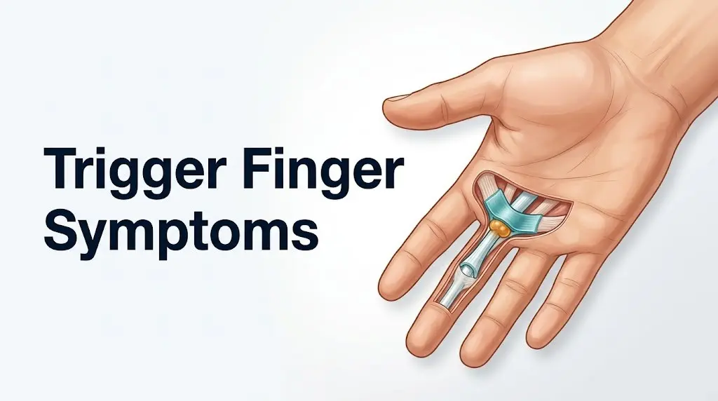
Think of it like a rope being pulled through a ring. When both the rope and the ring are the right size, movement is frictionless. When the rope develops a knot and the ring shrinks at the same time, pulling the rope through that ring produces a catching sensation and, eventually, the rope gets stuck.
The American Academy of Orthopaedic Surgeons (AAOS) confirms that the ring finger and thumb are the most commonly affected digits, though any finger can develop stenosing tenosynovitis. The condition usually begins with localized pain and tenderness before the clicking and locking stages develop.
- Pain or tenderness at the base of the finger on the palm side
- Stiffness, particularly after sleep or rest
- A clicking, snapping, or catching sensation during finger movement
- A painful locking of the finger in a bent position
- A palpable nodule at the base of the affected finger
Older adults, people with diabetes, and people with inflammatory arthropathy are more likely to present with multiple affected fingers simultaneously rather than isolated single-finger involvement.
What Are the Symptoms of Trigger Finger?
The symptoms of trigger finger are pain at the base of the finger on the palm side, a clicking or snapping sensation during movement, morning stiffness, catching of the finger mid-motion, and in more advanced stages, a finger that locks in a bent position and must be physically straightened with the other hand.
Each symptom maps directly to a specific stage of the underlying tissue pathology. Pain and tenderness appear first, produced by nociceptor activation as the swollen A1 pulley and early-stage tendon nodule begin to generate friction. Clicking appears once the nodule is large enough to catch on the proximal edge of the A1 pulley but still small enough to force its way through. Locking appears when the nodule can no longer pass through the narrowed tunnel at all.
A 2019 study published in the Journal of Hand Surgery examined histopathological specimens from patients undergoing surgical trigger finger release and found consistent evidence of fibrocartilaginous metaplasia in the A1 pulley tissue. This is not simple inflammation. The pulley tissue actually changes its cellular composition toward a cartilage-like structure, making the narrowing a structural rather than purely inflammatory problem. This distinction matters clinically because it explains why anti-inflammatory measures alone often do not fully resolve advanced trigger finger.
| Symptom | When It Typically Appears | Primary Mechanism |
|---|---|---|
| Pain at base of finger | Early (Grade 1) | Nociceptor activation in inflamed A1 pulley |
| Morning stiffness | Early to moderate (Grade 1-2) | Reduced synovial fluid mobility after overnight rest |
| Clicking or snapping | Moderate (Grade 2) | Tendon nodule forcing past narrowed A1 pulley |
| Catching that self-releases | Moderate (Grade 2) | Intermittent nodule impingement |
| Locking requiring passive correction | Advanced (Grade 3) | Nodule unable to pass through pulley without external force |
| Fixed locked position | Severe (Grade 4) | Complete mechanical blockade of tendon glide |
In people with rheumatoid arthritis, all of these symptoms can develop more rapidly than in idiopathic cases because synovial proliferation inside the tendon sheath adds a second mechanism of narrowing on top of any pulley changes.
What Does Trigger Finger Feel Like?
Trigger finger feels like a catch, snap, or pop in the finger during bending or straightening, accompanied by pain at the base of the finger that can radiate into the palm or up toward the fingertip. The sensation is distinct enough that most people describe it immediately and accurately when asked.
In the earliest stage, before any catching occurs, the finger simply feels achy and tender when you press on the base of it on the palm side. This is the point most people dismiss as a minor strain or overuse soreness. The tenderness is specific to one small area, directly over the A1 pulley, which sits roughly at the level of the crease where the finger meets the palm.
As the condition progresses, the catching sensation becomes the defining feature. Some people describe it as similar to cracking a knuckle, but happening during the middle of a movement rather than at the end. Others describe a feeling of the finger getting briefly stuck before releasing with a pop. The pop is the tendon nodule being forced through the narrowed A1 pulley under increasing muscle load.
At more advanced stages, the sensation shifts from a pop to a stuck feeling. The finger won’t straighten from a bent position without help from the other hand. When passive extension is applied, people often feel and hear an audible click as the nodule clears the pulley. This can be painful enough that some people avoid straightening the finger altogether, which over time risks developing a secondary flexion contracture of the proximal interphalangeal joint.
For children, particularly those under three years old presenting with what appears to be trigger thumb, the experience is different. Congenital trigger thumb in pediatric patients typically presents not as a locking-and-releasing phenomenon but as a fixed interphalangeal joint flexion deformity that does not actively trigger. The mechanism in pediatric cases involves a distinct nodule on the flexor pollicis longus tendon called Notta’s node, a different anatomical presentation from adult stenosing tenosynovitis.
Key Takeaway: Trigger finger produces a specific, localized catching or locking sensation at the base of the finger on the palm side because a tendon nodule and a narrowed A1 pulley are mechanically impinging on each other, not because of a problem inside the joint itself.
Trigger Finger Pain Location: Where It Hurts and Why
Trigger finger pain is located primarily at the base of the affected finger on the palm side, directly over the A1 pulley at the level of the metacarpophalangeal joint, though pain can radiate proximally into the palm or distally toward the fingertip during active triggering.
This specific location is anatomically precise. The A1 pulley for each finger sits just distal to the distal palmar crease. For the thumb, the A1 pulley is at the level of the metacarpophalangeal joint of the thumb, slightly more proximal than people expect. When a clinician presses on this exact point during a physical examination, tenderness there is one of the most reliable physical signs for confirming the diagnosis.
The pain is typically described as a dull ache at rest that sharpens to a more intense discomfort during the moment of triggering. When the tendon nodule is forced past the A1 pulley, the nociceptors in the compressed tendon sheath fire simultaneously, producing a brief, sharper pain layered onto the background ache.
A practical way to think about the pain geography: press the pad of your opposite thumb firmly along the palm side of your finger, starting from the fingertip and moving toward the palm. The A1 pulley is exactly at the point where your finger meets your palm at the deepest part of that crease. If that spot is specifically tender, that’s your A1 pulley.
People with diabetes may notice that their trigger finger pain is less intense than expected relative to the degree of mechanical impingement, because peripheral sensory neuropathy associated with long-standing diabetes can blunt pain perception in the hands. This means diabetic patients sometimes present at more advanced stages because they haven’t had strong pain as an early warning signal.
Quick Tip:
- Pain directly over the base of the finger on the palm side, not inside the finger joint, is the key locating feature of trigger finger
- Pain that spreads up into the palm during active gripping is consistent with A1 pulley inflammation
- People with pre-existing peripheral neuropathy, including diabetic neuropathy, may underreport pain severity despite having advanced mechanical changes
Trigger Finger Stiffness in the Morning: Why It’s Worse After Sleep
Trigger finger stiffness in the morning is worse than at other times of day because overnight immobility reduces the circulation of synovial fluid through the tendon sheath, increasing the viscosity of that fluid and making the inflamed tendon-pulley interface even more resistant to smooth gliding.
The synovial fluid inside the tendon sheath serves as both a lubricant and a nutrient delivery system for the avascular tendon tissue. During activity, the mechanical pumping action of tendon gliding circulates this fluid continuously. During sleep, with the hand still, fluid circulation slows dramatically. Inflammatory mediators produced by the inflamed synovium, including prostaglandin E2, accumulate locally. The thickened, stagnant fluid increases the resistance to tendon movement when you first begin using your hand in the morning.
This is why most people with trigger finger notice that the clicking, catching, or stiffness is worst in the first fifteen to thirty minutes after waking, and then gradually improves as hand use warms and circulates the synovial fluid. The phenomenon is clinically analogous to the gel phenomenon seen in rheumatoid arthritis, though the underlying tissue is the tendon sheath rather than the joint synovium.
According to the AAOS, morning stiffness that gradually improves with activity is one of the characteristic clinical features that helps distinguish trigger finger from other causes of finger pain where stiffness worsens with activity rather than improving with it.
Occupational and hand therapists often recommend gentle active range of motion exercises in warm water first thing in the morning to accelerate the warming and circulation of synovial fluid before demanding gripping tasks. This does not treat the underlying structural change but can reduce the functional impact of morning symptoms.
Postmenopausal women tend to report more pronounced morning stiffness with trigger finger than premenopausal women. The decline in estrogen after menopause is associated with broader changes in tendon and ligament hydration and elasticity, which may amplify the viscosity-related stiffness effect on already compromised tendon sheaths.
Trigger Finger Clicking or Snapping Sensation
The clicking or snapping sensation in trigger finger occurs when the fibrous nodule on the flexor tendon is forced past the narrowed edge of the A1 pulley, producing a palpable and sometimes audible pop as the tendon snaps through the constriction.
This is Grade 2 on the clinically recognized Green’s classification of trigger finger severity. At this stage, the triggering occurs during active movement but resolves spontaneously. The person can complete the flexion or extension arc, but there is a distinct, reproducible click somewhere in the motion.
The biomechanics are precise. The tendon nodule sits just proximal to the A1 pulley when the finger is in extension. As the finger flexes, the nodule is dragged distally against the proximal edge of the A1 pulley. If the nodule is large enough to be impeded but still small enough to pass through with sufficient force, the tendon suddenly snaps through the narrowed aperture. That snap is the click.
Some people hear the sound. Many more feel it without hearing it. A few describe it happening most reliably when they make a fist, others when they try to extend a finger that was relaxed in a partially bent position. The variability depends on the exact size ratio between the nodule and the residual pulley aperture, which is why two people with Grade 2 trigger finger can describe the sensation differently.
A 2021 study published in the Journal of Hand Surgery noted that ultrasound imaging can visualize the tendon nodule and confirm its size and location relative to the A1 pulley with high diagnostic accuracy, providing objective confirmation of the mechanical impingement that produces the clicking sensation.
People who perform repetitive gripping at work, including surgeons, musicians, and manual laborers, often notice the clicking worsens significantly during workdays and is milder on rest days, because the frequency of nodule-pulley impingement events is directly proportional to the number of gripping cycles performed. This pattern is itself diagnostically useful.
Trigger Finger Locking Sensation: When the Finger Gets Stuck
The trigger finger locking sensation occurs when the flexor tendon nodule is too large to pass through the narrowed A1 pulley during active finger movement, causing the finger to become stuck in a flexed or extended position until external force is applied to push the nodule through.
Locking represents Grade 3 on Green’s classification. The finger can still be moved from the locked position, but only by using the other hand to passively extend or flex it through the point of impingement. When the nodule finally clears the pulley, the person feels and often hears the characteristic pop of tendon release.
The locking occurs preferentially in flexion rather than extension for a mechanically important reason. The flexor muscles of the forearm (flexor digitorum superficialis and flexor digitorum profundus) are substantially stronger than the extensor muscles acting on the same finger. This means the nodule can be pulled into the palm-side of the pulley with enough force to clear it on the way into flexion. But when trying to straighten the finger, the weaker extensors cannot generate the force needed to pull the nodule back through from the distal side. The finger gets stuck bent.
This asymmetry explains why people with trigger finger can usually make a fist but struggle to straighten their fingers fully, rather than the reverse.
| Locking Pattern | Mechanical Cause | Grade |
|---|---|---|
| Catching that releases on its own | Nodule intermittently impinges, resolves spontaneously | Grade 2 |
| Locking requiring passive correction | Nodule fully impinges, requires external force | Grade 3 |
| Fixed locked position | Complete mechanical block, cannot be passively corrected | Grade 4 |
Older adults, particularly those over 65, are more likely to develop a secondary proximal interphalangeal (PIP) joint flexion contracture if Grade 3 locking persists without treatment. When the finger remains held in flexion repeatedly, the volar plate and collateral ligaments of the PIP joint adaptively shorten. This contracture is a separate and potentially permanent complication that requires its own treatment, independent of the trigger finger release.
Key Takeaway: The locking in trigger finger happens because the extensor muscles are not strong enough to pull the tendon nodule back through the narrowed A1 pulley, which is why fingers get stuck in flexion and must be manually straightened.
Trigger Finger Nodule Symptoms: The Bump You Can Feel
A palpable nodule at the base of the affected finger on the palm side, directly over the A1 pulley, is a hallmark finding of trigger finger and represents a fibrous thickening that has developed on the flexor tendon at the point of maximum mechanical friction.
Most people don’t realize this bump is there until a clinician points it out or they begin specifically palpating their palm. The nodule is typically three to five millimeters in diameter, firm but not rock-hard, and mobile. It moves with the tendon when the finger is flexed and extended. This mobility is the feature that clinically distinguishes a trigger finger nodule from a palmar ganglion cyst, which is attached to deeper structures and does not move with tendon motion.
The nodule itself is not the narrowing. The narrowing is in the A1 pulley. The nodule is the enlargement on the tendon side. It’s the combination of a narrower tunnel and a larger rope that creates the problem. Research published in the Journal of Hand Surgery using histopathological analysis has characterized the nodule tissue as composed of fibroblast-rich fibrous tissue with areas of myxoid degeneration and, in some cases, fibrocartilaginous transformation.
Pressing directly on the nodule typically reproduces or intensifies the patient’s pain. This response, pain on direct palpation of the A1 pulley region, is one of the most consistent physical examination findings in trigger finger and forms a core part of the clinical diagnosis.
People with diabetes often develop nodules earlier in the disease course and with greater fibrotic density than people with idiopathic trigger finger. Research suggests this is related to advanced glycation end products (AGEs) accumulating in collagen-rich structures like tendons, accelerating the fibrotic remodeling process. This biochemical difference may explain why trigger finger in diabetic patients responds less well to corticosteroid injection alone and has higher recurrence rates after injection compared to the general population.
Trigger Finger Early Symptoms: Recognizing It Before It Gets Worse
The earliest symptom of trigger finger is localized tenderness at the base of the affected finger on the palm side, without any clicking, locking, or catching, which corresponds to Grade 1 on the Green’s classification and represents the optimal window for conservative intervention.
Grade 1 is called the “pretriggering” stage. The tendon is beginning to develop surface changes and the A1 pulley is starting to narrow and stiffen, but the mechanical mismatch between the nodule and the pulley has not yet become large enough to produce a catching sensation. The only symptom is pain. Specific, localized palm-side pain at the base of the finger.
At this stage, many people attribute the discomfort to a minor hand strain, a bruise, or general overuse soreness. The tenderness can be mild enough to ignore during the day but noticeable when gripping something tightly, using the affected hand for fine motor tasks, or pressing on the palm. Morning stiffness may be barely perceptible or absent.
Recognizing Grade 1 symptoms matters because early-stage trigger finger responds better to conservative treatment than advanced-stage trigger finger. According to a review published in the Archives of Physical Medicine and Rehabilitation, corticosteroid injection into the tendon sheath at Grade 1 or Grade 2 produces full resolution in approximately 60 to 90 percent of cases with a single injection. That resolution rate declines substantially as the condition advances to Grade 3 and 4.
To assess early symptoms at home before a medical appointment:
- Place the affected hand palm-side up on a flat surface
- Use the tip of your opposite index finger to press firmly along the palm side of the base of each finger
- Identify whether one specific spot is distinctly more tender than the surrounding area
- Flex and extend the finger slowly five to ten times and notice whether any catching or click occurs
- Note whether the stiffness or tenderness is worse in the first fifteen minutes after waking than it is mid-afternoon
Early symptoms presenting without triggering warrant evaluation by a primary care physician, who can confirm the diagnosis clinically and refer to a hand surgeon or hand therapist for conservative management planning.
Trigger Finger Stages and Grading: How Severe Is Your Presentation?
Trigger finger severity is classified using the Green’s classification system, which ranges from Grade 1 (pain and tenderness only) through Grade 4 (fixed locked finger that cannot be passively extended), with each grade representing a distinct stage of mechanical and structural deterioration that guides treatment decisions.
This grading system is the framework clinicians use to determine whether a patient is a candidate for splinting, corticosteroid injection, ultrasound-guided injection, percutaneous needle release, or open surgical release of the A1 pulley. Understanding your grade is the most important step in understanding your treatment options.
| Grade | Clinical Name | Symptoms | Typical Treatment Path |
|---|---|---|---|
| Grade 1 | Pretriggering | Palm-side pain and tenderness, no catching | Activity modification, splinting, NSAID therapy |
| Grade 2 | Active triggering | Clicking or catching that self-releases | Corticosteroid injection, splinting |
| Grade 3 | Locked with passive correction | Finger locks, requires other hand to straighten | Corticosteroid injection, percutaneous release, or surgery |
| Grade 4 | Fixed deformity | Finger permanently locked, cannot be passively straightened | Surgical A1 pulley release |
The American Society for Surgery of the Hand (ASSH) notes that the distinction between Grade 3 and Grade 4 is clinically significant because Grade 4 patients often have developed secondary joint changes that may require additional treatment beyond simple pulley release.
Progression through the grades is not inevitable or uniform. Some people remain at Grade 1 or 2 for months or years with minimal worsening. Others progress from Grade 1 to Grade 3 within weeks, particularly people with uncontrolled diabetes, active rheumatoid arthritis, or those whose occupation involves continuous repetitive gripping. Age is also a factor. People over 60 tend to progress more quickly through grades because of the broader age-related decline in tendon tissue elasticity and synovial fluid quality.
Key Takeaway: Green’s classification Grades 1 through 4 directly determine the recommended treatment pathway for trigger finger, making self-assessment of your grade a practical first step before a medical appointment.
Trigger Finger Locked Finger Symptoms: Grade 3 and 4 Presentation
A locked trigger finger at Grade 3 means the finger becomes stuck in a bent position during normal use and requires the other hand to straighten it, while Grade 4 means the finger is permanently fixed in flexion and cannot be straightened even with passive assistance.
Grade 3 locking is often the presentation that finally brings people to a clinician. The catching and clicking of earlier grades can be tolerated as a nuisance, but a finger that locks closed when making a fist and won’t open without manual help is functionally limiting and frightening enough to prompt action.
When a Grade 3 finger locks, the specific sequence of events is predictable. The person flexes the finger into the palm, the nodule passes through the A1 pulley into the distal tendon sheath, and then the nodule cannot be pulled back through the pulley by the relatively weaker extensor mechanism. The person applies the thumb and index finger of the opposite hand to the locked finger and straightens it with force. The pop felt when the finger releases is the nodule clearing the proximal edge of the A1 pulley.
Grade 4, the locked finger that cannot be straightened even passively, is less common at initial presentation and usually represents either very advanced disease or a case in which Grade 3 locking was left untreated for long enough that a secondary flexion contracture of the PIP joint developed. At this stage, even after successful surgical release of the A1 pulley, physical therapy or occupational therapy is required to regain full passive range of motion at the PIP joint.
People who have been living with a Grade 3 locked finger and avoiding straightening it to spare themselves the pain of the pop are inadvertently accelerating their risk of PIP joint contracture. A hand therapist or certified hand therapist (CHT) can design a splinting protocol specifically to prevent PIP contracture while the underlying trigger finger is being treated.
Symptoms of Trigger Thumb: How the Thumb Differs
Symptoms of trigger thumb involve pain, clicking, and locking at the thumb’s metacarpophalangeal joint on the palmar surface, but the clinical picture, the relevant tendon (the flexor pollicis longus), and the functional consequences of thumb locking differ substantially from trigger finger because the thumb’s opposition function makes thumb locking disproportionately disabling.
The A1 pulley of the thumb is anatomically positioned slightly differently from the finger A1 pulleys, sitting over the thumb MCP joint at the level of the thenar crease rather than the deeper palmar creases. This means the point of tenderness in trigger thumb is in a slightly different location from what people expect, and it can be mistaken for thumb basal joint arthritis (carpometacarpal joint arthritis), which also produces pain at the base of the thumb but over a different anatomical structure.
The functional stakes of thumb locking are higher than for any individual finger. The thumb provides roughly 40 to 50 percent of total hand function through its ability to oppose the other fingers. A locked thumb makes pinching, writing, buttoning, and most fine motor activities extremely difficult or impossible. This is why trigger thumb is typically referred to a board-certified hand surgeon sooner than an equivalent grade of trigger finger in the ring or middle finger.
In children under three years old, trigger thumb presents differently from adult trigger thumb. Pediatric trigger thumb (congenital trigger thumb) is not a true stenosing tenosynovitis. It presents as a fixed flexion deformity of the thumb interphalangeal joint, caused by a distinct fibrous nodule called Notta’s node on the flexor pollicis longus tendon. Unlike adult trigger thumb, it does not typically catch and release. The thumb simply remains bent. Approximately 30 percent of congenital trigger thumb cases resolve spontaneously by age three, according to research published in the Journal of Pediatric Orthopaedics. Cases persisting beyond age three generally require surgical treatment.
Women develop trigger thumb at approximately the same elevated rate relative to men as they develop trigger finger, and the postmenopausal period appears to be the peak risk window for both conditions.
Trigger Finger Symptoms in Diabetics: Why It Presents Differently
Trigger finger symptoms in people with diabetes tend to be more severe at presentation, affect multiple fingers simultaneously, occur bilaterally more often than in the general population, and respond less completely to corticosteroid injection, because the underlying biochemical mechanism of A1 pulley and tendon change in diabetes differs from the idiopathic mechanism.
The National Institute of Arthritis and Musculoskeletal and Skin Diseases reports that people with diabetes have two to three times the rate of trigger finger compared to the general population. The reason lies in the metabolic effects of chronically elevated blood glucose on connective tissue. Advanced glycation end products (AGEs) accumulate in the collagen of tendons and pulley tissue when blood glucose is poorly controlled. AGE accumulation cross-links collagen fibers, making the tendon stiffer and the pulley tissue thicker without requiring the same mechanical repetition or inflammatory trigger that drives idiopathic trigger finger.
This means a person with poorly controlled Type 2 diabetes may develop trigger finger in multiple fingers simultaneously, including fingers that are not heavily used in their daily occupation. Multiple simultaneous trigger fingers in the absence of obvious repetitive strain or inflammatory arthropathy should prompt a clinician to assess for undiagnosed or poorly controlled diabetes.
The clinical implication for treatment is practical. Research published in the Journal of Hand Surgery found that the success rate of a single corticosteroid injection for trigger finger in diabetic patients is approximately 50 to 57 percent, compared to 60 to 90 percent in non-diabetic patients. Diabetic patients are also more likely to experience a transient spike in blood glucose levels in the 24 to 72 hours following a corticosteroid injection into the tendon sheath, even from the small doses typically used for this purpose. People with insulin-dependent diabetes should discuss this with their prescribing physician before undergoing injection treatment.
- Multiple fingers affected at once in the absence of repetitive strain
- Bilateral presentation (both hands affected)
- Less pronounced pain than expected given the degree of mechanical impingement
- Higher likelihood of progressing to Grade 3 or 4 before seeking care due to reduced pain sensitivity from peripheral neuropathy
Key Takeaway: Trigger finger in people with diabetes is driven partly by advanced glycation end product accumulation in tendon collagen, which is why multiple fingers are often affected simultaneously and why corticosteroid injection is less reliably effective.
Trigger Finger Symptoms with Rheumatoid Arthritis
Trigger finger symptoms in people with rheumatoid arthritis arise through a different primary mechanism than idiopathic trigger finger, driven by synovial proliferation inside the flexor tendon sheath rather than A1 pulley fibrosis alone, which produces a distinct clinical picture and has different treatment implications.
In rheumatoid arthritis (RA), the same autoimmune process that inflames joint synovium also targets the synovial lining of tendon sheaths. The synovium inside the flexor tendon sheath of the finger, the tenosynovium, proliferates under the influence of tumor necrosis factor alpha (TNF-alpha) and interleukin-6, producing a pannus-like tissue that fills the tendon sheath from within. This intrasynovial proliferation narrows the effective cross-sectional area available for tendon gliding, even if the A1 pulley itself has not yet undergone significant fibrotic change.
The clinical result is that trigger finger in RA patients often presents with more diffuse tendon sheath swelling, more warmth and redness along the entire tendon path on the palmar surface, and a softer, more fluctuant swelling at the base of the finger than the firm, localized nodule typical of idiopathic trigger finger. The triggering sensation may be less crisp and the stiffness may affect a longer segment of the finger than the isolated A1 pulley region.
According to research published in Annals of the Rheumatic Diseases, flexor tenosynovitis is present in up to 55 percent of patients with early RA, often before joint erosions are detectable on plain radiography. This means trigger finger or diffuse flexor tendon sheath swelling may be one of the earliest clinical signs of RA in some patients.
Treatment of trigger finger in RA requires attention to both the local tendon-pulley problem and the systemic inflammatory disease. A rheumatologist managing the underlying RA and a board-certified hand surgeon addressing the mechanical tendon problem work best as a team. Disease-modifying antirheumatic drugs (DMARDs) that control systemic RA activity often reduce the severity of tenosynovitis as a secondary effect.
RA-associated trigger finger is more likely to affect multiple fingers simultaneously, the tendon sheath swelling may be visible on inspection of the palm rather than requiring palpation, and the morning stiffness associated with RA tenosynovitis typically lasts longer than the fifteen to thirty minutes typical of idiopathic trigger finger morning stiffness.
When to See a Doctor for Trigger Finger
You should see a doctor for trigger finger when symptoms include a finger that catches or locks during normal activities, pain that interferes with daily tasks, morning stiffness lasting more than thirty minutes, or any finger that becomes locked and cannot be straightened without help from the other hand.
The appropriate type of provider depends on your symptom grade. A primary care physician can diagnose trigger finger clinically through physical examination alone (no imaging is typically required for an initial diagnosis), and can initiate first-line management including splinting and anti-inflammatory medication. For anything beyond Grade 1, or for Grade 1 symptoms in a person with diabetes or RA, a referral to a board-certified hand surgeon (a surgeon who has completed fellowship training accredited by the American Society for Surgery of the Hand) is appropriate.
To prepare for a medical appointment for trigger finger:
- Note which finger or fingers are affected and on which hand
- Describe when the catching or locking first began and whether it has worsened over time
- Identify the time of day when symptoms are worst and whether activity or rest changes the severity
- List any prior hand injuries, surgeries, or medical conditions including diabetes, thyroid disease, or arthritis
- Note any occupational or hobby activities involving repetitive gripping, vibrating tools, or extended instrument use
- Bring a list of all current medications, since some (including fluoroquinolone antibiotics and certain statins) are associated with tendon pathology
The American Society for Surgery of the Hand recommends that a finger locked in a fixed flexion deformity that cannot be passively straightened should be evaluated by a hand surgeon promptly, on a same-day or next-day basis, rather than waiting for a routine appointment.
People with sudden onset of finger swelling, extreme pain along the entire length of the finger’s palm-side tendon sheath, warmth, redness, and a posture of holding the finger in a semiflexed position that is exquisitely painful to move should not assume this is routine trigger finger. These are the Kanavel signs of septic flexor tenosynovitis, a surgical emergency that requires immediate evaluation, as described in the next section.
Emergency Symptoms: When to Seek Urgent Evaluation
Trigger finger itself is not a life-threatening emergency. However, one condition that can initially mimic trigger finger is a surgical emergency and requires recognition.
Seek same-day emergency evaluation at a hospital emergency department immediately if you notice any of the following:
- A finger that is uniformly swollen from base to tip on the palm side (not just at the base)
- Extreme tenderness along the entire length of the finger’s tendon sheath, not just at the A1 pulley
- The finger held in a slightly bent position that is intensely painful to move passively in any direction
- Redness, warmth, and swelling spreading toward the palm or wrist
- Fever accompanying finger swelling and pain
- Recent puncture wound, animal bite, or cut to the palm or finger in the days before symptoms began
These are the clinical features of septic flexor tenosynovitis, a bacterial infection of the flexor tendon sheath. Unlike stenosing tenosynovitis (trigger finger), which develops gradually over weeks to months, septic tenosynovitis develops within hours to days after a bacterial inoculation event. Untreated septic tenosynovitis can destroy the flexor tendon within 24 to 48 hours and result in permanent loss of finger function.
Go to the nearest hospital emergency department immediately. This condition requires surgical irrigation and debridement of the tendon sheath, intravenous antibiotics, and specialist hand surgery involvement. An urgent care center is not equipped to manage septic flexor tenosynovitis. Do not wait to see if it improves.
A locked fixed flexion deformity of a finger in a person who has had no prior trigger finger symptoms and no mechanism of injury should also prompt same-day evaluation by a hand surgeon, as conditions including flexor tendon rupture, locked joint foreign body, or other acute structural problems can mimic sudden-onset trigger finger.
Key Takeaway: Fever, warmth, uniform finger swelling, and extreme pain along the entire tendon sheath after a skin puncture or bite are signs of septic flexor tenosynovitis, a surgical emergency that requires immediate emergency department evaluation, not a routine hand surgery appointment.
Frequently Asked Questions About Trigger Finger Symptoms
What does trigger finger feel like at first?
At first, trigger finger feels like a specific, localized ache or tenderness at the base of the affected finger on the palm side, without any catching or clicking.
This early tenderness is directly over the A1 pulley at the MCP joint level and is typically noticeable when gripping firmly or pressing on that area.
Morning stiffness that eases within thirty minutes of activity is a second early feature that many people notice before any mechanical catching develops.
What are the first signs of trigger finger?
The first sign of trigger finger is pain and tenderness at the base of the finger on the palm side, corresponding to Grade 1 on the Green’s classification system.
This localized tenderness is produced by early inflammation and thickening of the A1 pulley and beginning surface changes on the flexor tendon.
A slight increase in morning stiffness or aching after prolonged hand use are common accompaniments at this early stage.
Can trigger finger go away on its own?
Some cases of trigger finger, particularly Grade 1 presentations, can improve with rest and activity modification, though complete spontaneous resolution without any treatment is not reliably predictable.
Grade 2 and above rarely resolve fully without intervention, and untreated moderate-to-advanced trigger finger tends to progress rather than spontaneously improve.
Evaluation by a primary care physician or hand surgeon is appropriate for any presentation that includes catching, locking, or symptoms persisting beyond two to four weeks.
What finger is most commonly affected by trigger finger?
The ring finger and the thumb are the most commonly affected digits in adults with trigger finger.
The American Academy of Orthopaedic Surgeons notes that any finger can be involved, and multiple fingers are affected simultaneously more often in people with diabetes or rheumatoid arthritis.
The middle finger is the third most commonly affected, followed by the index finger, with the little finger least commonly involved.
Is trigger finger worse in the morning?
Yes, trigger finger is characteristically worse in the morning because overnight immobility reduces the circulation of synovial fluid through the tendon sheath and allows inflammatory mediators to accumulate locally.
The increased viscosity of stagnant synovial fluid at the site of tendon-pulley friction makes the first movements of the day more resistant and more likely to trigger the catching or locking sensation.
Symptoms typically improve within fifteen to thirty minutes of hand use as synovial fluid warms and circulates, distinguishing trigger finger from conditions in which activity worsens rather than improves symptoms.
When should I worry about trigger finger?
You should seek evaluation when trigger finger symptoms include catching or locking that requires the other hand to straighten the finger, pain that interferes with daily activities, or symptoms that have been present and worsening over several weeks.
A finger that cannot be passively straightened (Grade 4), or symptoms accompanied by fever, warmth, and uniform swelling along the entire finger after a skin injury, require same-day evaluation.
People with diabetes who notice multiple fingers affected simultaneously, or people with rheumatoid arthritis who notice new tendon-side swelling in the palm, should contact a primary care physician or rheumatologist promptly.
Closing
The symptoms of trigger finger tell a specific mechanical story: a tendon that can no longer glide freely through a narrowed pulley produces pain, stiffness, clicking, and eventually locking in a precise, predictable sequence. Every symptom has a tissue-level explanation, and understanding that explanation helps you assess where you are in that progression.
The most practical action you can take right now is to identify your grade. If you have localized pain and morning stiffness without catching, you’re likely at Grade 1 and early intervention gives you the best odds of resolution. If you’re catching and clicking, you’re at Grade 2 and a single corticosteroid injection resolves the majority of cases at that stage. If your finger is locking and you’re using your other hand to straighten it, contact a board-certified hand surgeon rather than waiting to see if it resolves.
Your primary care physician can confirm the diagnosis, assess your overall health picture including diabetes and inflammatory arthritis risk, and refer you to the right specialist. You now have the clinical picture clearly enough to describe your symptoms precisely and ask the right questions at that appointment.