Blood Clot Behind Knee Symptoms: Signs Not to Ignore
Blood clot behind knee symptoms include swelling, deep aching pain, warmth, and redness in the posterior knee and calf region, produced when a thrombus forms in the popliteal vein and obstructs venous blood flow out of the lower leg. This condition, called popliteal deep vein thrombosis (DVT), ranges from a completely silent presentation with no noticeable symptoms to a rapidly escalating emergency if the clot dislodges and travels to the lungs.
The National Heart, Lung, and Blood Institute (NHLBI) estimates that venous thromboembolism, which encompasses DVT and its complication pulmonary embolism, affects approximately 300,000 to 600,000 Americans each year. What makes popliteal DVT particularly deceptive is that its symptoms closely mimic a ruptured Baker’s cyst or a calf muscle strain, meaning many people dismiss warning signs for days before seeking care.
This article explains what each symptom of a blood clot behind the knee actually feels like and why it occurs at the physiological level, how to distinguish DVT from its most common clinical mimics, what the pulmonary embolism warning signs are, and when you need emergency evaluation rather than a scheduled appointment.
Blood Clot Behind Knee Symptoms: What This Condition Is and Why It Happens
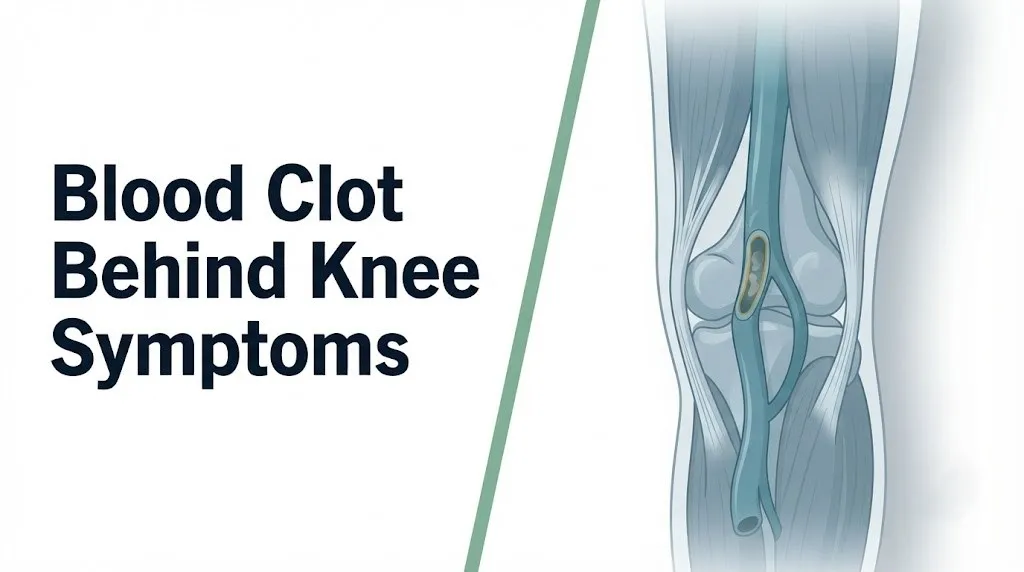
A blood clot behind the knee is a thrombosis within the popliteal vein, the major deep vein that runs through the popliteal fossa (the hollow space at the back of the knee), producing symptoms through two simultaneous mechanisms: venous outflow obstruction and local inflammatory activation.
The popliteal fossa is bounded superiorly by the hamstring muscle group and inferiorly by the two heads of the gastrocnemius muscle. The popliteal vein runs through this space alongside the popliteal artery and the tibial and common peroneal nerves. When a thrombus forms within the popliteal vein, it blocks blood from draining out of the calf and lower leg through the deep venous system.
The mechanism behind clot formation is described by Virchow’s triad, a three-part framework named after the 19th-century pathologist Rudolf Virchow. The three elements are venous stasis (slowed or pooled blood flow), endothelial injury (damage to the inner lining of the vein), and hypercoagulability (an abnormal tendency for the blood to clot). When any one or more of these conditions is present, the coagulation cascade can be triggered, generating thrombin from prothrombin, which then converts soluble fibrinogen to insoluble fibrin. The fibrin strands trap red blood cells and platelets, forming the thrombus that lodges in the popliteal vein.
| Virchow’s Triad Component | Common Clinical Triggers | How It Contributes to Popliteal DVT |
|---|---|---|
| Venous stasis | Prolonged immobility, long flights, bed rest, sedentary lifestyle | Slowed flow allows coagulation factors to concentrate and platelets to contact vein wall |
| Endothelial injury | Surgery, trauma to knee or leg, catheter placement, inflammation | Exposes subendothelial collagen and tissue factor, activating both coagulation pathways |
| Hypercoagulability | Inherited thrombophilia, malignancy, pregnancy, oral contraceptives, HRT | Shifts the coagulation/fibrinolysis balance toward thrombus formation |
People with prior DVT or known thrombophilia should be aware that a single triggering event, such as a long flight or a minor knee injury, may be sufficient to initiate popliteal DVT even when it would not cause a clot in a person without underlying risk factors.
The Popliteal Vein: The Specific Anatomy Behind a Blood Clot Behind the Knee
The popliteal vein is the primary deep vein of the posterior knee, formed by the confluence of the anterior tibial vein, posterior tibial vein, and peroneal vein at the venous trifurcation just below the knee joint, and it serves as the main drainage route for all venous blood returning from the lower leg.
Understanding this anatomy explains why a clot in the popliteal vein produces such pronounced symptoms in the calf and lower leg. When the popliteal vein is blocked, blood cannot escape upward through the normal deep venous route. The venous pressure in all the veins draining into the popliteal vein rises sharply, forcing fluid and plasma proteins out of the capillaries and into the surrounding tissues.
Think of the popliteal vein as the main drain of a bathtub. If you block that drain while the water is running, water backs up and overflows into the surrounding area. A popliteal DVT blocks the drain for all the blood returning from the lower leg. The overflow is interstitial fluid, and that fluid produces the swelling visible and palpable in the calf and behind the knee.
The popliteal vein transitions into the femoral vein as it passes through the adductor canal (also called Hunter’s canal) in the lower thigh. A thrombus that propagates proximally from the popliteal vein into the femoral vein, and then into the common femoral vein and iliac veins, represents a proximal extension that carries a meaningfully higher pulmonary embolism risk than an isolated popliteal clot.
The Mayo Clinic notes that the popliteal vein, because of its central position in the posterior knee alongside the tibial nerve and the posterior tibial artery, is surrounded by structures that can be compressed or irritated when the vein distends or when perivenous inflammation develops, which partly explains why DVT at this location can produce a broader range of referred symptoms than DVT in the calf veins alone.
What Does a Blood Clot Behind the Knee Feel Like
A blood clot behind the knee typically produces a combination of deep, aching pressure or cramping pain in the popliteal fossa and upper calf, accompanied by a feeling of fullness, heaviness, or tightness in the lower leg that does not resolve with rest the way muscle soreness would.
Most people with popliteal DVT describe the pain as a persistent, deep aching or throbbing that feels different from ordinary muscle soreness. It tends to be constant rather than positional, though many people notice it worsens when they lower their leg (because gravity increases venous pressure further when upright) and eases very slightly when the leg is elevated.
The tightness that many patients report is not from the clot itself pressing on surrounding structures. It comes from the edema filling the fascial compartments of the calf. As fluid accumulates in the gastrocnemius and soleus muscle compartments, the pressure builds within those compartments, creating a sensation of stretching or fullness that can feel like a severe cramp that never releases.
Quick Tip:
- DVT pain is typically present at rest and not exclusively triggered by movement. Muscle strain pain is usually worse with specific movements and eases when the muscle is not loaded.
- If squeezing the calf reproduces a deep, aching pain that is distinctly different from the surface tenderness of a bruise, that finding warrants same-day evaluation by a primary care physician or emergency medicine physician.
- People on anticoagulant therapy for other conditions can still develop DVT. A breakthrough DVT on anticoagulation is less common but does occur and should not be dismissed because someone is already on a blood thinner.
Key Takeaway: The characteristic feel of a blood clot behind the knee is a persistent deep aching or pressure in the posterior knee and upper calf that does not fully resolve with elevation, rest, or over-the-counter pain relief, and that is accompanied by swelling, warmth, or heaviness in the same leg.
Swelling as a Symptom of Blood Clot Behind the Knee
Swelling in the calf and behind the knee is one of the most clinically consistent symptoms of popliteal DVT, occurring because thrombus occlusion of the popliteal vein raises venous hydrostatic pressure in the distal veins, forcing plasma fluid and proteins through the capillary walls into the interstitial tissue of the lower leg.
The swelling in DVT is almost always unilateral (affecting one leg only), which distinguishes it from the bilateral leg swelling more commonly associated with cardiac or renal causes of edema. The asymmetry is one of the most clinically useful features a clinician looks for: a calf circumference difference of 3 centimeters or more between the two legs at the same measuring point is one of the criteria in the Wells DVT Clinical Prediction Score, the validated scoring tool widely used in emergency medicine and primary care to estimate DVT probability before imaging is obtained.
According to the Annals of Internal Medicine validation studies for the Wells score, unilateral calf swelling of 3 centimeters or greater is one of the strongest individual clinical predictors of DVT in outpatient settings.
The swelling from popliteal DVT typically involves the calf more than the foot, though the foot and ankle can swell if the venous pressure elevation is severe. Pressing the swollen calf with a finger may leave a temporary indentation (pitting edema) if the fluid accumulation is significant. The skin overlying the swollen area may appear stretched, shiny, or slightly discolored.
People with pre-existing chronic venous insufficiency (damaged venous valves from a prior DVT) may already have some baseline calf swelling, making the new acute swelling from a fresh DVT harder to recognize as distinctly abnormal. Any person with known chronic venous insufficiency who notices a sudden increase in one-sided leg swelling, particularly with associated pain or warmth, should seek same-day evaluation.
Pain and Tenderness as Symptoms of DVT Behind the Knee
Pain and tenderness in the posterior knee and upper calf are cardinal symptoms of popliteal DVT, arising from nociceptor sensitization in the perivenous tissue by inflammatory mediators released during the thrombotic process, including bradykinin, prostaglandin E2, and tumor necrosis factor-alpha.
The pain of DVT behind the knee is characteristically located in the popliteal fossa and the upper calf, directly over the course of the popliteal vein and its immediate tributaries. It is usually described as a deep, constant ache, a dull throb, or a cramping pressure rather than a sharp, stabbing, or shooting pain. Pressing directly on the popliteal fossa or squeezing the calf typically reproduces or worsens the pain.
Historically, clinicians used a bedside test called Homan’s sign (pain in the calf produced by dorsiflexion of the foot) as a screening indicator for DVT. The American Heart Association and multiple evidence-based clinical guidelines have since determined that Homan’s sign is unreliable: it is neither sensitive nor specific enough to include or exclude DVT and should not be used as a standalone assessment. Its presence does not confirm DVT; its absence does not exclude it.
The prostaglandin E2 and bradykinin released at the site of thrombus-related vascular inflammation directly activate and sensitize the free nerve endings (nociceptors) of the tibial nerve branches that run through the popliteal fossa. This perivenous inflammation is what produces the tenderness on palpation. It is separate from the mechanical compression the clot may exert on adjacent structures.
Older adults, particularly those with peripheral neuropathy from type 2 diabetes or chronic alcohol use, may have blunted pain sensation in the lower extremities. This means they can develop a significant popliteal DVT with very little reported pain, making swelling and skin changes the more reliable early indicators in this population.
Warmth, Redness, and Skin Changes as Symptoms of Blood Clot Behind the Knee
Warmth and redness over the posterior knee and calf in the context of DVT occur because the inflammatory mediators released during thrombosis, particularly prostaglandin E2 and bradykinin, cause local vasodilation in the skin microcirculation and increased permeability of the dermal capillaries overlying the thrombosed popliteal vein.
The warmth in DVT-affected tissue is measurable. Clinicians often compare the temperature of the symptomatic leg to the contralateral leg by placing the back of the hand against the posterior knee and calf of each leg sequentially. A perceptible temperature difference between the two legs, particularly combined with swelling and pain, increases the clinical likelihood of DVT and contributes to the Wells score calculation.
Redness (erythema) in popliteal DVT tends to be diffuse rather than sharply bordered, spreading across the posterior calf rather than tracking along a distinct linear path. A redness pattern that tracks in a thin line along the surface of the skin more closely suggests superficial thrombophlebitis (a clot in the superficial venous system rather than the deep veins), which carries a different risk profile than DVT and rarely causes pulmonary embolism unless it extends into the deep system.
In cases where DVT has been present for several days without treatment, the skin over the affected area can develop a bluish or purplish discoloration, particularly in the ankle and foot, reflecting severe venous congestion. A rare but serious form of this presentation, called phlegmasia cerulea dolens, involves extreme venous congestion causing the entire leg to turn blue-grey, with associated arterial compromise and a risk of limb loss. This represents a true vascular emergency.
People with darker skin tones should be aware that the classical redness of DVT may not be readily visible. Warmth and swelling take on greater diagnostic weight in these individuals. Asking specifically about the tactile warmth of the affected limb and comparing skin temperature between legs is the more reliable assessment approach.
Key Takeaway: In people with darker skin tones, warmth and swelling are more clinically reliable indicators of possible DVT than redness or erythema, which may not be visually apparent even with a significant thrombotic and inflammatory response in the popliteal vein.
Silent DVT: Blood Clot Behind the Knee Without Symptoms
Approximately 50% of DVT cases, including popliteal DVT, produce no symptoms at all, a presentation called silent or asymptomatic DVT, in which the clot forms and may even propagate proximally without any pain, swelling, or warmth that would prompt the person to seek medical evaluation.
This is one of the most clinically important facts about DVT: the absence of symptoms does not mean the absence of a clot. Asymptomatic DVT is most commonly discovered incidentally during imaging performed for other reasons, during routine DVT surveillance after major orthopedic surgery (total knee replacement, total hip replacement), or tragically after a pulmonary embolism prompts retrograde investigation.
Research published in the New England Journal of Medicine has documented that a substantial proportion of hospital-acquired DVTs detected on surveillance ultrasound after orthopedic surgery are asymptomatic at the time of detection, which is why clinical guidelines from the American College of Chest Physicians (ACCP) recommend pharmacological or mechanical DVT prophylaxis for high-risk surgical patients rather than relying on symptom development as the trigger for evaluation.
The reasons why some DVTs remain silent are not fully understood. Possible explanations include partial rather than complete venous occlusion (allowing enough collateral venous flow to prevent significant pressure buildup), individual variation in the inflammatory response to thrombus, and the gradual development of collateral venous channels around a slowly forming clot.
Silent DVT is particularly common in the calf veins (the veins below the popliteal trifurcation). A proportion of these distal silent clots propagate proximally into the popliteal vein over a period of days to weeks, at which point symptoms may develop. Any high-risk patient (postoperative, pregnant, recently immobilized) who has been told they are at elevated DVT risk should not interpret the absence of symptoms as reassurance that no clot is present.
Early Symptoms of Blood Clot Behind the Knee
The earliest detectable symptoms of blood clot behind the knee are often subtle: a vague sense of heaviness or fullness in the calf, mild tenderness on deep pressure over the popliteal fossa or upper calf, and a barely perceptible difference in calf circumference between the two legs.
Early popliteal DVT may not produce dramatic swelling or severe pain. In the first 24 to 48 hours after a thrombus forms, the inflammatory response is still building, and the venous pressure elevation may be insufficient to produce obvious edema. This is the period during which many people attribute their symptoms to standing too long, a mild muscle pull, or a bruise they don’t recall sustaining.
What distinguishes early DVT from general leg discomfort is the combination and pattern:
- Symptoms are in one leg only, not both
- The heaviness or aching persists or worsens over 24 to 48 hours rather than improving
- The location is specifically in the posterior knee and upper calf rather than in the ankle or foot
- There is no direct mechanical explanation (no fall, no unusually intense workout, no direct blow to the area)
- Elevation provides only partial and temporary relief
Early DVT symptoms sometimes develop only after a triggering event such as rising from a long car journey, walking after a period of bed rest, or exerting the leg in a way that increases blood flow velocity and breaks up a soft partial thrombus enough to increase local inflammation. This temporal pattern of “felt fine sitting, noticed pain and heaviness when I stood up” is clinically consistent with early popliteal DVT.
A primary care physician evaluating early possible DVT will calculate the Wells DVT Clinical Prediction Score and use the result to guide whether a D-dimer blood test, immediate compression ultrasound, or both are warranted. Do not delay evaluation because symptoms seem mild.
Progressive and Worsening Symptoms of Popliteal DVT
Untreated popliteal DVT typically progresses in one of three directions: the thrombus remains localized and may partially resolve through natural fibrinolysis, it propagates proximally into the femoral and iliac veins increasing PE risk substantially, or it leads over months to years to post-thrombotic syndrome from permanent venous valve damage.
When symptoms worsen over 48 to 72 hours, the clinical picture becomes more urgent. Progressive DVT is characterized by increasing calf swelling that spreads toward the ankle and foot, deepening pain that becomes more constant and less tolerable, skin discoloration progressing from redness to a dusky purple-red tone, and worsening heaviness with any attempt to stand or walk.
The risk of proximal thrombus extension, meaning the clot growing upward into the femoral vein and beyond, is what elevates isolated popliteal DVT from a localized problem to a systemic one. According to the Journal of Thrombosis and Haemostasis, studies examining the natural history of untreated isolated distal DVT (including popliteal DVT) have found proximal extension rates of approximately 15 to 25% when no anticoagulation is provided. Once the thrombus reaches the common femoral or iliac veins, the cross-sectional area of the vessel is large enough that a dislodged fragment can obstruct a major pulmonary artery branch.
Post-thrombotic syndrome (PTS) is the long-term complication of inadequately treated popliteal DVT. The thrombus damages and scars the venous valves as it partially recanalized. These valves, which normally prevent retrograde blood flow in the deep veins, become incompetent. Retrograde pooling of blood in the lower leg produces chronic venous hypertension, manifesting as persistent calf swelling, aching, heaviness, skin pigmentation changes (lipodermatosclerosis), and in severe cases venous ulceration of the lower leg. The American Heart Association estimates that PTS develops in 20 to 40% of people with proximal DVT, and lower but still clinically meaningful rates in isolated popliteal DVT.
Key Takeaway: Popliteal DVT that worsens over 48 to 72 hours without anticoagulation carries a 15 to 25% risk of proximal propagation into the femoral vein, at which point pulmonary embolism risk increases substantially and the situation requires urgent anticoagulant therapy initiated by a primary care physician or emergency medicine physician the same day.
Blood Clot Behind Knee vs Baker’s Cyst: How to Tell the Difference
A Baker’s cyst (popliteal cyst) and popliteal DVT produce nearly identical symptoms in the posterior knee region, including swelling, fullness, and pain, because both conditions affect structures within the anatomically confined popliteal fossa, but they arise from entirely different mechanisms and require different treatments.
A Baker’s cyst is a synovial fluid-filled cyst that forms at the posterior knee joint capsule, between the medial gastrocnemius head and the semimembranosus tendon. It develops when excess synovial fluid (often produced by knee arthritis or a meniscal tear) accumulates in the joint and pushes through a weak point in the posterior capsule. When the cyst is intact and non-ruptured, it produces a palpable posterior knee fullness and aching that can be difficult to distinguish from popliteal DVT on the basis of symptoms alone.
When a Baker’s cyst ruptures, it releases synovial fluid into the calf tissues, producing sudden-onset calf pain, swelling, warmth, and sometimes bruising that tracks down the medial aspect of the ankle (a finding sometimes called the crescent sign or Mondor’s sign). This presentation can be nearly indistinguishable from acute DVT in the clinic, which is why clinicians who see a patient with sudden posterior knee and calf symptoms in the context of known knee arthritis or previous meniscal injury still order a compression duplex ultrasound to exclude DVT before attributing the presentation to a ruptured cyst.
| Feature | Popliteal DVT | Baker’s Cyst (Ruptured) |
|---|---|---|
| Location of swelling | Posterior knee and calf | Posterior knee, may track to inner ankle |
| Onset speed | Hours to days | Often sudden (pop or give-way sensation) |
| Skin bruising | Absent or minimal | May develop medial ankle bruising within 24-48 hours |
| Associated knee pathology | Not typically related to joint disease | Often associated with osteoarthritis or meniscal tear |
| Palpable posterior knee mass before event | Usually absent | Often present (intact cyst felt as soft posterior mass) |
| D-dimer test | Elevated in active DVT | May be mildly elevated from local inflammation |
| Compression ultrasound | Shows venous thrombus | Shows cyst, no venous thrombus |
The clinical bottom line is that you cannot reliably distinguish these two conditions from symptoms alone. A compression duplex ultrasound performed by a radiologist or vascular medicine specialist is required to confirm or exclude DVT in either presentation.
Blood Clot Behind Knee vs Muscle Strain or Calf Injury
A calf muscle strain (typically involving the gastrocnemius or soleus muscle belly, or less commonly the plantaris tendon) produces posterior leg pain and swelling that overlaps substantially with popliteal DVT, but the two conditions differ in onset trigger, pain character, and physical examination findings in ways that can help direct urgent evaluation.
Calf muscle strains, particularly medial gastrocnemius tears, are common in adults aged 35 to 60 who participate in court sports (tennis, squash, pickleball) or who push off forcefully during running or jumping. The classic presentation is a sudden, sharp pain in the posterior calf during physical exertion, often described as feeling like being struck by a ball or hit from behind. This sudden onset during activity, with an immediate reduction in ability to push off or walk, distinguishes it clinically from DVT, which does not produce a sudden onset pain during a discrete physical moment.
However, the subsequent swelling, warmth, and aching from a gastrocnemius tear, as it develops over the following 12 to 48 hours, can look and feel nearly identical to DVT. This is compounded by the clinical reality that physical activity and muscle injury are themselves mild DVT risk factors because trauma to the muscle can produce local venous endothelial injury.
To describe your symptom pattern clearly to a clinician, use these steps:
- Identify the exact moment the pain started: was it sudden during activity, or did it develop gradually over hours to days without a precipitating event?
- Note whether there is visible bruising and where it is located. Bruising from a muscle tear typically appears within 24 to 48 hours at the injury site or tracks distally with gravity. DVT does not typically produce bruising unless anticoagulant therapy is started.
- Assess whether your range of ankle motion worsens the pain symmetrically (muscle strain) or whether the aching is constant regardless of ankle position (more consistent with DVT).
- Note any recent risk factors: prolonged sitting, recent travel, surgery within the past three months, or active cancer.
- Report all findings to the primary care physician or emergency medicine physician and allow compression duplex ultrasound to confirm or exclude DVT.
Athletes and physically active adults are a population in whom DVT and muscle injury can genuinely coexist. A muscle tear that causes local venous injury can trigger a DVT in the same limb simultaneously.
Pulmonary Embolism Symptoms: When a Blood Clot Behind the Knee Becomes a Chest Emergency
Pulmonary embolism (PE) occurs when a thrombus fragment dislodges from the popliteal vein, travels through the femoral vein, iliac vein, and inferior vena cava into the right ventricle, and lodges in the pulmonary arterial tree, obstructing blood flow to one or more segments of lung tissue.
PE is a life-threatening complication of DVT. According to data from the Centers for Disease Control and Prevention, PE kills approximately 60,000 to 100,000 Americans annually, making it one of the leading preventable causes of hospital-related death. The popliteal vein, because of its relatively smaller caliber compared to the femoral or iliac veins, tends to produce smaller thrombus fragments when they embolize. This means popliteal DVT-associated PE is more likely to produce a segmental or subsegmental PE than a massive saddle embolus, but it can still be fatal if it produces sufficient right ventricular pressure overload.
The symptoms of PE are distinct from the leg symptoms of DVT and require immediate recognition:
- Sudden shortness of breath at rest or with minimal exertion (dyspnea)
- Sharp or pleuritic chest pain (worse with breathing in)
- Rapid heart rate (tachycardia), typically above 100 beats per minute
- Lightheadedness, near-syncope, or fainting
- Coughing up blood (hemoptysis)
- A sense of impending doom or extreme anxiety
- Worsening hypoxia detectable as bluish discoloration of the lips or fingernails (cyanosis)
Some patients experience PE symptoms before they notice any leg symptoms. This is because, as noted in the silent DVT section, many popliteal DVTs produce no leg signs before embolization occurs. Anyone who develops sudden unexplained shortness of breath or chest pain, even without obvious leg symptoms, requires emergency evaluation to exclude PE.
Risk Factors and Populations Most Affected by DVT Behind the Knee
The risk factors for developing DVT in the popliteal vein reflect all three components of Virchow’s triad: conditions that promote venous stasis, conditions that damage the venous endothelium, and conditions that shift the blood toward a hypercoagulable state.
The NHLBI identifies the following as established risk factors for DVT:
- Prolonged immobility: bed rest of three or more days, long-haul air or car travel (flights over four hours)
- Major surgery, particularly orthopedic procedures involving the lower extremity (total knee replacement carries particularly high DVT risk because it involves direct manipulation of the popliteal vein’s anatomical neighbors and prolonged tourniquet use)
- Trauma to the leg, pelvis, or hip
- Active cancer (malignancy activates coagulation through multiple pathways including tissue factor overexpression and fibrinolysis suppression)
- Inherited thrombophilia, most commonly factor V Leiden mutation (present in approximately 5% of the population of European ancestry) and prothrombin gene mutation (G20210A)
- Prior DVT or PE (personal history approximately doubles the recurrence risk per decade)
- Obesity (BMI above 30)
- Age over 40 (incidence approximately doubles each decade after 40, per NHLBI epidemiological data)
- Heart failure, chronic kidney disease, and inflammatory bowel disease (through various mechanisms affecting flow, endothelium, and coagulation factor production)
People with active malignancy deserve specific note. Cancer-associated thrombosis, sometimes called Trousseau syndrome, involves a systemic pro-coagulant state in which tumor cells and their microenvironments continuously activate coagulation. Cancer patients have approximately a 4 to 7-fold elevated VTE risk compared to the general population, and DVT may be the presenting sign of an undiagnosed malignancy.
Blood Clot Behind the Knee in Pregnancy and Women
Pregnant women face a 5 to 10-fold elevated risk of DVT compared to non-pregnant women of the same age, driven by a physiologically induced hypercoagulable state, mechanical compression of the iliac veins by the growing uterus, and reduced venous flow velocity in the lower extremities throughout pregnancy.
Estrogen, elevated throughout pregnancy and in women using combined oral contraceptives or hormone replacement therapy, directly increases hepatic synthesis of coagulation factors II, VII, VIII, and X while simultaneously reducing levels of the natural anticoagulant protein S. This shifts the hemostatic balance substantially toward clot formation. According to data published in JAMA Internal Medicine, the DVT risk is highest in the third trimester and in the six weeks immediately postpartum, when the combination of pelvic venous compression, prolonged labor-related immobility, and vascular endothelial injury from delivery produces the perfect intersection of all three Virchow’s triad components.
Women using combined oral contraceptives (estrogen plus progestogen) have approximately a 3 to 4-fold elevated VTE risk compared to non-users. This risk is higher in women who smoke, who are over age 35, or who carry an underlying thrombophilia such as factor V Leiden. The American College of Obstetricians and Gynecologists (ACOG) recommends that women with a personal or family history of DVT or known thrombophilia discuss alternative contraceptive options with their obstetrician-gynecologist before starting combined hormonal contraceptives.
Women in general may experience DVT symptoms that are attributed to pregnancy-related leg discomfort (swelling and heaviness being common in normal third-trimester pregnancy) for longer than men before seeking evaluation, which delays diagnosis. Any pregnant woman with asymmetric unilateral calf or posterior knee swelling, particularly with pain or warmth, should be evaluated the same day by their obstetrician-gynecologist or an emergency medicine physician, not reassured that swelling is normal without imaging.
Postmenopausal women using oral hormone replacement therapy (HRT) also face elevated DVT risk. The Women’s Health Initiative study, published in the New England Journal of Medicine, found that conjugated equine estrogen combined with progestogen increased VTE risk approximately two-fold compared to placebo in postmenopausal women.
Key Takeaway: Pregnant women and women on combined oral contraceptives face a substantially elevated DVT risk that means any unilateral leg swelling or posterior knee pain in these populations warrants same-day evaluation rather than watchful waiting, because the symptom overlap with normal pregnancy or minor muscle ache is clinically unreliable as a basis for dismissal.
Diagnosing a Blood Clot Behind the Knee: Tests and Clinical Evaluation
The diagnosis of popliteal DVT is confirmed by compression duplex ultrasonography, a non-invasive imaging test that combines B-mode ultrasound visualization of the popliteal vein with Doppler flow analysis and direct manual compression to identify the presence and location of a thrombus.
The diagnostic process typically unfolds as follows:
- Clinical history: the clinician asks about onset, symptom character, laterality, prior DVT, recent surgery, immobility, medications, and thrombophilia history
- Wells DVT Clinical Prediction Score calculation: this validated scoring tool assigns points for specific clinical features (unilateral calf swelling greater than 3 cm, localized tenderness along the deep venous system, entire leg swelling, pitting edema, active cancer, immobilization, prior DVT, alternative diagnosis less likely than DVT). A total Wells score of 2 or above indicates high clinical probability.
- D-dimer blood test: the D-dimer assay measures fibrin degradation products in the blood. A negative D-dimer in a person with low Wells score probability effectively excludes DVT without the need for imaging. A positive D-dimer in any probability category requires compression ultrasound.
- Compression duplex ultrasonography: the primary imaging test. The sonographer places the ultrasound probe over the popliteal fossa and applies manual compression to the vein. A normal vein collapses completely under gentle pressure; a thrombosed vein does not compress. Doppler analysis additionally shows absent or reduced flow through the popliteal vein.
- Proximal vein evaluation: the ultrasound includes evaluation of the femoral and common femoral veins to assess for proximal propagation.
- CT pulmonary angiography (CTPA): ordered if PE symptoms accompany the DVT presentation.
The D-dimer test has an important limitation: it is elevated in many conditions other than DVT, including pregnancy, surgery, trauma, infection, and malignancy. This means a positive D-dimer result requires ultrasound to confirm, but a negative D-dimer in a low-probability clinical setting is highly reassuring. Pregnant women have physiologically elevated D-dimer levels throughout gestation, making the D-dimer test unreliable as a standalone negative predictor in this population. Imaging is typically required when DVT is clinically suspected in pregnancy regardless of D-dimer result.
Emergency Symptoms: When to Call 911 for a Blood Clot Behind the Knee
Certain symptoms associated with a blood clot behind the knee and its potential complication of pulmonary embolism require immediate emergency evaluation. Do not wait to see if these resolve on their own.
Call 911 or go to the nearest emergency room immediately if you experience:
- Sudden shortness of breath at rest or with minimal exertion, particularly if it developed within minutes and has no obvious explanation such as physical exertion (this pattern indicates possible pulmonary embolism from a DVT fragment reaching the pulmonary arterial system)
- Chest pain that is sharp, stabbing, or pleuritic (worsened by breathing in), occurring in the context of known or suspected leg DVT (this is the classic presentation of pulmonary infarction from PE)
- Rapid heart rate above 100 beats per minute combined with shortness of breath and leg symptoms
- Coughing up blood or blood-stained mucus alongside lower extremity DVT symptoms
- Lightheadedness, near-fainting, or loss of consciousness in the context of any of the above
- Sudden severe bluish or grey discoloration of the entire affected leg with extreme pain (this indicates phlegmasia cerulea dolens, a vascular emergency requiring immediate intervention)
- Worsening confusion, extreme agitation, or a sense of impending doom accompanying chest or leg symptoms
These presentations can indicate pulmonary embolism, massive venous occlusion with threatened arterial compromise, or hemodynamic instability from DVT-related PE, and they require emergency medical assessment in a facility capable of CT pulmonary angiography and urgent anticoagulation or thrombolytic therapy, not an urgent care appointment or a next-day callback.
Frequently Asked Questions About Blood Clot Behind Knee Symptoms
What does a blood clot behind the knee feel like compared to a pulled muscle?
A blood clot behind the knee produces a persistent deep aching or pressure that is present at rest and does not relate to a specific movement or exertion, while a pulled calf muscle typically begins with a sudden sharp pain during activity and then produces soreness that worsens with specific movements.
DVT pain tends to be constant and accompanied by swelling, warmth, and heaviness in the entire lower leg, while muscle strain soreness is more focal and tends to improve significantly with rest over 24 to 48 hours.
Any posterior knee or calf pain that persists beyond 24 to 48 hours, worsens rather than improves, and is accompanied by swelling or warmth warrants same-day evaluation by a primary care physician or emergency medicine physician rather than home rest.
Can a blood clot behind the knee go away on its own?
Some small distal calf vein clots may partially resolve through the body’s natural fibrinolytic mechanisms without anticoagulation, but popliteal vein DVT carries a meaningful risk of proximal extension and pulmonary embolism that makes watchful waiting without medical evaluation clinically inappropriate.
The American College of Chest Physicians guidelines recommend anticoagulation for confirmed symptomatic DVT involving the popliteal vein and above, and serial imaging surveillance or anticoagulation for distal isolated DVT depending on individual risk factors.
No suspected DVT should be managed at home without a confirmed diagnosis and a treatment plan from a primary care physician, vascular medicine specialist, or hematologist.
How long does it take for symptoms of a blood clot behind the knee to appear?
Symptoms of popliteal DVT can appear within hours of thrombus formation or may not appear at all in the approximately 50% of DVT cases that are clinically silent.
When symptoms do develop, they typically begin within 24 to 72 hours of thrombus formation as venous pressure builds and the perivenous inflammatory response escalates.
Symptoms that appear gradually over two to five days after a triggering event such as surgery, a long flight, or a period of immobility are the most common temporal pattern for symptomatic popliteal DVT.
Is a blood clot behind the knee always visible or swollen?
A blood clot behind the knee is not always associated with visible swelling, particularly in early-stage or partial thrombosis where venous outflow obstruction is incomplete.
Approximately 50% of DVT cases produce no obvious swelling or visual skin changes, and the absence of swelling does not exclude the diagnosis.
A difference in calf circumference of 3 centimeters or more between the two legs is a clinically meaningful finding, but smaller differences require compression duplex ultrasonography to confirm or exclude DVT.
What is the most reliable test for a blood clot behind the knee?
Compression duplex ultrasonography is the most reliable non-invasive diagnostic test for popliteal DVT, with sensitivity and specificity above 94% for proximal DVT according to meta-analyses published in the Annals of Internal Medicine.
The test uses manual compression of the vein with the ultrasound probe combined with Doppler flow imaging to identify thrombus that prevents the vein from collapsing and interrupts normal venous flow patterns.
Contrast venography is technically the historical gold-standard test but is rarely performed today because compression ultrasound is highly accurate, non-invasive, and widely available in most emergency departments and radiology departments.
Can a blood clot behind the knee cause a pulmonary embolism?
Yes, a blood clot in the popliteal vein can cause pulmonary embolism when a thrombus fragment dislodges and travels through the femoral vein, iliac vein, and inferior vena cava to obstruct the pulmonary arterial system.
Popliteal DVT carries a lower PE risk than more proximal ileofemoral DVT because the smaller vessel caliber tends to produce smaller emboli, but the risk is not negligible and PE from popliteal DVT can be fatal.
Call 911 immediately if sudden shortness of breath, chest pain, rapid heart rate, or lightheadedness develops in anyone with known or suspected DVT behind the knee.
Closing
The symptoms of a blood clot behind the knee are real, specific, and grounded in anatomy that you can now understand: swelling from venous outflow obstruction, pain from perivenous inflammatory mediators, warmth from vasodilation, and the ever-present risk of pulmonary embolism if the thrombus propagates or fragments. Knowing what each symptom means physiologically is the difference between dismissing posterior knee heaviness as a muscle ache and recognizing it as a reason to call your primary care physician today.
The single most actionable step you can take is this: if you have unilateral calf or posterior knee swelling with pain or warmth lasting more than 24 hours and no clear traumatic explanation, call your primary care physician or go to an emergency department for compression duplex ultrasonography. Do not wait for symptoms to worsen. DVT that is caught early and treated with anticoagulation under the supervision of a primary care physician or vascular medicine specialist carries a far better outcome than DVT discovered after a pulmonary embolism.
Your leg is telling you something worth listening to with clinical precision, not with reassurance.