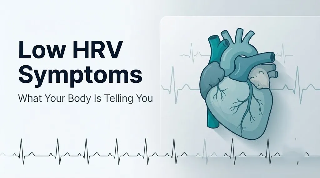
Low HRV Symptoms: What Your Body Is Telling You
Low HRV symptoms include persistent fatigue, disrupted sleep, difficulty concentrating, heightened anxiety, slow physical recovery, and an elevated resting heart rate. These are not arbitrary associations: each symptom traces directly to a specific disruption in how your autonomic nervous system regulates cardiac rhythm, immune function, and the stress response.
Heart rate variability is recognized as one of the most clinically informative non-invasive measures of cardiovascular and autonomic nervous system health available today. According to the landmark 1996 Task Force publication by the European Society of Cardiology and the North American Society of Pacing and Electrophysiology (ESC/NASPE), published in Circulation, reduced HRV is an independent predictor of cardiovascular mortality and identifies individuals with impaired autonomic cardiac regulation, a condition associated with significantly worse outcomes across multiple disease states.
This article covers what low HRV actually feels like at the symptom level, the physiological mechanisms that produce each symptom, how causes ranging from poor sleep to type 2 diabetes alter HRV, how the picture changes with age, sex, and fitness level, and the specific signs that mean your low HRV reading warrants professional medical evaluation rather than a lifestyle tweak.
What Is HRV and How Does It Work
Heart rate variability is the measurable variation in time between consecutive heartbeats, specifically between successive R-wave peaks on an electrocardiogram, which clinicians call R-R interval variability. A healthy heart does not beat with metronomic precision. The interval between beats changes subtly with every breath, movement, and demand on the cardiovascular system, and the degree of that variation is what HRV quantifies.
The sinoatrial (SA) node, located in the right atrium, sets the base firing rate of the heart. What determines how much that rate varies moment to moment is the balance between two competing branches of the autonomic nervous system: the parasympathetic system (operating primarily through the vagus nerve, cranial nerve X) and the sympathetic system (operating through thoracic cardiac sympathetic fibers). Parasympathetic activation via the vagus nerve releases acetylcholine onto cardiac muscarinic receptors at the SA node, slowing firing and increasing variability between beats. Sympathetic activation releases norepinephrine onto beta-1 adrenergic receptors at the SA node, accelerating firing and reducing variability.
Think of it like the tension between a car’s accelerator and brake. High HRV means both pedals are engaged responsively. Low HRV means the accelerator (sympathetic drive) is stuck partially down and the brake (parasympathetic tone) is disengaged.
HRV is measured using time-domain statistics. The two most clinically referenced are SDNN (standard deviation of all normal-to-normal R-R intervals over a 24-hour period) and rMSSD (root mean square of successive differences between adjacent R-R intervals), which reflects short-term vagal tone and is the metric most wearable devices use. Frequency-domain analysis separates HRV into high-frequency (HF) power, reflecting parasympathetic activity, and low-frequency (LF) power, reflecting a mix of sympathetic and parasympathetic influence.
| HRV Metric | What It Measures | Clinical Reference |
|---|---|---|
| SDNN | Total autonomic variability over 24 hours | ESC/NASPE: SDNN below 50ms associated with poor prognosis |
| rMSSD | Short-term vagal tone, beat-to-beat variation | Lower rMSSD = reduced parasympathetic activity |
| HF Power | High-frequency band: pure parasympathetic activity | Reduced HF = parasympathetic withdrawal |
| LF Power | Low-frequency band: mixed autonomic influence | Elevated LF/HF ratio = sympathetic dominance |
For individuals with pre-existing cardiac arrhythmias such as atrial fibrillation, standard HRV metrics are unreliable because R-R intervals are inherently irregular from the arrhythmia itself, not from genuine autonomic variability. Any HRV reading from a wearable device in someone with known atrial fibrillation requires clinical interpretation by a cardiologist.
What Counts as Low HRV
Low HRV is not a single absolute threshold: it is always interpreted relative to an individual’s age-matched reference range, their baseline trend, and the context in which the measurement was taken. This is one of the most misunderstood aspects of HRV in consumer health technology.
According to the ESC/NASPE Task Force standards, clinical HRV measured from a 24-hour Holter ECG recording uses SDNN as the primary reference. An SDNN below 50 milliseconds is associated with poor cardiovascular prognosis. An SDNN of 50 to 100 milliseconds falls in a borderline range warranting monitoring. An SDNN above 100 milliseconds is considered normal.
Consumer wearable devices use rMSSD derived from photoplethysmography (PPG) rather than ECG, which introduces measurement variability. Published population data from several large device-based studies suggests that average rMSSD values from wearables range from approximately 20 to 40 ms in middle-aged adults with typical fitness levels, rising to 50 to 80 ms or higher in aerobically trained individuals, and declining progressively with age. A value significantly below your own personal 30-day rolling average on a wearable is clinically more meaningful than comparison to any population norm.
| HRV Context | What “Low” Means Practically | Clinical Weight |
|---|---|---|
| Single night below your average | Likely reflects poor sleep, alcohol, or acute stress | Low: monitor, do not act |
| 5 to 7 consecutive days below average | Suggests sustained physiological stress or early illness | Moderate: assess causes |
| Weeks to months consistently low | May indicate chronic disease, overtraining, or cardiac condition | High: clinical evaluation warranted |
| Low SDNN on 24-hour Holter ECG | Established clinical marker with documented outcome data | Very High: interpret with cardiologist |
Wearable accuracy also varies by device. A 2021 analysis in the Journal of Medical Internet Research found that wrist-based PPG devices had acceptable accuracy for rMSSD during sleep but showed greater error during movement or in individuals with darker skin tones, due to optical signal interference. People using wearables should treat their HRV data as a directional trend, not a clinical measurement equivalent to an ECG-based recording.
Low HRV Symptoms: The Complete Clinical Picture
Low HRV symptoms represent the measurable consequences of sustained sympathetic nervous system dominance and parasympathetic withdrawal across multiple body systems simultaneously. They are not random: each symptom has a specific physiological origin.
The core symptom cluster includes:
- Persistent fatigue that does not resolve with adequate sleep, produced by impaired autonomic regulation of mitochondrial energy metabolism and reduced parasympathetic-driven cellular recovery processes
- Sleep disturbances, particularly difficulty entering or maintaining slow-wave sleep and REM sleep, caused by inadequate parasympathetic activation during sleep onset
- Difficulty concentrating and brain fog, produced by reduced cerebral blood flow variability and blunted prefrontal cortex activity under sustained sympathetic drive
- Elevated resting heart rate, a direct consequence of reduced vagal tone on the SA node allowing sympathetic tone to drive heart rate upward
- Heightened anxiety and emotional reactivity, produced by reduced vagal brake function on the amygdala and limbic system
- Slow physical recovery from exercise or illness, because parasympathetic tone drives protein synthesis, anti-inflammatory signaling, and glycogen replenishment
- Increased susceptibility to illness, because vagal activity directly suppresses inflammatory cytokine release through the cholinergic anti-inflammatory pathway
- Palpitations or awareness of heartbeat, because reduced beat-to-beat variability makes the heart’s rhythm feel more regular and noticeable
- Orthostatic symptoms (lightheadedness when standing), seen particularly when autonomic dysregulation affects blood pressure responses
The severity and number of symptoms depend heavily on how long HRV has been suppressed, the underlying cause, and the individual’s baseline autonomic reserve.
What Does Low HRV Feel Like Physically
The physical experience of low HRV is most often described as a persistent low-grade exhaustion combined with a sense that the body is running at higher than normal effort for routine tasks. People who track HRV consistently through wearables often notice that on low HRV days, they feel “off” before they can articulate why, reflecting that the autonomic state is physically experienced even when not consciously analyzed.
Physically, the dominant sensations associated with sustained low HRV include:
Fatigue that feels different from ordinary tiredness. It does not improve substantially after a night of sleep. This quality reflects impaired parasympathetic-driven restorative processes rather than simple sleep debt. The parasympathetic nervous system drives cellular repair via its influence on the hypothalamic sleep centers and its role in modulating growth hormone secretion during slow-wave sleep.
A sense of chest tightness or awareness of heartbeat at rest. When beat-to-beat variability is low, the rhythm of the heart becomes more uniform and more perceptible. This is not the same as a pathological arrhythmia, but it can feel alarming and produce secondary anxiety. People with pre-existing anxiety disorders may misattribute these sensations and experience a feedback loop that further reduces vagal tone.
Difficulty with cognitive tasks that require sustained attention. The prefrontal cortex, which governs working memory, impulse control, and executive function, requires adequate cardiac vagal activity for optimal performance. A 2018 study published in Frontiers in Neuroscience found that rMSSD positively predicted prefrontal cortex activation during sustained attention tasks, consistent with the known role of vagal tone in top-down cognitive regulation.
For older adults over 65, low HRV physical symptoms may be more subtle and overlap substantially with age-related fatigue and cognitive slowing, making the pattern harder to attribute specifically to autonomic dysfunction without clinical evaluation.
Quick Tip:
- Track your resting heart rate alongside HRV: a rising resting heart rate combined with a falling HRV trend over five or more days is a more meaningful signal than HRV alone.
- Note whether symptoms are worst in the morning (suggesting overnight sympathetic activation) or persistent throughout the day (suggesting chronic baseline autonomic imbalance).
- For those with hypothyroidism or taking beta-blocker medications, HRV readings may be artificially elevated or suppressed independent of true autonomic tone, and interpretation requires physician guidance.
Low HRV and the Autonomic Nervous System
Low HRV and the autonomic nervous system are inseparable: a reduced HRV reading is, at its core, a direct measurement of reduced autonomic nervous system flexibility, specifically a dominance of sympathetic tone over parasympathetic (vagal) tone.
The autonomic nervous system regulates every involuntary function your body manages: heart rate, blood pressure, digestion, immune response, pupil diameter, glandular secretion, and thermoregulation. Its two primary divisions pull in opposite directions. The sympathetic division mobilizes the body for action, raising heart rate, redirecting blood flow to skeletal muscle, releasing cortisol and epinephrine from the adrenal glands, and suppressing digestive and immune functions. The parasympathetic division, dominated by vagal activity, reverses this pattern: slowing the heart, promoting digestion, modulating immune inflammatory responses, and restoring tissue homeostasis.
The vagus nerve is the primary driver of parasympathetic cardiac activity. Its fibers synapse directly onto the SA node and AV node of the heart, and the moment-to-moment variation in vagal firing rate produces the beat-to-beat interval variability that HRV measures. When vagal tone is high, HRV is high. When the hypothalamic-pituitary-adrenal (HPA) axis is chronically activated by stress, illness, or disease, cortisol and sympathetic drive suppress vagal tone, and HRV drops.
According to the National Institutes of Health National Heart, Lung, and Blood Institute (NHLBI), the autonomic nervous system plays a central role in cardiovascular homeostasis, and impaired autonomic modulation is recognized as a predictor of adverse cardiac outcomes across multiple disease categories. This is the clinical foundation for using HRV as a health metric beyond fitness tracking.
The autonomic nervous system also operates a mechanism called the cholinergic anti-inflammatory pathway, in which vagal acetylcholine release suppresses macrophage production of inflammatory cytokines including interleukin-6 (IL-6) and tumor necrosis factor-alpha (TNF-alpha). When vagal tone drops and HRV falls, this pathway is less active, and low-grade systemic inflammation tends to rise. This mechanistically connects low HRV to the inflammatory symptoms discussed in later sections.
Key Takeaway: Low HRV is a direct window into how well your autonomic nervous system is balancing sympathetic and parasympathetic activity, and when that balance tips toward sustained sympathetic dominance, the downstream effects touch nearly every major organ system in the body.
Low HRV and Fatigue
Low HRV and fatigue are physiologically linked through at least three distinct mechanisms, none of which involves simple overexertion. Understanding which mechanism is driving fatigue in a given person changes both what they should do about it and how seriously they should take it.
Mechanism 1: Impaired parasympathetic-driven recovery. During rest and sleep, the parasympathetic nervous system drives tissue repair, glycogen replenishment, and protein synthesis. When vagal tone is reduced and HRV is low, these repair processes are less efficiently activated. The result is fatigue that persists despite rest, which is clinically distinct from fatigue caused by inadequate sleep or caloric deficit. A 2019 study published in the International Journal of Environmental Research and Public Health found that lower rMSSD values were independently associated with perceived fatigue scores in working adults, even after controlling for sleep duration and physical activity levels.
Mechanism 2: Sustained HPA axis activation and cortisol burden. Chronically elevated cortisol from HPA axis hyperactivity directly suppresses parasympathetic tone. It also promotes catabolic metabolism (breaking down tissue rather than building it) and dysregulates the sleep-wake cycle. The fatigue produced by this mechanism has a characteristic pattern: moderate energy in the morning, a pronounced energy crash in the early afternoon, and difficulty feeling genuinely rested even after a full night of sleep.
Mechanism 3: Inflammatory cytokine-driven fatigue. With reduced vagal anti-inflammatory activity, circulating levels of IL-6 and TNF-alpha tend to rise. Both cytokines act centrally in the brain to produce “sickness behavior,” a conserved biological response that includes fatigue, reduced motivation, and cognitive slowing. This mechanism is particularly active in individuals whose low HRV accompanies a chronic illness or persistent low-grade infection.
People with fibromyalgia, chronic fatigue syndrome (myalgic encephalomyelitis), or lupus often have measurably reduced HRV alongside their characteristic fatigue, reflecting shared autonomic and inflammatory dysregulation rather than a coincidental association.
Low HRV and Sleep Problems
Low HRV and disrupted sleep share a bidirectional relationship: poor sleep lowers HRV, and low HRV impairs sleep quality, creating a cycle that is more difficult to break than either problem in isolation.
The mechanism starts at the level of sleep architecture. Healthy sleep cycles alternate between non-REM stages (including slow-wave or deep sleep) and REM sleep. Both slow-wave sleep and REM sleep are parasympathetically dominant states. The heart rate slows, beat-to-beat variability increases (HRV rises), and the body devotes maximum resources to physical and neural restoration. When parasympathetic tone is chronically reduced, this shift into parasympathetic dominance during sleep is blunted. The result is fragmented sleep with reduced time in slow-wave and REM stages.
Research published in Sleep Medicine found that healthy subjects showed significantly higher rMSSD during slow-wave sleep compared to wakefulness or light sleep, confirming that the depth and restorative quality of sleep are directly tied to vagal activation. Subjects with chronically low nocturnal HRV showed less total slow-wave sleep and reported poorer subjective sleep quality even when total sleep duration appeared adequate.
Sleep apnea is a specific condition that dramatically suppresses HRV through repeated sympathetic surges triggered by apneic episodes (brief cessations of breathing during sleep). Each apneic event triggers a cortisol and catecholamine release as the brain responds to falling oxygen saturation. According to the American Heart Association, obstructive sleep apnea is associated with measurably reduced 24-hour HRV, elevated nocturnal heart rate, and increased cardiovascular risk. Individuals with chronically low HRV who also experience snoring, morning headaches, or excessive daytime sleepiness should discuss sleep apnea evaluation with their primary care physician.
People who use alcohol as a sleep aid should note that alcohol acutely suppresses REM sleep and sympathetically activates the cardiovascular system during the second half of the night, producing a characteristic HRV dip visible on wearables the following morning.
Key Takeaway: Poor sleep and low HRV reinforce each other through parasympathetic withdrawal during sleep stages. Addressing sleep quality directly, rather than monitoring HRV scores alone, is one of the fastest routes to measurable autonomic improvement.
Low HRV and Anxiety and Stress
Low HRV and anxiety have a well-documented, mechanistically grounded relationship that runs in both directions. Chronic psychological stress suppresses vagal tone and reduces HRV. And low vagal tone removes what researchers call the “vagal brake,” the parasympathetic inhibition that normally dampens emotional reactivity in the amygdala and limbic system.
The amygdala, the brain’s threat-detection center, is tonically inhibited by vagal afferent signals transmitted through the nucleus tractus solitarius in the brainstem to prefrontal structures that regulate emotional response. When vagal tone is high, this inhibitory pathway is active: the prefrontal cortex can effectively downregulate amygdala reactivity, and the physiological stress response is dampened and well-timed. When vagal tone drops and HRV falls, this top-down inhibitory control weakens. The amygdala becomes more reactive to perceived threats, the threshold for triggering the sympathetic stress response drops, and anxiety symptoms intensify.
A 2019 meta-analysis published in Neuroscience and Biobehavioral Reviews examining 172 studies found that lower resting HRV was consistently associated with greater anxiety and reduced emotional regulation capacity across multiple populations and anxiety disorder types, including generalized anxiety disorder (GAD), social anxiety disorder, and post-traumatic stress disorder (PTSD). The association held even when accounting for depression comorbidity.
The physical symptoms that overlap between low HRV and anxiety are worth identifying:
- Elevated resting heart rate (both share sympathetic dominance as the mechanism)
- Fatigue (both share HPA axis activation and poor sleep quality as drivers)
- Cognitive difficulty (both share prefrontal cortex impairment under sustained stress)
- Palpitation awareness (both share increased cardiac sympathetic tone)
Because the symptom overlap is substantial, it is not always clear from self-assessment alone whether anxiety symptoms are driving low HRV or whether autonomic dysregulation from another cause (illness, cardiovascular disease, metabolic condition) is producing the anxiety-like picture. A primary care physician can help distinguish these patterns and refer to the appropriate specialist: a cardiologist for cardiac causes, or a psychiatrist or clinical psychologist for anxiety or mood disorder evaluation.
For anyone who experiences intrusive thoughts of self-harm or suicide alongside these symptoms, this is not a sign of weakness and does not need to be faced alone. Contact the 988 Suicide and Crisis Lifeline by calling or texting 988 at any time. This service is free, confidential, and available 24 hours a day, 7 days a week.
Low HRV Causes
Low HRV has a range of causes spanning the spectrum from completely normal and temporary to clinically serious and requiring medical management. The physiological mechanism by which each cause suppresses HRV is distinct, and knowing which cause applies to a given person changes both the timeline for recovery and the appropriate response.
| Cause | Mechanism | Duration | Clinical Urgency |
|---|---|---|---|
| Acute psychological stress | HPA axis cortisol release suppresses vagal tone | Hours to days | Low: monitor |
| Alcohol consumption | Sympathetic rebound in second half of night, REM suppression | 12 to 24 hours | Low: resolves with abstinence |
| Acute illness or infection | Inflammatory cytokines (IL-6, TNF-alpha) suppress vagal activity | Days to weeks | Low to moderate: monitor recovery |
| Sleep deprivation | Reduced parasympathetic overnight recovery, cortisol elevation | Days | Low: resolves with sleep |
| Overtraining | Sustained sympathetic drive without adequate recovery | Weeks | Moderate: rest and recovery protocol |
| Chronic psychological stress | Sustained HPA axis hyperactivation | Weeks to months | Moderate: clinical assessment warranted |
| Type 2 diabetes | Peripheral autonomic nerve damage (cardiovascular autonomic neuropathy) | Progressive | High: endocrinologist evaluation |
| Cardiovascular disease | Cardiac autonomic remodeling post-MI, heart failure autonomic dysregulation | Persistent | High: cardiology evaluation |
| Major depressive disorder | Chronic HPA activation, reduced vagal tone via limbic-autonomic pathways | Variable | High: psychiatric and medical evaluation |
| Medications (beta-blockers, anticholinergics) | Direct pharmacological alteration of autonomic tone | Duration of use | Low to moderate: physician guidance |
Medications deserve specific attention. Beta-adrenergic blocking agents (beta-blockers) reduce sympathetic drive and can artificially alter HRV readings in ways that do not reflect true autonomic health. Anticholinergic medications (including some antihistamines, bladder medications, and tricyclic antidepressants) reduce parasympathetic tone and can suppress HRV. Anyone taking these medication classes should discuss the interpretation of their HRV readings with their prescribing physician before drawing clinical conclusions from wearable data.
Low HRV and Inflammation
Low HRV and systemic inflammation are connected through a specific biological pathway called the cholinergic anti-inflammatory pathway, first described in detail by Dr. Kevin Tracey and colleagues in research published in Nature in 2002. This pathway establishes a direct mechanistic link between vagal tone, acetylcholine, and the regulation of inflammatory cytokine production.
Here is how it works. Vagal nerve fibers innervate the spleen and other immune organs. Acetylcholine released at these sites binds to alpha-7 nicotinic receptors on macrophages and dendritic cells. This binding suppresses intracellular activation of nuclear factor kappa B (NF-kB), a master transcription factor that drives production of IL-6, TNF-alpha, and other pro-inflammatory cytokines. When vagal tone is high and HRV is elevated, this anti-inflammatory brake is consistently active. When vagal tone falls and HRV drops, the brake releases, and inflammatory cytokine production rises.
According to research published in the Journal of the American Medical Association (JAMA), lower HRV is significantly associated with elevated high-sensitivity C-reactive protein (hs-CRP), a systemic inflammatory marker, in population-based studies. This association holds after adjusting for body mass index, smoking status, and physical activity level, suggesting the autonomic-inflammatory connection is not simply mediated by lifestyle factors.
The practical clinical implication is that chronically low HRV may contribute to a state of low-grade systemic inflammation that compounds risk for conditions including atherosclerosis, type 2 diabetes, rheumatoid arthritis, and possibly certain cancers. This is not proven causation from a single direction: inflammation itself suppresses vagal tone through central mechanisms involving cytokine signaling to the brainstem, creating a bidirectional loop.
Individuals with autoimmune inflammatory conditions (rheumatoid arthritis, lupus, inflammatory bowel disease) frequently show lower HRV during disease flares compared to remission periods, a clinically consistent pattern that may eventually serve as a non-invasive monitoring tool under physician guidance.
Key Takeaway: Low HRV is not just a fitness metric. Through the cholinergic anti-inflammatory pathway, reduced vagal tone has measurable effects on immune regulation and inflammatory status that can compound cardiovascular and metabolic disease risk over time.
Low HRV in Athletes and Overtraining
In aerobically trained athletes, HRV typically runs higher than population norms because sustained endurance training increases vagal tone through cardiac autonomic adaptation, an effect well-documented in the sports medicine literature. This makes athletes both better positioned to use HRV as a training guide and more vulnerable to misinterpreting a drop in their HRV.
Overtraining syndrome is a clinical condition in which training load chronically exceeds the body’s capacity for recovery, producing a sustained sympathetic nervous system dominance (the sympathetic or “Basedow” form of overtraining) or, less commonly, a parasympathetic dominance pattern (“Addison” form). The sympathetic form is characterized by:
- HRV falling progressively below the athlete’s own 30-day baseline
- Elevated resting heart rate (often 5 to 10 beats per minute above normal baseline)
- Persistent fatigue that does not resolve with normal rest periods
- Reduced performance despite maintained or increased training effort
- Irritability, mood instability, and increased injury rate
- Disturbed sleep with difficulty falling asleep despite fatigue
A 2020 systematic review published in the European Journal of Sport Science concluded that daily morning HRV monitoring over periods of four or more weeks reliably detected overreaching (a precursor to overtraining syndrome) before performance decrements were subjectively apparent, supporting HRV-guided training as a practical tool in competitive athletes.
The challenge is distinguishing functional overreaching (short-term, resolves with one to two weeks of reduced training) from non-functional overreaching (resolves over weeks to months) and true overtraining syndrome (may require months of reduced activity and clinical evaluation). A sports medicine physician should evaluate any athlete whose HRV remains consistently and significantly suppressed for more than two to three weeks despite appropriate rest, or who develops systemic symptoms including unexplained weight loss, hormonal irregularities, or immune suppression.
Female athletes should note that HRV is particularly sensitive to the physiological stress of Relative Energy Deficiency in Sport (RED-S), a condition in which chronic caloric restriction relative to training load suppresses reproductive hormones and autonomic function simultaneously. A board-certified sports medicine physician or sports dietitian should be consulted if low HRV accompanies irregular or absent menstrual cycles in female athletes.
Low HRV and Chronic Disease: Diabetes, Heart Disease, and Depression
Three chronic disease states are associated with the most clinically severe and mechanistically distinct forms of low HRV: type 2 diabetes mellitus with cardiovascular autonomic neuropathy, established cardiovascular disease, and major depressive disorder.
Type 2 Diabetes and Cardiovascular Autonomic Neuropathy (CAN). In type 2 diabetes, chronic hyperglycemia causes oxidative stress and advanced glycation end-product accumulation that damage small autonomic nerve fibers throughout the body, including the cardiac autonomic fibers that regulate HRV. The result is cardiovascular autonomic neuropathy (CAN), a condition characterized by markedly reduced HRV, resting tachycardia, orthostatic hypotension, exercise intolerance, and increased risk of sudden cardiac death. According to the American Heart Association, individuals with diabetes and confirmed CAN have a significantly elevated risk of silent myocardial infarction and cardiovascular mortality compared to diabetic individuals without autonomic neuropathy. CAN is evaluated clinically through a battery of autonomic function tests including standing-to-lying R-R interval ratio, Valsalva maneuver ratio, and blood pressure response to standing, administered by a cardiologist or neurologist specializing in autonomic disorders.
Cardiovascular Disease. Post-myocardial infarction, HRV is dramatically reduced due to cardiac autonomic remodeling: the infarcted myocardium disrupts sympathetic and parasympathetic innervation patterns, and the surviving myocardium often operates under elevated sympathetic drive. The ESC/NASPE Task Force in its landmark 1996 publication established that SDNN below 50ms in post-MI patients carries a relative risk of mortality more than five times that of patients with SDNN above 100ms, making 24-hour HRV one of the strongest non-invasive predictors of post-MI outcomes in clinical cardiology.
Major Depressive Disorder. Depression is consistently associated with reduced HRV across multiple prospective studies. The mechanism involves sustained HPA axis activation, limbic-to-brainstem pathways that suppress the vagal cardiac efferents, and inflammatory cytokine burden from depressive illness. A meta-analysis published in Biological Psychiatry found that patients with major depressive disorder showed significantly lower rMSSD compared to matched healthy controls, with severity of depression correlating inversely with HRV magnitude.
Low HRV in Women Versus Men
The relationship between biological sex and HRV is more clinically nuanced than most health content acknowledges. Women and men show systematically different HRV patterns across the lifespan, driven by distinct hormonal, anatomical, and physiological factors.
Premenopausal women, on average, show slightly lower absolute HRV than age-matched men in some time-domain metrics, but higher HF power (parasympathetic activity) in frequency-domain analysis. The predominant reason is that estrogen has direct parasympathomimetic effects on cardiac autonomic regulation, enhancing vagal tone through estrogen receptor pathways in the autonomic nuclei of the brainstem. Progesterone has modest sympathoexcitatory effects and may partly counterbalance estrogen’s vagal enhancement during the luteal phase of the menstrual cycle.
This means that female HRV naturally fluctuates across the menstrual cycle in a predictable pattern: higher rMSSD during the follicular phase when estrogen is rising, and a measurable dip in the luteal phase when progesterone is dominant. A drop in HRV during the luteal phase is not a sign of illness. It is a normal hormonal effect. Women who track HRV continuously should interpret their readings in the context of their cycle phase to avoid misattributing normal hormonal variation as a health problem.
At menopause, estrogen declines sharply, and with it, the parasympathomimetic protection it conferred. Post-menopausal women show substantially reduced HRV compared to their premenopausal baseline, and this reduction is partially responsible for the well-documented increase in cardiovascular risk that occurs after menopause. A 2021 study published in Menopause: The Journal of The Menopause Society found that postmenopausal women with lower HRV had significantly higher rates of hypertension and adverse lipid profiles compared to postmenopausal women with preserved HRV.
Men show higher absolute HRV in some metrics during middle age but also demonstrate a steeper age-related HRV decline and higher rates of overt cardiovascular disease, which contributes additional autonomic suppression. For both sexes, chronically low HRV in the context of cardiovascular risk factors warrants discussion with a primary care physician.
Low HRV and Aging
HRV declines with age in virtually all studied populations, making age the single most predictable and universal biological modifier of HRV. This decline is not random: it reflects progressive changes in autonomic nervous system structure and function that occur across the human lifespan.
The primary mechanisms of age-related HRV decline include:
- Progressive reduction in SA node intrinsic firing flexibility due to fibrotic changes in the cardiac conduction system
- Reduced vagal efferent firing rate from age-related neurodegeneration in the dorsal motor nucleus of the vagus and nucleus ambiguus in the brainstem
- Blunted baroreflex sensitivity, meaning the arterial baroreceptors become less responsive to blood pressure changes, reducing the dynamic autonomic adjustments that produce HRV
- Elevated baseline systemic inflammation in older adults (a state sometimes called “inflammaging”) that tonically suppresses vagal tone through central cytokine signaling
- Higher prevalence of comorbidities (hypertension, type 2 diabetes, coronary artery disease, sleep apnea) that independently suppress HRV
According to data from the Framingham Heart Study, published in Circulation, mean SDNN declines by approximately 3 to 5 milliseconds per decade from age 40 onward, with sharper declines in individuals with metabolic risk factors. A 70-year-old healthy adult will typically have an HRV substantially below that of a healthy 30-year-old, and interpreting an older adult’s HRV against population-wide averages or against their younger self without age-adjustment is a common and clinically misleading error.
For older adults over 65, symptoms that in a younger person might simply reflect a transient HRV suppression (persistent fatigue, orthostatic lightheadedness, exercise intolerance) require more prompt clinical evaluation because the threshold for underlying cardiovascular or autonomic disease is lower. A primary care physician should evaluate persistent fatigue, new orthostatic symptoms, or unexplained resting tachycardia in this age group, with cardiology referral if autonomic testing or cardiac imaging is indicated.
Key Takeaway: HRV declines predictably with age through well-understood mechanisms in the cardiac conduction system and autonomic brainstem centers, meaning older adults need age-adjusted HRV interpretation and a lower threshold for clinical evaluation of symptoms.
When Is Low HRV Dangerous
Low HRV is dangerous, in the clinical sense, when it reflects an underlying condition that carries independent cardiovascular mortality risk or when it accompanies acute symptoms suggesting cardiac or neurological compromise. The key is distinguishing normal, transient HRV suppression (which is common and self-resolving) from pathological chronic or acute low HRV (which requires medical evaluation).
Clinically serious patterns include:
- HRV that has been consistently and severely suppressed for more than three to four weeks without a clear, reversible cause (illness, travel, heavy training load)
- Low HRV accompanied by new resting tachycardia (heart rate above 100 beats per minute at rest) in someone without a prior history of this
- Low HRV in a person with known type 2 diabetes, coronary artery disease, or heart failure, where autonomic suppression carries documented mortality implications
- Low HRV accompanying orthostatic hypotension (blood pressure dropping more than 20 mmHg systolic on standing) with dizziness or near-fainting
- Low HRV in an athlete who is not recovering despite adequate rest reduction over two or more weeks
- Low HRV accompanied by unexplained weight loss, night sweats, or systemic symptoms suggesting an occult illness
According to research published in The Lancet, reduced 24-hour HRV (SDNN below 70ms) was an independent predictor of all-cause mortality and specifically cardiac mortality in prospective cohort studies of middle-aged adults, even in the absence of clinically apparent heart disease at baseline.
The most important clinical fact is this: low HRV on a wearable device is not a diagnosis, and it is not an emergency alert. It is a signal that warrants attention. What makes it dangerous is not the number itself but the clinical context surrounding it. When in doubt, a primary care physician is the appropriate first contact for a conversation about persistent low HRV in the context of symptoms.
Emergency Symptoms: When to Call 911 or Go to the ER
Certain symptoms associated with low HRV, specifically those reflecting severe autonomic dysregulation, acute cardiac arrhythmia, or cardiovascular decompensation, require immediate emergency evaluation. Do not wait to see if these resolve on their own.
Call 911 or go to the nearest emergency room immediately if you experience:
- Chest pain or chest pressure at rest, particularly if radiating to the arm, jaw, neck, or back
- Severe palpitations with dizziness, lightheadedness, or near-fainting, which may indicate a serious cardiac arrhythmia requiring immediate ECG evaluation
- Sudden loss of consciousness or syncope, even briefly, without a clear non-cardiac cause
- Shortness of breath at rest that is new, worsening, or accompanied by chest tightness
- A sensation that your heart is racing or skipping beats combined with cold sweats, nausea, or pallor
- Severe orthostatic symptoms (profound dizziness or fainting upon standing) that do not resolve within seconds, particularly in someone with known diabetes or cardiovascular disease
- Heart rate persistently above 130 beats per minute at rest without obvious cause such as fever or acute emotional distress
These presentations can indicate acute myocardial infarction, life-threatening cardiac arrhythmia, or severe autonomic cardiovascular crisis, all of which require emergency medical assessment and cardiac monitoring, not an urgent care appointment or a call back from a nurse line.
How to Improve Low HRV
Improving low HRV is possible through specific, evidence-grounded interventions that target the underlying physiological mechanism: restoring parasympathetic tone and reducing sustained sympathetic drive. The speed and degree of improvement depend heavily on the cause.
Evidence-supported approaches:
1. Slow, paced breathing at approximately 5 to 6 breaths per minute. This respiratory rate produces resonance with the cardiovascular system’s natural baroreflex frequency, maximally amplifying vagal cardiac activity. A 2017 randomized controlled trial in Frontiers in Physiology found that six weeks of daily slow-paced breathing (5 breaths per minute, 20 minutes daily) produced statistically significant increases in rMSSD and SDNN compared to control. This is the single most accessible, evidence-backed intervention for acutely and chronically increasing vagal tone.
2. Aerobic exercise at moderate intensity, consistently over weeks to months. Endurance training increases vagal tone through cardiac autonomic adaptation. The effect requires sustained, regular training over at least six to eight weeks before robust HRV improvements appear. Acute intense exercise temporarily suppresses HRV before improving it through recovery.
3. Adequate and consistent sleep. Targeting seven to nine hours of sleep nightly in a cool, dark environment restores nocturnal parasympathetic activity. Treating underlying sleep apnea (via CPAP or mandibular advancement device, guided by a pulmonologist or sleep medicine physician) produces measurable HRV improvements in affected individuals.
4. Reducing alcohol consumption. Even moderate alcohol consumption acutely suppresses HRV for 12 to 24 hours after ingestion. Elimination or substantial reduction produces measurable HRV recovery within one to two weeks in most individuals.
5. Cold water exposure (cold showers or cold face immersion). The diving reflex, activated by cold water on the face, produces an acute vagal surge that transiently increases HRV. While the chronic effect is less robustly studied, preliminary research supports regular cold exposure as a vagal tone training tool.
6. Biofeedback therapy with HRV-specific protocols, delivered by a trained psychologist or physiatrist, is well-studied for autonomic regulation in anxiety disorders, post-MI recovery, and chronic pain conditions. The Association for Applied Psychophysiology and Biofeedback recognizes HRV biofeedback as an evidence-based treatment for stress-related conditions.
For individuals whose low HRV reflects an underlying medical condition (diabetic autonomic neuropathy, cardiovascular disease, major depressive disorder, sleep apnea), lifestyle interventions alone are insufficient. Medical management of the underlying condition is the primary pathway to HRV improvement, and the appropriate specialist should lead that care.
Frequently Asked Questions About Low HRV Symptoms
What does it mean if your HRV is low?
Low HRV means your heart’s beat-to-beat interval variability is reduced, reflecting reduced parasympathetic (vagal) activity and relatively increased sympathetic nervous system dominance over cardiac regulation.
This pattern is associated with increased physiological stress, slower recovery, poorer sleep quality, and, in chronic form, higher risk for cardiovascular and metabolic disease.
A single low HRV reading is usually not clinically meaningful; a sustained trend below your personal baseline over days to weeks warrants attention and, if accompanied by symptoms, physician evaluation.
What symptoms can you feel when your HRV is low?
When HRV is low, the most commonly reported physical symptoms include persistent fatigue, poor or unrefreshing sleep, difficulty concentrating, elevated resting heart rate, heightened anxiety, and slow recovery after exercise or illness.
These symptoms trace directly to the physiological consequences of reduced vagal tone and increased sympathetic drive on cardiac, immune, and neurological function.
Not everyone with low HRV experiences noticeable symptoms, particularly if the suppression is mild and transient.
Is a low HRV score dangerous?
A low HRV score is not inherently dangerous in isolation, but chronically low HRV measured over weeks or months, particularly in the context of cardiovascular disease, type 2 diabetes, or significant cardiac symptoms, carries documented clinical risk supported by prospective cohort data.
The ESC/NASPE Task Force established that 24-hour SDNN below 50ms is associated with significantly poor cardiovascular prognosis in clinical populations.
A single low HRV reading from a wearable device during a week of poor sleep or illness does not carry the same clinical weight as persistently reduced HRV on clinical ECG monitoring.
Can low HRV cause anxiety?
Low HRV does not directly “cause” anxiety in the traditional sense, but reduced vagal tone removes the parasympathetic brake on amygdala reactivity, which lowers the threshold for the physiological stress response and worsens anxiety symptoms.
A 2019 meta-analysis in Neuroscience and Biobehavioral Reviews found consistent inverse associations between resting HRV and anxiety severity across 172 studies.
The relationship is bidirectional: anxiety disorders also reduce vagal tone and HRV through sustained HPA axis activation and sympathetic nervous system engagement.
What is a dangerously low HRV number?
In clinical cardiac medicine, a 24-hour SDNN below 50 milliseconds measured on a Holter ECG is the established threshold associated with significantly poor cardiovascular prognosis, per ESC/NASPE standards.
On consumer wearables using rMSSD, there is no universal “dangerous” absolute number because population norms vary substantially by age, sex, and fitness level, making personal baseline trend analysis more meaningful than absolute values.
Any wearable-derived HRV reading that is consistently well below your personal rolling average, accompanied by cardiac symptoms, should be discussed with a primary care physician who can order clinical autonomic and cardiac testing.
How quickly can you improve your HRV?
Transient HRV suppression from alcohol, poor sleep, or acute illness typically resolves within 24 to 72 hours after the triggering factor is removed.
Structural HRV improvement from consistent aerobic training, slow-paced breathing practice, or sleep optimization typically appears within six to twelve weeks of consistent daily application.
HRV associated with underlying chronic disease (cardiovascular autonomic neuropathy, heart failure, major depressive disorder) improves primarily through treatment of the underlying condition under physician supervision, and the timeline depends on disease severity and treatment response.
Closing
Low HRV symptoms, from fatigue and brain fog to disrupted sleep and anxiety, are not vague wellness complaints. They are the measurable downstream consequences of a specific physiological imbalance: parasympathetic withdrawal and sympathetic dominance in the autonomic nervous system. Understanding the mechanism behind each symptom gives you a far more useful framework for interpreting your body’s signals than any single HRV number on a wearable screen.
The practical steps from this article are these: track your HRV as a trend rather than an absolute score, pair it with resting heart rate and sleep quality data, address the most common modifiable causes first (alcohol, poor sleep, chronic stress), and apply slow-paced breathing daily for vagal tone support. If your low HRV is persistent, accompanied by cardiac symptoms, or occurs in the context of diabetes or cardiovascular disease, this warrants evaluation by a primary care physician, not continued self-monitoring.
Your HRV is one of the most sensitive, non-invasive windows into how your autonomic nervous system is functioning right now. Use it as a signal, not a diagnosis, and bring the trend data to your next physician appointment if the pattern concerns you.