Heel Fracture Symptoms: Signs You Should Never Ignore
Heel fracture symptoms range from immediate, severe pain with an inability to bear weight after a traumatic fall to a slow-building, activity-triggered ache that many people mistake for plantar fasciitis for weeks before a diagnosis is made. Understanding which pattern applies to your situation determines whether you need an emergency room right now or an orthopedic appointment this week.
The calcaneus, the largest of the 26 bones in the foot, is the most commonly fractured tarsal bone. According to the American Academy of Orthopaedic Surgeons, calcaneal fractures account for approximately 60 percent of all tarsal bone fractures, and high-energy injuries to this bone carry complication rates significantly higher than other foot fractures. Recognizing the specific symptom pattern early changes outcomes in measurable ways.
This article covers every clinically recognized symptom of both acute calcaneal fractures and calcaneal stress fractures, explains the anatomical and physiological reason behind each symptom, distinguishes the two injury types clearly, addresses which populations face elevated risk, and identifies the specific threshold at which heel symptoms require emergency evaluation rather than watchful waiting.
Heel Fracture Symptoms: What the Clinical Picture Actually Looks Like
Heel fracture symptoms depend on injury type. Acute calcaneal fractures from high-energy trauma produce immediate, severe, and unmistakable symptoms. Calcaneal stress fractures from repetitive loading produce a gradual, insidious pattern that worsens over days to weeks.
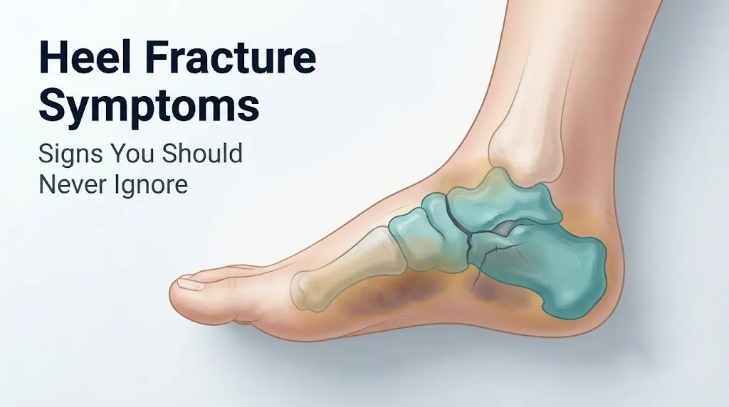
Both injury types involve the same bone: the calcaneus, a dense, roughly rectangular bone that absorbs the majority of ground reaction force with every step. Its internal structure is trabecular bone, a lattice-like cancellous network surrounded by a cortical shell. When this network fails, either suddenly or progressively, it produces a specific constellation of symptoms tied directly to the anatomy of the heel and surrounding soft tissue.
The core symptom in both types is pain localized to the heel. Beyond that shared feature, the pattern diverges in ways that a careful clinical history can usually distinguish. Pain that began the moment you landed on a hard surface from height is a different clinical picture than pain that appeared gradually during a new running program or military training.
The symptoms covered in the sections below apply specifically to the calcaneus and the structures it directly affects: the subtalar joint (the joint between the calcaneus and the talus above it), the plantar fascia (which originates at the calcaneal tuberosity), the calcaneal fat pad (the specialized tissue that cushions the heel), and the three major nerve branches that pass through or near the heel.
Key clinical point: the location of maximum tenderness and the pattern of any bruising provide the strongest early diagnostic signal before imaging is performed.
| Injury Type | Onset Pattern | Primary Symptom | Weight-Bearing Status |
|---|---|---|---|
| Acute calcaneal fracture | Immediate, after trauma | Severe heel pain, deformity possible | Usually impossible |
| Calcaneal stress fracture | Gradual, over days to weeks | Aching heel pain worse with activity | Usually preserved initially |
| Calcaneal stress reaction (pre-fracture) | Very gradual | Diffuse posterior heel ache | Preserved, worsens late in activity |
The Calcaneus: Why This Bone Fractures the Way It Does
The calcaneus fractures in predictable patterns because of its anatomy, and understanding these patterns explains exactly why the symptoms look the way they do.
The calcaneus is the primary weight-bearing bone of the heel. It sits directly beneath the talus and articulates with it at the subtalar joint, specifically at the posterior facet, the largest and most weight-bearing of the three subtalar joint facets. This joint is responsible for the inversion and eversion motion of the hindfoot. When an acute fracture displaces the posterior facet, subtalar joint pain and mechanical dysfunction become the dominant clinical features.
The bone’s internal architecture helps explain stress fracture mechanics. The trabecular network inside the calcaneus is organized into two primary compression trabeculae systems. When repetitive loading from running, marching, or jumping accelerates osteoclastic bone resorption faster than osteoblastic bone formation can replace it, micro-damage accumulates in these trabeculae. The posterior calcaneal body and the inferior cortex are the most common locations for this accumulation, which is why heel stress fracture pain typically centers under and behind the heel rather than at the plantar surface like plantar fasciitis.
Think of the trabecular structure like the internal honeycomb framework of a bridge. The bridge holds enormous loads under normal cycling because its engineers designed for gradual repair between load cycles. Exceed the load frequency before repairs can happen, and the internal framework begins to fail from the inside before any external cracking is visible.
According to the National Institute of Arthritis and Musculoskeletal and Skin Diseases, stress fractures occur most commonly in the weight-bearing bones of the lower extremity, with the calcaneus representing one of the most frequent stress fracture sites in both military and athletic populations. The Bohler angle, a measurement of calcaneal shape visible on a lateral X-ray, decreases when the trabecular architecture collapses inward under an acute fracture load, and this angle reduction is one of the earliest radiographic signs of injury.
Broken Heel Symptoms: The Immediate Signs After a Traumatic Injury
Broken heel symptoms from an acute calcaneal fracture typically begin within seconds of the injury mechanism, most commonly a fall from height, a motor vehicle accident, or a direct crush force applied to the heel.
The most immediate symptom is severe pain in the heel that is described by most patients as sudden, intense, and worsening with any attempt at weight-bearing. This pain originates from periosteal nociceptor activation: the thin membrane covering the calcaneus, the periosteum, is richly supplied with pain-sensing nerve fibers. Fracture of the cortical bone tears these nerve endings and triggers an immediate release of prostaglandins E2 and I2 along with substance P, which together produce the acute pain signal that reaches the brain within milliseconds via the medial plantar nerve, lateral plantar nerve, and sural nerve pathways.
Within minutes to hours, marked swelling develops around the entire heel and spreading toward the ankle. The calcaneal soft tissue envelope is the first structure to fill with fluid: this is the layer of subcutaneous tissue surrounding the calcaneus. Bleeding from torn periosteal blood vessels flows into this space almost immediately after fracture.
Visible deformity of the heel is present in severe fractures, particularly the tongue-type calcaneal fracture and severely displaced Sanders type III or IV intra-articular fractures. The heel may appear flattened, widened, or displaced laterally compared to the uninjured side. The heel width increases because the lateral calcaneal wall blows outward when the bone fails under axial load, impinging on the peroneal tendons that run in a groove just lateral to the calcaneus.
Key observable broken heel symptoms immediately after injury:
- Sudden severe heel pain rated 8 to 10 out of 10 on the Visual Analog Scale in most patients
- Inability to bear any weight on the affected foot
- Rapid onset of swelling around the heel and into the ankle within the first hour
- Visible bruising that develops over the first 24 to 48 hours, typically extending onto the plantar arch
- Heel widening or lateral bulging visible when compared to the other foot
- Extreme point tenderness directly over the calcaneus when pressed from any direction
Older adults deserve specific attention here. A 2019 study published in Foot and Ankle International found that calcaneal fractures in adults over 65 occur with lower-energy mechanisms, including simple ground-level falls, at substantially higher rates than in younger adults. The initial pain severity may be perceived as lower in older adults with baseline peripheral neuropathy, which can lead to delayed presentation and underestimation of injury severity.
Fractured Heel Symptoms: How Swelling and Bruising Develop and Why
The swelling and bruising pattern in a fractured heel is not random. It follows specific anatomical pathways determined by fascial planes and the structural connections between the calcaneus and the surrounding soft tissue.
Swelling in a calcaneal fracture develops from two simultaneous processes: hemorrhage from torn periosteal and intraosseous blood vessels, and an inflammatory exudate driven by the release of histamine, bradykinin, and interleukin-1 beta from damaged cells. These mediators increase vascular permeability in the capillary network surrounding the calcaneus, allowing plasma proteins and fluid to leak into the interstitial space faster than lymphatic drainage can remove them. The calcaneal fat pad, which cushions the heel beneath the calcaneus, becomes edematous and loses its normal shock-absorbing architecture within hours of a fracture.
Bruising follows the fascial planes connecting the calcaneus to the foot and ankle. The most clinically recognized pattern is Mondor’s sign, also called the plantar bruise sign of calcaneal fracture. Blood from the fracture hematoma tracks along the deep fascia of the sole and appears as dark ecchymosis along the medial and lateral borders of the plantar surface, typically visible within 24 to 48 hours. This bruising pattern is considered a reliable clinical indicator of calcaneal fracture and has been described in orthopedic literature as a bedside diagnostic finding.
Bruising also commonly extends upward into the medial ankle and lower calf as blood spreads through the fascial connections between the calcaneal periosteum and the deep posterior compartment of the leg. This upward bruise extension can mislead a clinician or patient into thinking the ankle is injured when the primary injury is the heel.
| Symptom | Mechanism | Timeline | Clinical Significance |
|---|---|---|---|
| Heel swelling | Periosteal hemorrhage, inflammatory exudate | Minutes to hours | Universal in acute fracture |
| Plantar bruising (Mondor’s sign) | Hematoma tracking along plantar fascia | 24 to 48 hours | Strong calcaneal fracture indicator |
| Medial ankle bruising | Blood spreading via deep posterior fascial plane | 48 to 72 hours | Can mimic ankle sprain, indicates calcaneal origin |
| Heel widening | Lateral calcaneal wall blowout under axial load | Immediate | Indicates Sanders type III or IV |
| Calcaneal fat pad edema | Inflammatory exudate, impaired lymphatic drainage | Hours | Worsens weight-bearing pain dramatically |
In pregnant women, the inflammatory response may present with more pronounced swelling due to baseline physiological edema and increased circulating blood volume during pregnancy. Any significant heel injury in pregnancy requires orthopedic evaluation because standard imaging protocols and some pain management approaches require modification.
Key Takeaway: Bruising that appears along the bottom of the foot or tracks up toward the ankle after a heel injury is a known clinical sign of calcaneal fracture, not ankle sprain, and warrants imaging before any weight-bearing is attempted.
Heel Stress Fracture Symptoms: The Gradual Warning Pattern
Heel stress fracture symptoms develop over a timeline of days to weeks, not seconds to minutes. This is the defining feature that separates stress fractures from acute fractures, and it is also the reason stress fractures are so frequently misidentified as plantar fasciitis, Achilles tendinopathy, or general heel bruising.
The pain of a calcaneal stress fracture typically begins as a diffuse aching discomfort in the posterior heel that is present during activity and relieves with rest. Early on, the pain may not appear until 30 to 60 minutes into a run, march, or workout. As the stress fracture progresses, the threshold shifts: pain begins earlier in the activity, then appears within the first few minutes of activity, and eventually becomes present with normal walking even on flat surfaces.
The underlying physiology explains this progression precisely. Early stress fracture pain reflects periosteal irritation at the site of micro-damage accumulation without complete cortical crack propagation. At this stage, the pain represents a stress reaction, the pre-fracture stage in which bone marrow edema is visible on MRI but no discrete fracture line is yet present. As micro-damage progresses to a true cortical crack, the pain becomes more localized, more intense, and present with less provocation.
According to a systematic review published in the Orthopaedic Journal of Sports Medicine, calcaneal stress fractures account for approximately 2 percent of all stress fractures in athletes and up to 4 percent of stress fractures in military recruits during basic training. These numbers likely underrepresent the true incidence because early-stage calcaneal stress reactions are frequently misattributed to plantar fasciitis and go unimaged at the appropriate stage.
Key progressive heel stress fracture symptoms:
- Diffuse posterior or plantar heel ache that begins during and worsens throughout activity
- Pain that initially resolves completely with rest but progressively persists longer after activity ends
- Point tenderness directly over the posterior calcaneal body that is sharper and more localized than plantar fasciitis tenderness
- Positive calcaneal squeeze test: pain produced by compressing the calcaneus from both sides simultaneously between the examiner’s palms
- Mild swelling around the posterior heel and Achilles tendon insertion area, often subtle
- Morning stiffness and start-up pain that differs from plantar fasciitis: stress fracture morning pain is centered at the body of the heel, not the plantar fascial insertion point
Calcaneal Stress Fracture Symptoms: Recognizing the Insidious Onset
Calcaneal stress fracture symptoms are insidious by definition. The injury does not announce itself with a moment of acute pain or a specific traumatic event. Instead, the symptoms accumulate alongside a training error, a new footwear change, a rapid increase in activity duration, or a period of nutritional inadequacy that compromises bone remodeling capacity.
The most clinically reliable early symptom is point tenderness directly over the posteromedial calcaneus, the area just medial to the Achilles tendon insertion on the calcaneal tuberosity. This is distinct from the point tenderness of plantar fasciitis, which is maximum at the medial calcaneal tuberosity on the plantar surface. Pressing firmly with one finger over the posteromedial calcaneus in a calcaneal stress fracture reproduces exact-location pain that the patient confirms matches their training pain precisely.
A related clinical finding is the calcaneal squeeze test: when the examiner compresses the entire heel between both palms from the medial and lateral sides simultaneously, pain is reproduced at the fracture site in the majority of calcaneal stress fractures. This test is not diagnostic by itself, but a positive result in the context of an appropriate clinical history should prompt imaging.
According to the American Academy of Orthopaedic Surgeons, standard radiographs (X-rays) are frequently normal in early calcaneal stress fractures because the trabecular micro-damage and early cortical crack are below the resolution of plain film imaging. A periosteal reaction or a faint linear density across the posterior calcaneus may become visible on X-ray only after 2 to 4 weeks of injury. MRI is the preferred imaging modality for early stress fractures because it detects bone marrow edema, the earliest sign of stress reaction, before any cortical abnormality is visible.
Calcaneal stress fractures in adolescent athletes merit separate attention. Adolescents between ages 9 and 15 may develop a related condition called calcaneal apophysitis (Sever’s disease) rather than a true stress fracture. Calcaneal apophysitis involves inflammation at the growth plate in the calcaneal tuberosity and produces posterior heel pain in the same general location as a stress fracture. A bone scan or MRI is often necessary to distinguish the two in this age group.
Key Takeaway: Point tenderness over the posteromedial calcaneus combined with pain reproduction during the calcaneal squeeze test, in a person who has recently increased training load, is the strongest clinical indicator of calcaneal stress fracture before imaging is available.
Stress Fracture in Heel Symptoms: How Activity Changes the Pain
The relationship between physical activity and pain is the single most diagnostically informative feature of a calcaneal stress fracture. No other heel condition produces the same activity-pain curve.
Early in the stress fracture process, pain begins after a specific duration of activity, typically longer runs or extended walking on hard surfaces. The person may complete 30 to 45 minutes of a workout before the heel ache becomes noticeable. At this stage, stopping activity and resting for a few minutes often allows the pain to decrease enough to continue, though it returns with the next activity session.
As the fracture progresses through its stress reaction stage toward a true cortical crack, this threshold shrinks. Pain that once appeared at 40 minutes now appears at 15 minutes, then at 5 minutes, then at the very start of activity. Some patients describe a specific “warm-up period” during which the heel hurts intensely for the first several minutes of running, then eases slightly as blood flow increases, then returns with greater intensity as fatigue accumulates. This pattern, sometimes described as the “warm-up phenomenon” of stress fractures, is mechanistically explained by temporary prostaglandin clearance during increased local blood flow and its re-accumulation at lower activity intensity.
The Journal of Bone and Joint Surgery has published case series documenting that calcaneal stress fractures, if activity is not modified, can progress from stress reaction (bone marrow edema only on MRI) to a complete cortical fracture within 4 to 8 weeks of continued loading. Complete fractures of the calcaneus from a pre-existing stress fracture line are not theoretical, they occur in runners who push through worsening heel pain without imaging.
Activity-related pain pattern for heel stress fractures:
- Stage 1: Pain begins late in activity, resolves fully with rest
- Stage 2: Pain begins earlier in activity, some post-activity soreness persisting for hours
- Stage 3: Pain present from start of activity, persists significantly after rest
- Stage 4: Pain present during normal walking, present at rest, worsening with any weight-bearing
Footwear change is a specific risk factor. Switching from well-cushioned training shoes to minimalist footwear dramatically increases calcaneal loading per stride. Research published in the Orthopaedic Journal of Sports Medicine found that the transition to minimalist footwear without an adequate adaptation period increased the incidence of foot stress injuries, including calcaneal stress reactions, compared to gradual adaptation protocols.
What Does a Heel Stress Fracture Feel Like: Describing the Pain Accurately
A heel stress fracture produces a specific type of pain that most people describe as a deep, aching throb located in the body of the heel bone itself, not on the surface, not in the tendons, and not at the plantar sole where plantar fasciitis pain concentrates.
The pain is typically localized rather than diffuse. Patients with a calcaneal stress fracture can usually point with one finger to the exact spot that hurts most, and that spot is reproducible on repeated pressing. This differs from the more diffuse “whole heel hurts” description typical of a severe plantar fasciitis flare. The depth of the pain is described as “inside the heel,” “in the bone itself,” or “like a bruise deep inside,” reflecting that the pain originates from periosteal nociceptors surrounding damaged trabecular bone rather than from soft tissue structures on the surface.
Morning pain character helps differentiate the two conditions further. Plantar fasciitis pain is maximal with the very first steps after sleep, improves after 5 to 10 minutes of walking, and worsens again after prolonged standing. Calcaneal stress fracture morning pain, if present at an advanced stage, improves less reliably with a few minutes of walking, and patients sometimes report that their heel aches throughout the night if the fracture is severe enough to cause resting bone pressure.
Intensity description on the Visual Analog Scale for calcaneal stress fractures typically ranges from 3 to 5 out of 10 at rest in early stages, escalating to 6 to 8 out of 10 with activity in moderate stages, and 7 to 9 out of 10 with any weight-bearing in advanced stages. For comparison, acute calcaneal fractures from trauma are rated at 8 to 10 out of 10 at rest immediately post-injury in most patients.
A practical self-assessment a person can perform before their appointment:
- Sit with your foot off the ground in a non-weight-bearing position.
- Note whether your heel aches continuously or only when touched.
- Press firmly with your thumb over the back and sides of your heel bone.
- Mark the spot where pressure reproduces pain that matches your training pain exactly.
- Have someone press both sides of your heel simultaneously (calcaneal squeeze test) and note whether this reproduces your pain.
- Walk 10 steps and report whether your pain starts immediately or after some walking distance.
- Record how your pain has changed over the past two to four weeks.
Key Takeaway: Pain that feels like it originates “inside the bone” of the heel, worsens with activity in a progressively shorter time window, and is reproducible with direct pressure or heel squeeze is the hallmark description of a calcaneal stress fracture that distinguishes it from plantar fasciitis.
Calcaneus Stress Fracture Symptoms vs. Acute Calcaneal Fracture
The clinical distinction between calcaneus stress fracture symptoms and acute calcaneal fracture symptoms is one of the most practically important comparisons in musculoskeletal medicine for active adults. Both injuries involve the same bone, but the mechanism, symptom onset, severity, and urgency are entirely different.
An acute calcaneal fracture is a structural failure event. It happens at a defined moment, produces immediate and severe symptoms, and leaves little ambiguity about whether something serious has happened. A calcaneal stress fracture is a progressive fatigue failure. It develops over weeks, produces symptoms that are easily rationalized as training soreness, and requires deliberate clinical assessment to identify before complete cortical failure occurs.
| Feature | Acute Calcaneal Fracture | Calcaneal Stress Fracture |
|---|---|---|
| Mechanism | High-energy trauma, fall from height, crush | Repetitive loading, training error, low bone density |
| Symptom onset | Immediate, at moment of injury | Gradual over days to weeks |
| Initial pain severity | 8 to 10 out of 10 | 2 to 5 out of 10 early |
| Weight-bearing at onset | Usually impossible | Usually preserved initially |
| Swelling pattern | Immediate, marked, entire heel | Mild, often subtle |
| Bruising | Yes, Mondor’s sign, 24 to 48 hours | Rarely present |
| Point tenderness | Diffuse over entire heel | Localized, posteromedial calcaneal body |
| Calcaneal squeeze test | Positive, severe | Positive, moderate |
| X-ray findings | Usually visible | Often normal initially |
| MRI findings | Fracture line, hematoma | Bone marrow edema, possible crack |
| Urgency | Emergency or same-day evaluation | Orthopedic appointment within 1 to 2 weeks |
The clinical overlap zone is the area of greatest diagnostic difficulty: a person with an acute stress fracture who sustains a milder traumatic event (twisting the foot, stepping off a curb) may present with the apparent characteristics of both. This presentation is called an acute-on-chronic stress fracture and requires imaging to evaluate correctly. The Cleveland Clinic orthopedic department notes that patients who present with what appears to be a minor ankle injury mechanism but have disproportionate pain and point tenderness over the calcaneus should be evaluated for pre-existing stress fracture.
Heel Fracture Symptoms by Population: Age, Sex, and Activity Level
Heel fracture symptoms and the circumstances that produce them vary substantially across different populations. The same symptom pattern carries different clinical weight depending on who is experiencing it.
Postmenopausal women represent one of the highest-risk groups for calcaneal insufficiency fractures, a subtype of stress fracture that occurs when normal or near-normal activity loads exceed the reduced structural capacity of bone with low mineral density. As estrogen levels decline after menopause, osteoclastic bone resorption outpaces osteoblastic formation in trabecular bone, reducing bone mineral density throughout the calcaneus. Insufficiency fractures in this population can occur with activities as low-impact as extended walking on hard floors. The National Institutes of Health reports that osteoporotic fractures affect an estimated 10 million Americans, with fracture risk increasing significantly with each decade after age 50.
Adolescents between ages 9 and 15 are more likely to develop calcaneal apophysitis than a true stress fracture because the calcaneal growth plate (apophysis) is the mechanical weak point in this age group. However, competitive adolescent athletes in high-impact sports can develop true calcaneal stress fractures, particularly in the context of the female athlete triad (low energy availability, menstrual dysfunction, and low bone mineral density). Symptoms in this group are often attributed to growing pains or overuse soreness, delaying diagnosis.
Older adults over 65 sustain acute calcaneal fractures from lower-energy mechanisms than younger adults. A ground-level fall is sufficient in someone with pre-existing osteoporosis. Initial pain severity may be underreported in this group due to baseline pain tolerance changes, neuropathy, or cognitive factors, which means family members and caregivers should be alert to sudden inability to walk or weight-bear after any fall.
| Population | Primary Risk Factor | Fracture Type Most Common | Symptom Note |
|---|---|---|---|
| Postmenopausal women | Low estrogen, reduced bone density | Insufficiency stress fracture | May occur with low-impact activity |
| Female athletes (RED-S) | Low energy availability, hormonal disruption | Calcaneal stress fracture | Menstrual irregularity is a risk flag |
| Military recruits | Rapid load increase, inadequate recovery | Stress fracture | Bilateral presentation possible |
| Distance runners | Training error, surface change | Stress fracture | Symptoms mimic plantar fasciitis initially |
| Adults over 65 | Osteoporosis, fall risk | Acute fracture from low-energy mechanism | Pain may be underreported |
| Young male adults | High-energy trauma, MVA, falls | Acute calcaneal fracture (Sanders II-IV) | Highest rate of complication |
Stress Fracture Heel Symptoms in Runners and Military Personnel
Runners and military personnel represent the two most studied populations for calcaneal stress fractures, and their symptom patterns have specific characteristics that deserve clinical attention.
In distance runners, calcaneal stress fractures most commonly develop during periods of rapid mileage increase, after surface changes from soft trails to hard pavement, or following transitions to minimalist footwear without an adequate adaptation period. Research published in Military Medicine consistently identifies calcaneal stress fractures among the most common overuse injuries during basic training, with incidence rates during intensive training periods estimated at 1 to 5 per 1,000 person-weeks of training.
The symptom pattern in runners develops over a predictable arc. The first symptom is typically posterior heel ache that begins in the last quarter of a long run and resolves within an hour of stopping. The runner may describe the heel as “sore” or “tender” the morning after a long run but attribute it to the training load. The correct attribution is periosteal irritation from accumulating trabecular micro-damage. Without a training load reduction, the irritation progresses to a full stress fracture.
Military recruits present a unique symptom profile because the training schedule does not allow voluntary rest. Recruits who develop calcaneal stress fractures often continue training through escalating pain until the pain becomes severe enough to force evaluation. By this stage, imaging often reveals a complete cortical stress fracture rather than an early stress reaction. Bilateral calcaneal stress fractures occur in military personnel at a rate not seen in most other populations, because both feet experience identical training loads simultaneously.
Specific warning signs for runners and military personnel:
- Heel pain that begins progressively earlier in a run over consecutive training days
- Pain that persists for more than 2 hours after training cessation (indicates progression beyond early stress reaction)
- Point tenderness that is sharper after a training session than before it started
- Positive calcaneal squeeze test on self-examination
- Swelling or puffiness around the posterior heel visible or palpable the morning after training
- Any bilateral heel pain with the above features during intensive training periods
Key Takeaway: A runner or military recruit who notices heel pain starting progressively earlier during training sessions over consecutive days should treat this as a calcaneal stress fracture until proven otherwise by imaging, not a soft tissue soreness to train through.
Can You Walk on a Fractured Heel
Whether you can walk on a fractured heel depends entirely on the type and severity of the fracture. The answer is never the same for an acute calcaneal fracture as it is for an early calcaneal stress fracture.
With an acute calcaneal fracture from high-energy trauma, walking is typically impossible immediately after injury. The combination of severe pain, rapid swelling, structural disruption of the calcaneal architecture, and potential subtalar joint involvement makes weight-bearing functionally and mechanically impossible in most patients. Attempting to walk on a severely fractured heel risks further fracture displacement, additional damage to the subtalar joint cartilage, and potentially triggering or worsening a developing compartment syndrome.
With a calcaneal stress fracture, early-stage injury preserves the ability to walk on flat surfaces, particularly at rest or low intensity. However, this preserved weight-bearing capacity is frequently misinterpreted as evidence that the injury is minor. The calcaneal stress fracture patient can typically walk slowly on a flat surface because the trabecular network retains partial structural integrity even as a cortical crack propagates. Walking with some heel pain is possible, but walking through a true calcaneal stress fracture without offloading carries the risk of progressing to a complete cortical fracture, which then becomes an injury requiring significantly more complex management.
The American Academy of Orthopaedic Surgeons states clearly in its clinical guidance that any suspected calcaneal stress fracture should be managed with weight-bearing restriction until imaging confirms the fracture grade and the treating physician establishes an appropriate offloading protocol. Walking through heel pain with a suspected fracture without professional evaluation is not appropriate self-management for any population.
Three clinical situations where weight-bearing on a suspected heel fracture must stop immediately:
- Any traumatic mechanism produced severe sudden heel pain and immediate swelling: this is an acute fracture until proven otherwise by imaging
- Stress fracture heel pain is now present at the start of every step rather than after extended activity
- Any neurological symptom (numbness, tingling, burning) has developed in the toes or plantar foot alongside heel pain after injury
Emergency Symptoms: When to Call 911 or Go to the ER
Certain symptoms associated with heel fractures require immediate emergency evaluation. Do not wait to see if these resolve on their own.
Call 911 or go to the nearest emergency room immediately if you experience:
- Severe heel and foot pain after a fall from height, a motor vehicle accident, or a crush injury, even if you believe the injury looks mild: high-energy calcaneal fractures have a high rate of associated compartment syndrome that is not visible on the skin surface
- Progressively worsening pain in the foot and heel that becomes disproportionate to the apparent injury, described as a deep, burning, pressure-type pain that is not relieved by elevation or pain medication: this is the cardinal symptom of acute compartment syndrome of the foot, a surgical emergency requiring fasciotomy
- Numbness, tingling, or complete loss of sensation in the toes or sole of the foot after a heel injury: indicates compression of the medial plantar nerve, lateral plantar nerve, or sural nerve within a swollen compartment, requiring emergency decompression
- A wound or skin break over the heel after trauma: any fracture with overlying skin injury is an open fracture, which carries a high risk of infection of the bone and requires emergency surgical debridement
- A foot or ankle that appears visibly deformed, angulated, or rotated abnormally after trauma to the heel: indicates significant fracture displacement that may compromise blood supply to the foot
- Heel and foot pain after trauma accompanied by the foot feeling cold, pale, or without a palpable pulse at the dorsum of the foot or ankle: indicates vascular injury requiring emergency vascular evaluation
These presentations can indicate acute compartment syndrome, open fracture, vascular injury, or severe nerve compression, all of which require emergency surgical evaluation, not an urgent care appointment or a phone call to a primary care office.
Compartment syndrome of the foot following calcaneal fracture deserves specific emphasis. The calcaneum is surrounded by five fascial compartments. When fracture hemorrhage fills these compartments faster than the tissue can accommodate it, compartment pressure rises and cuts off blood flow to the muscles and nerves inside. According to a study published in the Journal of Bone and Joint Surgery, compartment syndrome occurs in approximately 10 percent of severe calcaneal fractures. The diagnosis window for effective fasciotomy is narrow, generally within 6 hours of compartment pressure reaching a critical threshold. Missing this window results in permanent muscle necrosis and nerve damage.
Heel Fracture Diagnosis: Imaging and Clinical Evaluation
Diagnosing a heel fracture accurately requires a combination of clinical examination and imaging, and the imaging sequence differs depending on whether an acute fracture or stress fracture is suspected.
For an acute calcaneal fracture, plain radiographs (X-rays) in three views (lateral, axial calcaneal, and anteroposterior foot) are the first imaging step. The lateral view allows measurement of the Bohler angle, which normally measures 20 to 40 degrees. Angles below 20 degrees indicate calcaneal compression and fracture of the posterior facet. The Sanders classification system, which categorizes calcaneal fractures from type I (non-displaced) through type IV (highly comminuted), is applied using a coronal CT scan view through the posterior facet. This classification directly guides surgical versus non-surgical management decisions.
For a calcaneal stress fracture, plain X-rays are frequently negative in the first 2 to 4 weeks of injury. The preferred imaging is MRI, which detects bone marrow edema (the earliest pathological change) and any visible fracture line. A technetium bone scan can also detect stress fractures at an early stage through increased tracer uptake at the fracture site, but MRI provides superior anatomical detail and soft tissue visualization. CT scanning is reserved for cases where MRI is unavailable or contraindicated and where detailed cortical anatomy needs to be visualized.
Clinical examination findings that prompt imaging include:
- Positive calcaneal squeeze test
- Point tenderness over the posteromedial or inferior calcaneal body
- Inability to complete a single-leg heel raise on the affected side due to pain
- History of acute trauma to the heel with immediate pain and swelling
- Progressive activity-related heel pain over two or more weeks in a runner, military recruit, or person with osteoporosis
The healthcare provider most appropriate for initial evaluation of suspected calcaneal fractures is a primary care physician or emergency medicine physician for initial imaging and referral. Definitive management, including classification of intra-articular fractures and surgical planning, should involve a board-certified orthopedic surgeon with fellowship training in foot and ankle surgery. For stress fractures in athletes, a sports medicine physician with musculoskeletal imaging expertise is well-suited for management.
| Imaging Type | What It Detects | Best For | Limitations |
|---|---|---|---|
| Plain X-ray (3 views) | Fracture displacement, Bohler angle reduction | Acute fractures | Misses early stress fractures |
| CT scan (coronal cuts) | Posterior facet involvement, Sanders classification | Surgical planning for acute fractures | Radiation, no marrow edema |
| MRI | Bone marrow edema, early stress reaction, cortical cracks | Stress fractures, early stress reaction | Cost, availability |
| Bone scan (technetium) | Increased metabolic activity at fracture | Stress fractures when MRI unavailable | Low specificity, radiation |
Key Takeaway: A normal X-ray does not rule out a calcaneal stress fracture. MRI is the gold standard for detecting early stress fractures before they reach the stage of complete cortical crack, and any runner or military recruit with progressive heel pain should be evaluated with MRI even when X-rays appear normal.
Calcaneal Stress Fracture Recovery and Returning to Activity
Recovery from a calcaneal stress fracture follows a staged process tied to bone healing biology, and the timeline is not negotiable regardless of how well the patient feels subjectively.
Bone healing in a calcaneal stress fracture proceeds through three overlapping phases. The inflammatory phase (first one to two weeks) involves cessation of micro-damage accumulation, resorption of damaged trabeculae by osteoclasts, and initiation of new bone formation by osteoblasts at the fracture margins. The reparative phase (weeks 2 through 6) involves woven bone formation bridging the fracture site. The remodeling phase (weeks 6 through 12 or longer) converts the woven bone to organized lamellar bone and restores calcaneal structural integrity. Pain often resolves before the remodeling phase is complete, which is why clinical criteria for return to activity, not pain freedom alone, should guide progression.
Standard management of a calcaneal stress fracture includes a period of non-weightbearing or protected weightbearing in a walking boot, typically 4 to 8 weeks depending on fracture grade. Grade 1 (bone marrow edema only on MRI without visible fracture line) typically requires 4 to 6 weeks of activity modification. Grade 2 (bone marrow edema with visible fracture line on MRI) typically requires 6 to 10 weeks of protected weightbearing.
Return to running or impact activity before bone mineral consolidation at the fracture site carries a high risk of re-fracture. According to the National Institute of Arthritis and Musculoskeletal and Skin Diseases, stress fracture recurrence rates are significantly elevated in individuals who return to full activity before imaging confirms fracture resolution.
Signs that recovery is on track and that return to activity is progressing appropriately:
- Pain-free walking on flat surfaces without a boot (confirmed by the treating physician before discontinuing the boot)
- Negative calcaneal squeeze test at rest
- Pain-free single-leg heel raise on the affected side
- MRI showing resolution of bone marrow edema (where repeat imaging is ordered by the treating physician)
- Gradual return to impact with no recurrence of point tenderness or swelling after each activity increment
Women with the female athlete triad or relative energy deficiency in sport (RED-S) require assessment of nutritional status, bone mineral density via DEXA scan, and hormonal status alongside fracture management. A sports dietitian and sports medicine physician familiar with RED-S should be part of the recovery team for female athletes with calcaneal stress fractures, because fracture without addressing the underlying energy deficit carries very high recurrence risk.
Frequently Asked Questions About Heel Fracture Symptoms
What are the first signs of a heel stress fracture?
The first signs of a heel stress fracture are a diffuse, deep aching pain in the posterior or inferior heel that begins during activity and resolves with rest.
Point tenderness directly over the posteromedial calcaneal body, reproduced by pressing firmly with one finger, is the most specific early clinical sign.
If the calcaneal squeeze test (pressing both sides of the heel simultaneously) reproduces your training pain, a stress fracture is the most likely diagnosis until imaging proves otherwise.
How do you know if you have a broken heel or just a bad bruise?
A broken heel produces immediate, severe pain rated 7 to 10 out of 10, followed by rapid swelling and an inability to bear weight, which a bad bruise does not.
Bruising that tracks along the bottom of the foot (Mondor’s sign) within 24 to 48 hours of a heel injury is a known clinical indicator of calcaneal fracture, not a soft tissue bruise.
Any heel injury that prevents weight-bearing requires emergency or same-day imaging to rule out fracture before any walking is attempted.
Can you still walk with a stress fracture in your heel?
Yes, walking is often preserved in early calcaneal stress fractures because the trabecular structure retains partial integrity before a complete cortical crack develops.
The ability to walk does not mean the fracture is minor or that continued walking is safe: calcaneal stress fractures can progress to complete cortical fractures if activity is not modified.
A board-certified orthopedic surgeon or sports medicine physician should establish a specific weight-bearing protocol based on MRI findings, not on pain tolerance alone.
How long does a heel stress fracture take to heal?
A calcaneal stress fracture typically requires 6 to 10 weeks of protected weight-bearing followed by a graduated return-to-activity protocol over an additional 4 to 6 weeks.
Grade 1 stress reactions detected by MRI before a visible fracture line forms can resolve in as few as 4 to 6 weeks with complete rest from impact activity.
Individuals with low bone mineral density, the female athlete triad, or RED-S may require longer healing times and nutritional intervention to support bone repair.
What does a calcaneal fracture feel like compared to plantar fasciitis?
A calcaneal fracture feels like a deep, internal bone ache or throbbing in the body of the heel, while plantar fasciitis pain is located at the plantar surface at the fascial insertion point on the medial calcaneal tuberosity.
Plantar fasciitis pain is classically worst with the first few steps after sleeping and improves with 5 to 10 minutes of walking. Calcaneal stress fracture pain worsens progressively throughout activity and does not improve with a brief warm-up period.
Point tenderness over the posterior or lateral calcaneal body, rather than the plantar heel, and a positive calcaneal squeeze test distinguish stress fracture from plantar fasciitis with high clinical reliability.
When is heel pain after a fall a medical emergency?
Heel pain after a fall is a medical emergency when it is accompanied by inability to bear weight, progressive worsening pain out of proportion to the apparent injury, numbness or tingling in the toes, visible deformity of the heel, or a break in the skin over the injury site.
Burning, pressure-type pain that worsens with elevation and is not relieved by pain medication is the hallmark of compartment syndrome, which requires emergency surgical evaluation within hours.
Call 911 or go to the nearest emergency room immediately if any of these features are present: do not drive yourself and do not wait for a primary care appointment.
Closing
Heel fracture symptoms tell a specific anatomical story. Whether the injury is an acute calcaneal fracture from a traumatic fall or a calcaneal stress fracture built up over weeks of repetitive loading, the symptoms reflect damage to the same bone and the same surrounding structures in ways that are clinically distinct and diagnostically informative.
The most actionable takeaway is this: heel pain that begins with or immediately after a traumatic mechanism, prevents weight-bearing, and is accompanied by rapid swelling or bruising along the bottom of the foot requires emergency evaluation today, not a scheduled appointment next week. Heel pain that has been building gradually during athletic or military training, is located at the body of the heel rather than the plantar sole, and is reproducible with a squeeze test requires imaging at an orthopedic or sports medicine appointment within one to two weeks, not a wait-and-see approach.
If your heel symptoms match any of the patterns described here, your next step is a conversation with a primary care physician for initial evaluation and imaging referral, or a board-certified orthopedic surgeon or sports medicine physician for definitive assessment. Bring a detailed description of your symptom timeline, your recent activity changes, your footwear history, and the location and character of your pain. That information, combined with appropriate imaging, gives the clinician everything they need to give you an accurate diagnosis and a clear path back to full activity.