Spinal Fracture Symptoms: What Your Back Is Telling You
Spinal fracture symptoms range from localized back pain and gradual height loss in stable osteoporotic compression fractures to sudden severe pain, leg weakness, and loss of bladder control in unstable fractures that threaten the spinal cord or cauda equina nerve roots. The symptom pattern depends almost entirely on which type of fracture has occurred, which region of the spine is involved, and whether the bony injury has compromised the spinal canal or the nerve structures it protects.
According to the American Academy of Orthopaedic Surgeons (AAOS), vertebral compression fractures alone affect approximately 700,000 Americans each year, making them among the most frequent consequences of osteoporosis. Yet a clinically important proportion of these fractures produce no acute pain at all, which means the absence of immediate severe pain does not rule out a fracture, particularly in older adults with reduced bone mineral density.
This article covers every recognized symptom of spinal fractures with the anatomical mechanism behind each one, explains why different fracture types and spinal levels produce completely different symptom patterns, identifies the specific presentations that require emergency evaluation, and provides a clear clinical framework for distinguishing a stable compression fracture from an unstable injury with neurological risk.
Spinal Fracture Symptoms: The Complete Clinical Picture
Spinal fracture symptoms are produced by three distinct injury mechanisms: direct nociceptive pain from periosteal disruption at the fracture site, mechanical instability pain from pathological motion between fractured vertebral segments, and neurological symptoms from compression or contusion of the spinal cord, conus medullaris, or cauda equina nerve roots.
The spectrum runs from completely asymptomatic (discovered incidentally on imaging obtained for another reason) to catastrophic neurological injury requiring emergency surgical decompression. Where a given fracture falls on that spectrum depends on the Denis three-column classification of the injury.
The Denis three-column model, published in Spine in 1983 by Francis Denis, divides the spinal column into three longitudinal structural zones. The anterior column contains the anterior longitudinal ligament (ALL) and the anterior two-thirds of the vertebral body and disc. The middle column contains the posterior third of the vertebral body, the posterior annulus fibrosus, and the posterior longitudinal ligament (PLL). The posterior column contains the pedicles, facet joints, lamina, spinous processes, and the posterior ligamentous complex (PLC).
Core spinal fracture symptoms by injury severity:
- Localized, point-tender back or neck pain at the fracture level
- Pain that worsens with standing, walking, or any axial loading and relieves with lying flat
- Height loss of 1 centimeter or more, often cumulative across multiple fractures
- Progressive kyphotic curve (forward rounding) of the thoracic or thoracolumbar junction
- Paraspinal muscle spasm and stiffness surrounding the injury level
- Radiating pain into the buttocks, thighs, or legs from nerve root irritation
- Numbness, tingling, or weakness in the limbs from spinal cord or cauda equina compromise
- Loss of bladder or bowel control in cauda equina syndrome or conus medullaris injury
People with ankylosing spondylitis face a uniquely dangerous fracture pattern. Their spines are fused into a rigid column by the disease process, which means even minor falls produce high-energy fracture patterns at multiple levels through the rigid, chalk-stick-like ossified spine, often without the person feeling the fracture at the moment of injury.
Types of Spinal Fractures and Their Symptoms
The four major types of spinal fractures, compression, burst, chance, and fracture-dislocation, produce distinct symptom patterns based on which of the three spinal columns has failed and whether the spinal canal has been compromised.
Think of the spine like a load-bearing column inside a protective tube. The column provides structural support; the tube protects the spinal cord inside. A compression fracture crushes the front of the column without touching the tube. A burst fracture crushes the column so hard that pieces punch into the tube. A chance fracture pulls the column apart from back to front. A fracture-dislocation twists the column so severely that the tube is sheared entirely.
| Fracture Type | Columns Involved | Canal Compromise Risk | Neurological Deficit Risk | Typical Mechanism |
|---|---|---|---|---|
| Compression | Anterior only | Very low | Rare (below 5%) | Axial load, osteoporosis, low-energy fall |
| Burst | Anterior and middle | Moderate to high | 15 to 60% depending on canal compromise | High-energy axial load: motor vehicle crash, fall from height |
| Chance | Middle and posterior | Low unless displaced | Less than 15%, visceral injury common | Flexion-distraction: seat belt injury |
| Fracture-Dislocation | All three | High | 75 to 90% | High-energy rotation and shear |
According to the AAOS, burst fractures account for approximately 14% of all spinal column injuries and carry a substantially higher neurological complication rate than simple compression fractures. The distinction is not academic: a burst fracture that looks like a simple compression fracture on plain X-ray can have retropulsed bone significantly narrowing the spinal canal on CT scan, making CT imaging necessary for any suspected fracture with an axial loading mechanism.
Young men between 20 and 40 years old sustain the majority of traumatic burst fractures and fracture-dislocations, reflecting their higher rates of high-energy trauma from motor vehicle crashes, diving accidents, and contact sports injuries. Postmenopausal women over 65 sustain the majority of compression fractures, reflecting their disproportionately higher rates of osteoporosis.
Back Pain From a Spinal Fracture
Back pain from a spinal fracture is produced by periosteal nociceptor activation at the fracture site, inflammatory mediator release including prostaglandins and substance P into the surrounding soft tissue, and secondary paraspinal muscle spasm in the erector spinae group as the body attempts to splint and protect the injured vertebral segment.
The pain character differs meaningfully from the diffuse, movement-related pain of muscle strain or the radiating, dermatomal pattern of a herniated disc. Fracture pain is typically localized to a specific vertebral level, reproducible with direct palpation over the spinous process of the fractured vertebra, and dramatically worsened by any activity involving axial loading, including standing upright, walking, bending forward, and coughing or sneezing (which increase intradiscal pressure through Valsalva mechanism).
The relationship between pain and position is one of the most clinically useful distinguishing features. Fracture pain relieves substantially with lying flat, because recumbency eliminates the gravitational axial load on the fractured vertebral body. Herniated disc pain, by contrast, is often worse in certain lying positions and may not fully relieve with recumbency.
According to the Mayo Clinic, vertebral compression fractures in people with osteoporosis may cause pain severe enough to require significant activity restriction during the acute phase, typically the first 4 to 6 weeks after the fracture, before the natural healing process reduces the acute inflammatory nociception sufficiently to allow gradual mobilization.
People taking long-term corticosteroids for conditions like rheumatoid arthritis, inflammatory bowel disease, or asthma face an elevated fracture risk because corticosteroids suppress osteoblast activity and accelerate RANKL-mediated osteoclast activation, reducing bone mineral density over months to years. Their fracture pain may be attributed to their underlying inflammatory condition rather than recognized as a new vertebral fracture, delaying diagnosis.
Key Takeaway: Fracture-level back pain that worsens sharply with standing or walking and relieves substantially when lying flat is a pattern that strongly suggests vertebral body involvement and warrants imaging evaluation rather than a presumptive diagnosis of muscle strain.
Height Loss and Kyphosis From Vertebral Fractures
Progressive height loss and kyphotic deformity are the long-term structural consequences of vertebral compression fractures, produced by the cumulative anterior wedge collapse of one or more vertebral bodies that shifts the mechanical axis of the thoracic and lumbar spine anteriorly, reducing overall vertebral column height and increasing thoracic flexion curvature.
A healthy vertebral body is roughly cylindrical, contributing equal height to the anterior and posterior spine. In an osteoporotic compression fracture, the anterior portion collapses preferentially under axial load and flexion stress, producing a wedge shape that is lower in front than in back. Each millimeter of anterior height lost at one vertebral level contributes directly to the kyphotic angle at that segment. When multiple vertebrae fracture sequentially, the cumulative kyphotic curve can become the stooped posture classically associated with advanced osteoporosis.
The National Institute of Arthritis and Musculoskeletal and Skin Diseases (NIAMS) notes that each vertebral compression fracture produces an average height loss of approximately 1 to 3 centimeters. An individual who has sustained five or more vertebral compression fractures may lose 10 to 15 centimeters of standing height over time, which has functional consequences beyond appearance: severe thoracic kyphosis reduces the volume of the thoracic cage, compressing the lungs and reducing respiratory capacity, and can cause the lower rib margin to contact the iliac crests of the pelvis, producing abdominal pain and reduced gastric capacity.
Women with postmenopausal osteoporosis are the population most affected by cumulative vertebral height loss, because their bone mineral density decline after estrogen withdrawal accelerates both the rate of individual fractures and the likelihood of the sequential, cascade-type fracturing pattern. A woman who sustains one vertebral compression fracture has a roughly five-fold higher risk of sustaining another fracture within the following year compared to a woman with osteoporosis who has not yet fractured.
Muscle Spasm and Stiffness With a Spinal Fracture
Muscle spasm and stiffness with a spinal fracture are produced by involuntary co-contraction of the erector spinae muscle group (including the multifidus, iliocostalis, and longissimus muscles) surrounding the injury site, a protective reflex arc mediated through mechanoreceptor and nociceptor afferents from the fractured periosteum and adjacent ligamentous structures feeding into the spinal cord’s interneuron circuits.
The protective spasm serves a physiological purpose: by stiffening the muscles around the fracture, the body limits motion at the injury site and reduces the mechanical stress on the fractured vertebra. The clinical consequence is a patient who holds their spine rigidly in one position and finds any movement, particularly rotation and lateral bending, acutely painful. This pattern of painful motion restriction is sometimes misinterpreted as a musculoligamentous injury rather than a fracture, particularly when the mechanism of injury seems minor.
Stiffness is often worst in the morning, reflecting the accumulation of inflammatory edema around the fracture site during overnight recumbency and the relative gel-like state of the paraspinal soft tissue after prolonged rest. Unlike the morning stiffness of inflammatory arthritis (which eases over 30 to 60 minutes of gentle movement), fracture-related morning stiffness persists and does not reliably improve with gentle mobilization.
Quick Tip:
- Midline spinal tenderness on direct palpation of the spinous process at a specific level is a key clinical examination finding suggesting a fracture at that level, not a muscle or ligament injury.
- Paraspinal spasm that is asymmetric (worse on one side) can indicate that a nerve root is irritated by the fracture, adding a radicular component to the mechanical pain.
- In people with known osteoporosis, new severe back spasm after even a minor event like stepping off a curb or lifting a grocery bag warrants same-day imaging evaluation rather than a presumptive diagnosis of muscle strain.
Older adults may underreport the severity of their muscle spasm and stiffness because they attribute it to normal aging or pre-existing arthritis. A sudden change in the character or intensity of back stiffness, particularly after a fall or unusual exertion, should not be dismissed as routine arthritis flare in this population without imaging evaluation.
Neurological Symptoms of a Spinal Fracture
Neurological symptoms of a spinal fracture occur when fractured bone fragments, displaced vertebral segments, or traumatic hemorrhage compresses or contuses the spinal cord, conus medullaris, or cauda equina nerve roots, producing deficits in motor function, sensory function, or autonomic regulation that correspond precisely to the anatomical level of neural injury.
The specific neurological deficit pattern is determined by two variables: the spinal level of the fracture and the degree of neural compression or disruption. Fractures at the cervical level (C1-C7) threaten the entire cord below the injury, with high cervical injuries (C3-C5) potentially disrupting the phrenic nerve roots that control diaphragmatic breathing. Thoracic fractures (T1-T12) produce paraplegia without arm involvement. Fractures at the thoracolumbar junction (T12-L2) threaten the conus medullaris, producing a mixed injury picture with both upper and lower motor neuron features and invariable bowel and bladder dysfunction. Lower lumbar fractures (L3 and below) compress only cauda equina nerve roots, producing lower motor neuron deficits (flaccid weakness, hyporeflexia, saddle anesthesia).
The distinction between an upper motor neuron (UMN) injury pattern and a lower motor neuron (LMN) injury pattern is clinically important. UMN injuries from cord damage above the conus produce spastic paralysis, hyperreflexia, and an upgoing plantar response (positive Babinski sign). LMN injuries from cauda equina compression produce flaccid paralysis, diminished or absent deep tendon reflexes, and perineal numbness.
The American Association of Neurological Surgeons (AANS) uses the ASIA Impairment Scale (AIS) to classify spinal cord injury severity from Grade A (complete: no sensory or motor function below the injury level) through Grade E (normal function). This classification guides prognosis and surgical decision-making.
People with pre-existing spinal stenosis who sustain even a mild compression fracture face a disproportionately higher risk of neurological deficit than those with a normal-caliber spinal canal, because the pre-existing narrowing reduces the reserve space available before displaced bone contacts the cord.
Key Takeaway: New weakness, numbness, or tingling in the arms or legs following any back injury, no matter how the injury seemed at the time, requires emergency evaluation because these symptoms can indicate spinal cord compression that worsens with movement if the spine is not properly immobilized.
Bowel and Bladder Symptoms From a Spinal Fracture
Bowel and bladder dysfunction from a spinal fracture indicates injury to the sacral parasympathetic nerve roots (S2-S4) or the conus medullaris, which coordinates bladder detrusor contraction, internal urethral sphincter relaxation, and rectal motility through a combination of parasympathetic, sympathetic, and somatic neural pathways.
Cauda equina syndrome is the clinical emergency produced when the cauda equina nerve roots below the L2 vertebral level are compressed, typically by a burst fracture with significant canal compromise, a large herniated disc, or an epidural hematoma. The defining features are urinary retention (or incontinence from overflow), fecal incontinence or retention, saddle anesthesia (numbness in the perineum, inner thighs, and perianal region corresponding to the S3-S5 dermatomes), and bilateral leg weakness.
Bladder dysfunction is the most reliable early symptom of cauda equina involvement. The detrusor muscle of the bladder receives its primary motor innervation from the S2-S4 parasympathetic nerve roots via the pelvic splanchnic nerves. When these roots are compressed, the detrusor loses its ability to contract effectively, producing urinary retention. The person may report an inability to initiate voiding despite a full bladder sensation, or paradoxically, the loss of sensation of bladder fullness altogether.
The clinical timeline for cauda equina syndrome is not negotiable. Surgical decompression within 24 to 48 hours of symptom onset is associated with better neurological recovery than delayed surgery, according to research published in Spine. Every hour of continued compression of the cauda equina roots worsens the prognosis for recovery of bladder and bowel function. This is why cauda equina syndrome is classified as a spinal surgical emergency, not an elective evaluation.
Pregnant women in the third trimester who develop new-onset urinary retention or saddle anesthesia after a fall or during delivery should be evaluated urgently by an emergency medicine physician and spine surgeon, because the differential diagnosis includes both cauda equina syndrome and a pregnancy-related mechanical complication.
Compression Fracture Symptoms
Compression fracture symptoms are produced by isolated failure of the anterior spinal column, specifically the collapse of the anterior and superior endplate of the vertebral body under axial compressive load, producing localized back pain, tenderness at the fracture level, and a characteristic positional pain pattern without neurological deficits in the majority of cases.
The anterior column failure in a pure compression fracture does not compromise the middle or posterior columns, meaning the spinal canal retains its structural integrity and the cord or cauda equina is not threatened by bone displacement. This is the defining feature that separates a compression fracture from a burst fracture and explains why most compression fractures are treated non-operatively.
Symptoms develop along a consistent pattern. Acute onset is typically marked by sudden severe back pain at the fracture level, often at a specific moment of flexion or loading (lifting, bending, coughing). The pain is localized, not diffuse. Lying flat provides substantial relief within minutes. Standing and forward bending reproduce the pain immediately.
Within days to weeks, the acute inflammatory pain begins to subside as the fracture site undergoes the early phases of bone healing (hematoma organization, fibrovascular tissue invasion, woven bone formation). A dull, persistent ache may replace the acute sharp pain. Activity tolerance gradually improves, though loading activities continue to provoke discomfort for 6 to 12 weeks in most cases.
According to the NIAMS, approximately two-thirds of vertebral compression fractures in people with osteoporosis produce minimal or no acute symptoms, with the fracture discovered incidentally on imaging obtained for an unrelated reason. This clinically important subgroup underscores why a single back pain event or episode of sudden height loss in a postmenopausal woman or older man should prompt a conversation with a primary care physician about spine imaging and bone mineral density assessment.
Burst Fracture Symptoms
Burst fracture symptoms are more severe and more variable than compression fracture symptoms because the injury involves both the anterior and middle spinal columns, with retropulsion of the posterior vertebral body wall into the spinal canal producing canal compromise that ranges from mild (no neurological effect) to severe (complete spinal cord or cauda equina compression).
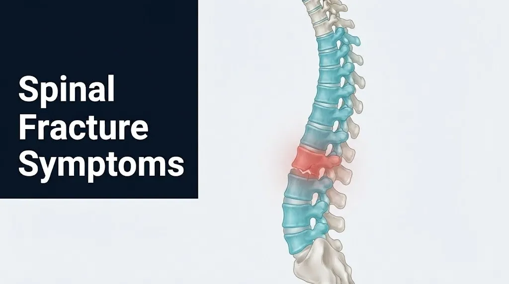
The pain from a burst fracture is typically immediate and severe. The mechanism is almost always a high-energy axial load: a motor vehicle crash, a fall from a height of more than six feet, a diving accident, or a heavy axial impact in contact sports. The person usually knows something serious has happened the moment of impact. That immediate severe central back or neck pain is qualitatively different from the muscular ache of a soft tissue injury.
What makes burst fractures clinically dangerous is the variability of the neurological picture. Some burst fractures with 40 to 50% canal compromise on CT scan produce no neurological deficit at presentation, because the cord or cauda equina has displaced to accommodate the retropulsed fragment. Others with similar imaging findings present with complete paraplegia. The canal compromise percentage on CT imaging does not reliably predict neurological status at presentation or recovery potential, which is why neurological examination by a spine surgeon or neurosurgeon is an essential component of burst fracture evaluation.
The Journal of Bone and Joint Surgery has published research documenting that approximately 15 to 20% of thoracolumbar burst fractures present with complete or partial neurological deficits, with the rate rising to over 50% in fractures involving severe translation or rotation at the injury level.
Men between 20 and 40 years old sustain the majority of burst fractures because high-energy trauma (motor vehicle crashes, sports injuries, military explosive blast injuries) is the primary mechanism, and young men disproportionately experience high-energy trauma events.
Key Takeaway: A burst fracture and a compression fracture can look similar on plain X-ray but carry completely different neurological risk, which is exactly why CT scan imaging is necessary for any spinal fracture with an axial loading mechanism before the patient is allowed to stand or be transported without spinal precautions.
Spinal Fracture Symptoms in Older Adults and Osteoporosis
Spinal fracture symptoms in older adults with osteoporosis often differ fundamentally from traumatic fracture presentations in younger people: the onset may be insidious rather than acute, the precipitating event may be trivially minor or unidentifiable, and the absence of severe immediate pain can lead both the patient and the clinician to attribute the fracture to pre-existing arthritis or normal aging.
Osteoporosis reduces bone mineral density (BMD) below a T-score of minus 2.5 on dual-energy X-ray absorptiometry (DEXA) scanning, reflecting a degree of trabecular bone loss that allows vertebral bodies to fracture under loads that healthy bone would tolerate easily. Activities as routine as bending to pick up an object, stepping off a curb, coughing forcefully, or turning over in bed can produce an osteoporotic compression fracture.
The clinical presentation often does not fit the typical fracture narrative. The person may report a gradual onset of midback pain over several days without recalling any specific event. They may notice increasing difficulty standing fully upright. A family member may observe new progressive rounding of the upper back. The gradual height loss that accumulates with multiple silent fractures may only become apparent at a routine medical appointment when height is measured and compared to a prior record.
According to the NIAMS, osteoporosis-related vertebral fractures disproportionately affect postmenopausal women, with an estimated 25% of postmenopausal women in the United States having at least one vertebral compression fracture. The risk rises steeply with age: women over 80 have a substantially higher fracture prevalence than those in their 60s.
Men with osteoporosis represent an underdiagnosed population. Male osteoporosis often results from hypogonadism, long-term alcohol use, corticosteroid therapy, or primary bone disease, and the clinical diagnosis is frequently delayed because both patients and clinicians less frequently consider osteoporosis in men presenting with back pain.
Pathological Spinal Fracture Symptoms in Cancer Patients
Pathological spinal fracture symptoms in cancer patients arise when tumor invasion of the vertebral body weakens its trabecular architecture sufficiently that normal physiological loads produce structural failure, and the symptom pattern differs from both traumatic and osteoporotic fractures in its onset quality, pain character, and associated systemic features.
The most common primary cancers that metastasize to the spine are breast, prostate, lung, kidney, and thyroid cancers. Spinal metastases occur in up to 40% of all cancer patients, with the thoracic spine affected most frequently, followed by the lumbar spine and cervical spine. Tumor-mediated activation of the RANK/RANKL osteoclast pathway, driven by parathyroid hormone-related protein (PTHrP) and other osteolytic factors secreted by tumor cells, produces localized bone destruction that reduces the vertebral body’s load-bearing capacity.
The distinguishing symptom features of pathological fracture from metastatic disease include:
- Night pain that is not relieved by positional changes, unlike mechanical back pain that typically eases with recumbency
- Constant pain that does not follow the predictable load-dependency of osteoporotic compression fracture pain
- Systemic features including unintentional weight loss, fatigue, reduced appetite, or night sweats
- Pain at an atypical level for osteoporotic fracture: mid-thoracic fractures (above T8) are rare in pure osteoporosis but common in metastatic disease
- Progressively worsening neurological symptoms as the tumor or fractured bone progressively compresses the adjacent spinal cord
The Spinal Instability Neoplastic Score (SINS), developed by the AO Spine Knowledge Forum Tumor group and validated in the spine oncology literature, is a clinical scoring tool used by spine surgeons to evaluate the degree of mechanical instability in metastatic spinal lesions, incorporating fracture location, pain quality, bone lesion type, spinal alignment, vertebral body collapse, and posterolateral involvement.
People with a known cancer diagnosis who develop new back pain should be evaluated by their oncologist with spinal imaging, even if the pain seems consistent with a muscle strain, because a pathological fracture missed and treated as a benign musculoskeletal condition can progress to irreversible spinal cord injury.
Stress Fracture of the Spine Symptoms
Stress fracture of the spine, most commonly spondylolysis (a fatigue fracture of the pars interarticularis of the vertebral arch, typically at L4 or L5), produces activity-related low back pain in young athletes without the acute onset, severe pain, and height loss that characterize traumatic or osteoporotic spinal fractures.
The pars interarticularis is the narrow isthmus of bone connecting the superior and inferior articular processes of the vertebral arch. It bears repetitive mechanical stress during combined extension and rotation movements, making it particularly vulnerable to fatigue fractures in athletes who perform repetitive hyperextension: gymnasts, football linemen, fast bowlers in cricket, weightlifters, and divers.
Spondylolysis symptoms develop gradually rather than acutely. The characteristic presentation is unilateral or bilateral low back pain that begins during activity, particularly during extension movements, and eases with rest. The pain may radiate slightly into the buttocks but typically does not produce true radiculopathy (electrical shooting pain down the leg in a dermatomal pattern), which helps distinguish it from a disc herniation.
A one-legged hyperextension test (standing on one leg and extending the lumbar spine backward) that reproduces ipsilateral back pain is the most clinically useful physical examination provocative test for spondylolysis at L5, because this position maximally stresses the pars interarticularis on the weight-bearing side.
Plain radiographs (oblique views showing the “Scotty dog” sign with a collar at the pars) can detect established spondylolysis but often miss early stress reactions. The AAOS recommends MRI with STIR sequences as the preferred imaging modality for early-stage pars stress reaction before a complete fracture is visible on CT or radiograph.
Adolescent athletes aged 10 to 18 represent the highest-risk group for spondylolysis, because the pars interarticularis reaches peak biomechanical vulnerability during the rapid growth phase of adolescence when bone mineral density has not yet fully matured to match the mechanical demands of intensive sport training.
Key Takeaway: Activity-related low back pain in a teenage or young adult athlete that is worse with back extension and hyperextension movements, but absent at rest, fits the clinical pattern of spondylolysis until proven otherwise by MRI with STIR sequences.
Spinal Fracture Symptoms in the Lumbar Region
Lumbar spinal fracture symptoms are centered in the lower back and can radiate into the buttocks, hips, and legs along the course of the lumbar nerve roots (L1 through L5) or the sacral roots of the cauda equina, with neurological symptoms ranging from single-root radiculopathy to the complete cauda equina syndrome if the fracture produces significant canal compromise.
The lumbar spine (L1-L5 vertebrae) is the most mobile and most axially loaded segment of the spine, bearing the combined weight of the head, trunk, and upper extremities with every upright activity. Burst fractures at the thoracolumbar junction (T12-L2) are the most biomechanically vulnerable zone, because this region transitions from the relatively rigid, rib-supported thoracic spine to the more mobile lumbar spine, concentrating stress at the junction.
An L1 fracture with posterior wall retropulsion threatens the conus medullaris, which resides at approximately the L1-L2 vertebral level in adults. Conus medullaris injury produces a mixed picture of upper and lower motor neuron findings, saddle anesthesia, bladder and bowel dysfunction, and sexual dysfunction, but with preserved proximal leg function in partial injuries.
Fractures at L3 and below compress only the cauda equina nerve roots, not the spinal cord itself. Each lumbar nerve root exits through a specific intervertebral foramen and controls a specific dermatomal and myotomal distribution. L4 root compression produces weakness of the quadriceps (knee extension) and medial calf sensory loss. L5 root compression produces weakness of the extensor hallucis longus (great toe extension) and dorsal foot sensory loss. S1 root compression produces weakness of the gastrocnemius-soleus complex (plantarflexion) and lateral foot sensory loss.
People who have undergone prior lumbar spine surgery for disc herniation or stenosis have altered mechanical properties at the operated level, including reduced disc height and altered load distribution. They face a different fracture risk profile than those with a surgically naive spine, and their symptoms may be confounded by pre-existing neurological deficits from the original condition.
Spinal Fracture Symptoms in the Thoracic Region
Thoracic spinal fracture symptoms present differently from lumbar fractures because the thoracic spinal canal is narrower relative to the cord diameter than at other levels, making even moderate bone retropulsion more likely to produce spinal cord compression, and because the thoracic cord (T1-T12) controls the intercostal muscles critical for breathing in addition to trunk and leg motor function below the injury level.
The thoracic spine is protected from fracture by the structural reinforcement of the rib cage, which dramatically increases the rigidity and load-distribution capacity of the T1-T10 segments. This protection means that fractures at T1-T10 typically require high-energy trauma (motor vehicle crashes, falls from height, direct blunt trauma) to occur in individuals with normal bone density. When they do occur, they are disproportionately associated with spinal cord injury because the thoracic canal’s narrow diameter provides little protective space.
A fracture at the T6 level, for example, produces paraplegia with complete motor and sensory loss below the T6 dermatome (a band across the mid-chest), while preserving full arm and hand function. Beevor’s sign is a clinical examination finding useful for localizing thoracic cord injury level: when the patient does a partial sit-up, the umbilicus moves toward the intact cord side because the lower abdominal muscles are paralyzed while the upper are preserved, indicating a T10 level injury.
Thoracic fractures from osteoporosis most commonly occur in the lower thoracic region (T7-T12), where the biomechanical stress concentration at the thoracolumbar junction is highest. They are less frequently associated with neurological deficit than burst fractures at the same level, because osteoporotic wedge compression does not typically produce the retropulsion of bone into the canal that characterizes high-energy burst injuries.
According to the AAOS, women with severe osteoporosis can sustain thoracic compression fractures from the mechanical stress of prolonged forward flexion posture alone, without any discrete traumatic event, as the kyphotic loading progressively exceeds the vertebral body’s load-to-failure threshold.
How Spinal Fractures Are Diagnosed
Spinal fractures are diagnosed through a combination of clinical history and physical examination establishing the injury mechanism and neurological status, followed by imaging beginning with plain radiographs and typically confirmed and characterized by computed tomography (CT) scanning, with magnetic resonance imaging (MRI) added when neurological symptoms, ligamentous injury, or pathological fracture is suspected.
Plain radiographs (anteroposterior and lateral views) are the initial imaging modality and can identify vertebral body height loss, kyphotic angulation, and obvious fracture lines. Their limitation is that they cannot visualize bone retropulsion into the spinal canal, the degree of canal compromise, or injury to the posterior ligamentous complex.
CT scanning (computed tomography) provides the definitive bony anatomy of a spinal fracture. Axial CT images at the fracture level directly visualize the degree of posterior wall retropulsion and canal compromise, allowing classification of the fracture type (compression versus burst), measurement of kyphotic angulation, and assessment of whether posterior element disruption is present.
MRI adds critical information that CT cannot provide: ligamentous injury (particularly posterior ligamentous complex disruption indicating instability), spinal cord contusion or edema (appearing as increased T2 signal within the cord on MRI), disc herniation at the fracture level, and the presence of bone marrow edema indicating an acute versus chronic fracture. In pathological fractures from metastatic disease, MRI is essential for identifying the full extent of tumor infiltration and assessing the degree of epidural cord compression.
To prepare for a spinal fracture evaluation:
- Do not attempt to stand or walk if you have experienced a high-energy mechanism of injury or have new neurological symptoms: wait for emergency personnel to assess and immobilize your spine.
- Tell the emergency team the exact mechanism of injury: fall from height, motor vehicle crash, direct impact, or onset without clear trauma.
- Report any change in neurological function since the injury: any new weakness, numbness, tingling, or difficulty controlling bladder or bowel.
- Bring a list of all current medications including bisphosphonates, corticosteroids, anticoagulants, and osteoporosis medications.
- Report any prior spinal surgery, known osteoporosis diagnosis, DEXA scan results, or known cancer diagnosis.
DEXA scan and bone density measurement are appropriate follow-up investigations for any person over 50 who sustains a low-trauma vertebral fracture, to establish the degree of osteoporosis and guide secondary prevention of further fractures.
Emergency Symptoms: When to Call 911 or Go to the ER
Certain spinal fracture symptoms require immediate emergency evaluation. Do not wait to see if these resolve on their own.
Call 911 or go to the nearest emergency room immediately if you experience:
- New weakness, paralysis, or inability to move the legs or arms after any back or neck injury: this can indicate spinal cord compression requiring emergency surgical decompression, and movement without spinal immobilization can worsen cord injury irreversibly.
- Inability to urinate, inability to feel when the bladder is full, or sudden loss of urinary control after back trauma or new severe back pain: these symptoms can indicate cauda equina syndrome, a surgical emergency where delay beyond 24 to 48 hours worsens recovery of bladder function.
- Loss of bowel control or inability to feel the perianal region (saddle anesthesia) after back injury or new severe back pain: this is a defining feature of cauda equina syndrome and requires the same emergency timeline.
- Severe neck pain with any weakness, tingling, or numbness in the arms or legs after trauma: a cervical spinal fracture with cord involvement can produce respiratory compromise at C3-C5 levels, and the person must not be moved without cervical immobilization.
- Any loss of consciousness, confusion, or unresponsiveness following a fall or traumatic event with associated back or neck pain: possible concurrent traumatic brain injury with spinal injury requires emergency evaluation for both conditions simultaneously.
- Difficulty breathing after a high thoracic injury (above T6): injury to the thoracic cord above the level controlling the intercostal muscles can require ventilatory support.
- Progressive worsening of neurological symptoms over hours: even if the initial presentation was mild, a deteriorating neurological examination indicates expanding cord compression and requires immediate emergency evaluation.
These presentations can indicate spinal cord injury, cauda equina syndrome, or unstable fracture with ongoing neural compression, all of which require emergency medical and surgical assessment, not an urgent care appointment or a primary care phone call.
Frequently Asked Questions About Spinal Fracture Symptoms
What does a spinal fracture feel like?
A spinal fracture typically feels like severe, localized back pain at a specific vertebral level that worsens sharply with standing, walking, or any axial loading and relieves substantially with lying flat.
High-energy traumatic fractures produce immediate severe pain at the moment of injury, while osteoporotic compression fractures can develop over hours to days after a minor precipitating event, or may even occur without a remembered event.
Any back pain that is reproducible by pressing directly on a specific spinal level, especially in the context of trauma or known osteoporosis, warrants imaging evaluation rather than presumptive treatment as muscle strain.
Can you have a spinal fracture without knowing it?
Yes, approximately two-thirds of osteoporotic vertebral compression fractures in older adults produce minimal or no acute pain and are discovered only on imaging obtained for another reason, according to the National Institute of Arthritis and Musculoskeletal and Skin Diseases (NIAMS).
These silent fractures still produce cumulative height loss and progressive kyphotic deformity over time, even when acute pain is absent.
Any postmenopausal woman or older man who has lost more than 2 to 3 centimeters of standing height or who shows progressive thoracic rounding should have spinal imaging and a bone mineral density assessment discussed with their primary care physician.
What are the first signs of a vertebral fracture?
The first signs of a vertebral fracture are typically sudden, localized back pain at a specific spinal level, often triggered by a specific movement, combined with pain that worsens immediately with upright posture and significantly relieves with lying flat.
In osteoporotic fractures, first signs can be subtler: a new aching quality to back pain that was previously absent, increased difficulty standing fully upright, or new back stiffness that is qualitatively different from prior arthritic discomfort.
New weakness, numbness, or bladder or bowel changes accompanying back pain are never early signs to wait and watch: they require same-day emergency evaluation.
Can a spinal fracture heal on its own?
Stable compression fractures without neurological deficit can heal through natural bone repair mechanisms over 6 to 12 weeks, with appropriate pain management and activity modification supervised by an orthopedic spine surgeon or physiatrist.
The caveat is that “healing” in an osteoporotic compression fracture means the fracture consolidates at the collapsed height, preserving any kyphotic deformity that developed, not a restoration of normal vertebral body height.
Unstable fractures (burst, chance, or fracture-dislocation patterns) and any fracture with neurological involvement do not heal safely without surgical stabilization and cannot be managed with simple rest.
What is the difference between a compression fracture and a burst fracture?
A compression fracture involves failure of only the anterior spinal column, producing anterior vertebral body wedge collapse without threatening the spinal canal, and is typically stable and treated non-operatively.
A burst fracture involves failure of both the anterior and middle columns, with retropulsion of the posterior vertebral body wall into the spinal canal, and carries a 15 to 60% neurological deficit rate depending on the degree of canal compromise and the spinal level affected.
The distinction cannot be reliably made on plain X-ray alone: CT scanning is required to evaluate the middle column and measure the degree of canal compromise in any fracture with an axial loading mechanism.
When is a spinal fracture a medical emergency?
A spinal fracture is a medical emergency when it is accompanied by any neurological deficit: new weakness, numbness, tingling, or loss of bladder or bowel control.
Cauda equina syndrome (urinary retention, saddle anesthesia, bilateral leg weakness), spinal cord injury (paralysis, breathing difficulty after cervical or high thoracic fracture), and any unstable three-column fracture with ongoing neurological deterioration require emergency room evaluation and spine surgical consultation the same day.
Call 911 immediately if the injured person cannot be safely moved, has any neurological symptom, or experienced a high-energy mechanism of injury: do not transport them in a passenger vehicle without first establishing spinal immobilization with emergency personnel.
Reading the Warning Signs Before They Escalate
Spinal fractures do not always announce themselves with dramatic severity. The person with osteoporosis who notices a new aching quality to their midback pain after lifting groceries may have a compression fracture. The young athlete whose low back pain is specifically provoked by arching backward may have a pars interarticularis stress fracture. The cancer patient whose back pain is constant and unresponsive to position changes may have a pathological vertebral fracture.
The single most actionable clinical principle across all spinal fracture types is this: back pain that is precisely localized to a specific vertebral level, reproducible with direct pressure on the spinous process, and position-dependent in a characteristic way warrants imaging rather than watchful waiting. A plain X-ray and CT scan answer the question definitively within hours.
If you or someone with you has any neurological symptom alongside back pain following injury or the onset of severe back pain, do not wait for a scheduled appointment. Go to the nearest emergency room or call 911 now. The window for preventing permanent neurological injury in spinal cord compression is narrow, and the decision to act quickly is the one clinical action that cannot be undone once it is missed.