L5-S1 Pain Symptoms: Nerve Signs & When It’s Urgent
L5-S1 pain symptoms arise from the lumbosacral junction, the single most mechanically stressed segment in the human spine, and produce a distinctive pattern of lower back pain, radiating leg pain, numbness, tingling, and sometimes muscle weakness that follows the specific nerve root being compressed or chemically irritated. The symptoms you experience tell a precise anatomical story about which nerve root is affected and how severely.
The L5-S1 intervertebral disc is the most commonly herniated disc in the lumbar spine, according to data reviewed by the American Academy of Orthopaedic Surgeons (AAOS). This level bears the greatest mechanical load of any spinal segment and is the site of the steepest lumbosacral angle, making it inherently vulnerable to both acute disc herniation and progressive degenerative change. What many people do not realize is that the specific leg symptoms produced by L5-S1 pathology depend entirely on whether the L5 or S1 nerve root (or both) is being compressed, and these two roots produce completely different pain maps, weakness patterns, and reflex changes.
This article covers every recognized symptom of L5-S1 pathology with the specific anatomical mechanism behind each one, the critical difference between L5 and S1 nerve root symptom profiles, the conditions that produce these symptoms, which presentations require same-day evaluation, and the specific emergency symptoms that require calling 911 immediately.
L5-S1 Pain Symptoms: Overview
L5-S1 pain symptoms are caused by pathological processes at the lumbosacral junction that compress, stretch, or chemically irritate the L5 or S1 nerve roots, producing a combination of lower back pain, radiating leg pain along the sciatic nerve distribution, and variable degrees of sensory and motor dysfunction.
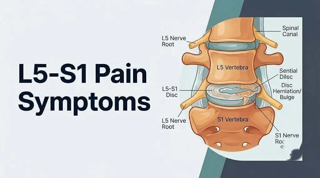
The term “L5-S1” refers to the joint formed between the fifth lumbar vertebra (L5) and the first sacral segment (S1). This segment includes the intervertebral disc between L5 and S1, the facet joints on both sides, the ligaments connecting these vertebrae, and the neural foramen through which the L5 nerve root exits the spinal canal. The S1 nerve root exits below this disc, through the first sacral foramen, but it travels through the spinal canal adjacent to the L5-S1 disc, making it vulnerable to compression from disc material that herniates toward the midline or central canal.
The clinical picture at L5-S1 is not monolithic. Symptoms depend on which structure is pathological (disc, facet joint, bony canal, or foramen), which nerve root is compressed (L5, S1, or both in severe cases), and whether the compression is purely mechanical or also involves chemical irritation from nucleus pulposus material.
| Symptom Category | L5 Root Involvement | S1 Root Involvement |
|---|---|---|
| Pain location | Lateral calf, dorsum of foot | Posterior calf, lateral foot, heel |
| Numbness area | First web space (between big and second toe) | Lateral foot, heel, little toe |
| Muscle weakness | Ankle dorsiflexion, big toe extension | Plantarflexion, standing on toes |
| Reflex change | Rarely affects a standard reflex | Achilles tendon reflex diminished or absent |
| Footdrop risk | Yes, if severe | No |
Older adults above age 65 are more likely to present with symptoms from degenerative lumbar stenosis at L5-S1 (progressive narrowing of the spinal canal from arthritic bone and thickened ligamentum flavum) rather than from acute disc herniation, producing a more bilateral, walking-dependent symptom pattern rather than the unilateral acute radiculopathy more typical of disc herniation in younger adults.
L5-S1 Anatomy and Why This Level Is Most Vulnerable
The L5-S1 segment is the most frequently affected level in lumbar spine pathology because its unique anatomical position at the base of the lumbar lordosis exposes it to the highest mechanical forces, the greatest range of sagittal motion, and the most acute angular loading of any intervertebral joint in the body.
The lumbar spine forms a lordotic (forward-curved) arc, and L5-S1 sits at the apex of the lumbosacral angle, the transition point where the mobile lumbar spine meets the fixed sacrum. Because the sacrum is rigidly fused and cannot move, all the mechanical stress from flexion, extension, lateral bending, and rotational loading that the lumbar spine absorbs must be transferred at this junction. The annulus fibrosus (the tough outer ring of the intervertebral disc) at L5-S1 is subjected to shear forces in multiple directions simultaneously during everyday activities including sitting, walking, and lifting.
Think of the L5-S1 disc like the hinge pin in a door that opens and closes thousands of times a day under heavy load. Every other disc in the lumbar spine has other segments above and below it sharing the load. L5-S1 is the last pin. It takes the full accumulated stress of the entire spine above it, transferred onto the fixed pelvis below.
The posterior annulus fibrosus at L5-S1 is inherently thinner and less well-supported than at higher lumbar levels, because the posterior longitudinal ligament (which reinforces the posterior disc wall) narrows as it descends and provides less coverage at L5-S1 than at L4-L5 or L3-L4. This anatomical thinning of posterior support concentrates the risk of posterior disc herniation precisely at this level.
Research published in Spine journal has confirmed that L4-L5 and L5-S1 together account for the overwhelming majority of lumbar disc herniations, with L5-S1 being the single most common individual level. People who smoke have accelerated disc degeneration at this level because smoking reduces blood flow through the vertebral endplates that provide nutrition to the avascular disc tissue, accelerating nucleus pulposus dehydration and annular weakening.
Lower Back Pain at the L5-S1 Level
Lower back pain at the L5-S1 level is produced by multiple concurrent pain generators, including disc-generated pain (discogenic pain), facet joint pain, and muscle guarding from segmental instability, and it is typically located in the central or bilateral lower lumbar region with a deep, aching quality.
The annulus fibrosus of the L5-S1 disc is innervated in its outer third by branches of the sinuvertebral nerve (a recurrent branch of the spinal nerve) and the gray rami communicantes from the sympathetic trunk. When the annulus develops tears from mechanical stress (annular fissures), inflammatory mediators including phospholipase A2 and prostaglandins from the leaking nucleus pulposus sensitize these nerve endings, producing deep, poorly localized lower back pain that is worsened by positions that load the disc (sitting, forward bending, coughing, sneezing) and relieved by positions that unload it (lying flat, walking with slight extension).
The L5-S1 facet joints (the paired zygapophyseal joints on each side of the posterior arch) have their own nerve supply through the medial branches of the dorsal rami of L4 and L5. Arthritic degeneration of these joints, called facet arthropathy or lumbar facet syndrome, produces a different pain pattern: deep aching in the lower back and buttocks, often worse with extension and rotation rather than with flexion, and often associated with morning stiffness.
People with obesity (body mass index above 30) face disproportionately higher discogenic and facet pain at L5-S1 because increased body mass elevates intradiscal pressure in the lumbar spine during all weight-bearing activities. According to a review in the journal Spine, each additional kilogram of body weight increases compressive load on the lumbar discs measurably, with the greatest proportional effect at L4-L5 and L5-S1.
Key Takeaway: Lower back pain at L5-S1 arises from disc, facet joint, and muscle sources simultaneously, and its character (worse with flexion vs. extension) helps a clinician identify which structure is most responsible.
L5-S1 Sciatica and Leg Pain
L5-S1 sciatica is radiating leg pain produced when disc herniation or bony stenosis at the L5-S1 level compresses or chemically irritates the L5 or S1 nerve root, causing pain to travel down the full length of that root’s dermatomal territory from the buttock to the foot.
The sciatic nerve is formed from the L4, L5, S1, S2, and S3 nerve roots, which join in the pelvis and exit through the greater sciatic foramen. An L5-S1 disc herniation primarily compresses the L5 or S1 roots before they join the sciatic nerve, so the pain follows the specific dermatomal path of whichever root is compressed rather than the full sciatic nerve territory.
The pain of L5-S1 sciatica is characteristically electric, burning, sharp, or shooting in quality. It travels from the lower back or buttock down the leg in a narrow band, not diffusely across the whole leg. This radiating quality distinguishes true radicular pain (from nerve root compression) from referred pain (from muscle or joint sources, which tends to be dull and diffuse rather than sharp and dermatomal in distribution).
The nucleus pulposus material that herniates through the annular tear does not only compress the nerve root mechanically. Research published in Spine journal has demonstrated that nucleus pulposus material released outside the disc directly sensitizes adjacent nerve roots through chemical irritation, primarily via phospholipase A2 and TNF-alpha released from disc cells and activated macrophages that infiltrate the herniation site. This chemical sensitization means that even a relatively small herniation can produce severe radicular pain if the extruded nucleus pulposus material contacts the nerve root directly.
Pregnant women in the second and third trimester are at elevated risk for L5-S1 sciatica because the anterior pelvic tilt that accompanies uterine growth increases lumbosacral lordosis and raises intradiscal pressure at L5-S1. Additionally, relaxin (a hormone produced in pregnancy) increases ligamentous laxity throughout the pelvis, contributing to segmental instability at L5-S1.
L5 Nerve Root Compression Symptoms
L5 nerve root compression produces a specific pattern of pain, sensory change, and motor deficit that affects the lateral calf, the top (dorsum) of the foot, and critically, the big toe and first web space, based on the L5 dermatome and myotome.
The L5 nerve root exits the spinal canal through the L5-S1 neural foramen (the bony tunnel between the L5 and S1 vertebrae on each side). A disc herniation at L5-S1 that extends posterolaterally (to the side of center) typically compresses the exiting L5 root in or near this foramen. The pain produced follows the L5 dermatome: it begins in the lower back or buttock, travels down the lateral thigh, continues down the lateral calf and shin, and terminates on the top of the foot and the space between the first and second toes.
The motor deficit specific to L5 root compression is weakness of ankle dorsiflexion (lifting the front of the foot upward), controlled primarily by the tibialis anterior muscle (L4-L5 innervation). A person with significant L5 compression may have difficulty walking on their heels or lifting the foot adequately during the swing phase of walking, producing a characteristic foot drop gait where the front of the foot drags or slaps the ground.
Big toe extension weakness is another specific L5 marker. The extensor hallucis longus muscle (exclusively innervated by L5) extends the big toe upward. Weakness in this specific movement, assessed by asking the person to raise the big toe against resistance, is one of the most specific clinical signs of L5 nerve root compression at the L5-S1 level, according to clinical examination guidelines from the American Academy of Orthopaedic Surgeons.
Standard deep tendon reflexes (patellar reflex at L3-L4, Achilles reflex at S1) are typically normal with isolated L5 root compression, because L5 does not significantly contribute to either standard reflex arc. This is why a person can have severe L5 radiculopathy with significant weakness and normal reflexes on examination.
S1 Nerve Root Compression Symptoms
S1 nerve root compression at the L5-S1 level produces a distinct and clinically identifiable symptom pattern affecting the posterior calf, the lateral foot, the heel, and the little toe, with the hallmark clinical finding of a diminished or absent Achilles tendon reflex.
The S1 nerve root exits the spinal canal through the first sacral foramen, which is below the L5-S1 disc level. However, the S1 root travels through the spinal canal adjacent to the L5-S1 disc as it descends from its origin (L1-S1 nerve roots form the cauda equina in the lumbar spinal canal). A large central or paracentral disc herniation at L5-S1 can compress the descending S1 root directly.
The S1 dermatome covers the posterior surface of the thigh and calf, the heel, and the lateral aspect of the foot including the little toe. Pain following S1 compression therefore travels down the back of the leg (posterior thigh and calf), wraps around the outer ankle, and radiates to the lateral foot and heel. Sitting typically aggravates S1 radicular pain more than walking, because seated posture increases tension on the posterior sciatic nerve.
The Achilles tendon reflex (the ankle jerk) is the most clinically reliable sign of S1 nerve root function. S1 provides the primary motor innervation to the gastrocnemius and soleus muscles (plantarflexion) through the tibial division of the sciatic nerve, and the reflex arc closes through the S1 segment. A diminished or absent Achilles reflex in the setting of posterior leg and lateral foot pain is a high-specificity indicator of S1 nerve root compromise. The National Institute of Neurological Disorders and Stroke (NINDS) identifies reflex changes as one of the most objective indicators of nerve root dysfunction in lumbar radiculopathy assessment.
S1 motor weakness manifests as difficulty with toe-walking and reduced plantarflexion strength. Clinically, asking the person to perform single-leg heel rises (standing on one foot and rising onto the toes repeatedly) tests plantarflexion endurance. A person with significant S1 motor involvement cannot perform this maneuver as many times on the affected side as on the healthy side.
Key Takeaway: The single most clinically useful way to distinguish L5 from S1 nerve root compression is the Achilles reflex: it is typically diminished or absent with S1 compression and typically normal with isolated L5 compression.
Numbness and Tingling From L5-S1
Numbness and tingling from L5-S1 pathology are sensory symptoms produced by disruption of normal afferent (sensory) signal transmission in compressed or chemically irritated nerve root axons, and their precise location on the leg and foot maps directly to the specific root involved.
Sensory nerve fibers in the L5 and S1 roots carry touch, temperature, pain, and vibration signals from the skin and underlying tissues of the leg and foot back to the dorsal horn of the spinal cord. When a disc herniation compresses these fibers against the bony edge of the neural foramen or the posterior vertebral body, the compressed axons begin to fire abnormally, producing the sensation of tingling (paresthesia) or, in more severe compression, complete sensory loss (anesthesia) in the affected dermatomal territory.
The dorsal root ganglion (DRG), a concentration of sensory neuron cell bodies located just proximal to the neural foramen, is particularly sensitive to compression. Research in Spine journal has shown that mechanical compression of the DRG produces more intense and more persistent pain and paresthesia than equivalent compression of the nerve root trunk distal to the ganglion, because the cell bodies are more metabolically active and more vulnerable to ischemia from pressure.
Tingling that follows a clear dermatomal strip (L5: top of foot and between big and second toe; S1: heel and lateral foot) is strongly suggestive of nerve root involvement. Diffuse tingling across the entire foot or leg without a clear dermatomal boundary is less specific for a single nerve root and may suggest peripheral neuropathy, vascular insufficiency, or involvement of multiple roots.
Postmenopausal women face a compounding diagnostic challenge in this context. Peripheral neuropathy from diabetic or metabolic causes, which becomes more prevalent with age and is exacerbated by hormonal changes that affect nerve cell metabolism, can produce diffuse foot tingling that overlaps with but is not caused by L5-S1 pathology. Any woman over 55 with foot tingling and lower back pain should have both lumbar spine imaging and a neurological evaluation including nerve conduction velocity (NCV) testing to distinguish the two causes.
Muscle Weakness and Foot Drop From L5-S1
Muscle weakness from L5-S1 pathology is a motor symptom produced by compression of efferent (motor) nerve fibers in the L5 or S1 root, reducing the neural signal strength reaching the muscles they innervate, and it represents a more serious neurological finding than pain or sensory symptoms alone.
Motor axons in compressed nerve roots sustain injury in two phases. The first phase involves ischemic dysfunction from compression-reduced blood flow to the root: the vasa nervorum (the small blood vessels that supply nerve roots) are compressed alongside the root axons, producing a metabolic insufficiency that impairs axonal conduction before structural damage occurs. This ischemic phase is reversible with timely decompression. The second phase, if compression is prolonged or severe, involves axonal demyelination and ultimately axonal death, which produces permanent motor deficit.
Foot drop is the most clinically dramatic motor deficit from L5-S1 pathology. It occurs when L5 motor compression reduces signal to the tibialis anterior muscle (primary ankle dorsiflexor) below the threshold for effective contraction. The person cannot lift the front of the foot during walking, causing the foot to drag or slap the floor. Foot drop from acute disc herniation can be rapid in onset, sometimes developing over hours during an acute disc extrusion event.
The American Academy of Orthopaedic Surgeons identifies progressive motor weakness, including any new foot drop, as a clinical indication for expedited surgical evaluation. A foot drop that has been present for weeks without improvement has a substantially worse prognosis for recovery than one addressed within days of onset, because prolonged axonal ischemia transitions into irreversible axonal death.
S1 motor weakness is more subtle. The person may report difficulty rising from a low chair, climbing stairs, or walking uphill, because all three require plantarflexion and hip extension strength that depends partially on S1-innervated musculature. Single-leg calf raises are the most sensitive clinical test for early S1 motor deficit.
People with diabetes mellitus face particular risk for persistent motor deficit from L5-S1 nerve root compression, because diabetic peripheral neuropathy compromises the metabolic reserve of nerve root axons, making them more susceptible to injury from mechanical compression and less capable of axonal regeneration after the compressive force is removed.
Key Takeaway: Any new foot drop or rapidly worsening leg weakness from L5-S1 pathology requires same-day evaluation by a board-certified orthopedic surgeon or neurosurgeon, because the window for recovery without permanent deficit narrows within days of onset.
L5-S1 Pain While Sitting, Standing, and Walking
The relationship between L5-S1 pain and specific postures and activities provides clinically useful information about the underlying cause, because different structural problems at L5-S1 are aggravated and relieved by different mechanical loading conditions.
Disc-related L5-S1 pain (from herniation or discogenic disease) is characteristically worse with sitting and forward bending. In a seated position, intradiscal pressure at L5-S1 increases substantially compared to standing or lying flat. A 2020 review in the journal Spine confirmed that intradiscal pressure measurements (from landmark studies using pressure transducers in vivo) are consistently highest during supported sitting and forward-flexed sitting. A person with discogenic L5-S1 pain who can walk comfortably but cannot sit for more than 15 to 20 minutes without increasing lower back or leg pain is displaying a classic disc-loading pattern.
Facet joint pain at L5-S1 behaves oppositely. Extension (backward bending of the spine) compresses the facet joints, and patients with facet arthropathy at L5-S1 typically report worse pain with prolonged standing (which requires slight lumbar extension), walking downhill, or arching the back backward. These patients often feel more comfortable sitting in a slightly forward-flexed position that unloads the posterior facets.
Neurogenic claudication from lumbar spinal stenosis at L5-S1 produces a walking-dependent pattern: pain, heaviness, and leg fatigue that begin after a specific walking distance and resolve with sitting or forward flexion. This pattern occurs because walking in an erect posture extends the lumbar spine, which narrows the already-stenotic spinal canal and increases pressure on the cauda equina or the L5 and S1 roots. Leaning forward on a shopping cart (the “shopping cart sign”) decompresses the canal and allows longer walking distances, which is a characteristic history in spinal stenosis patients.
Athletes who engage in repetitive lumbar hyperextension (gymnasts, wrestlers, rowers, offensive linemen in American football) develop isthmic spondylolisthesis at L5-S1 through stress fractures of the pars interarticularis, allowing the L5 vertebra to slip forward on S1. These individuals may report pain that is worse with extension and sport-specific loading, combined with L5 radicular symptoms from foraminal narrowing as L5 slips forward.
Conditions That Cause L5-S1 Symptoms
Multiple distinct pathological conditions produce L5-S1 pain symptoms, and differentiating between them requires clinical examination and imaging, because the treatment approach and urgency differ substantially depending on the underlying diagnosis.
The most clinically important conditions at L5-S1, ordered by clinical prevalence in the general adult population:
- Herniated nucleus pulposus (disc herniation): most common cause of acute L5-S1 radiculopathy in adults aged 30 to 50. The nucleus pulposus (the soft, gel-like inner core of the disc) extrudes through a tear in the annulus fibrosus and contacts the adjacent nerve root. Pain is typically acute in onset, severe, and associated with a specific precipitating event or position.
- Degenerative disc disease (DDD): progressive loss of disc height and hydration with age, causing discogenic pain and progressive foraminal narrowing from disc collapse.
- Disc bulge: diffuse outward bulging of the disc without complete annular rupture, producing a milder and often more chronic symptom pattern than frank herniation.
- Lumbar spinal stenosis: narrowing of the spinal canal or neural foramina at L5-S1 from arthritic bone spurs (osteophytes), thickened ligamentum flavum, and facet joint hypertrophy. Most prevalent in adults above age 60.
- Isthmic spondylolisthesis: forward slip of L5 on S1 from pars interarticularis defects. More common in athletes and younger adults.
- Degenerative spondylolisthesis: forward slip from facet joint degeneration allowing segmental instability. More common in older adults and postmenopausal women (due to estrogen-related ligamentous laxity changes).
- Facet joint arthropathy: arthritic degeneration of the L5-S1 zygapophyseal joints, producing axial low back pain and sometimes referred pain to the buttock and posterior thigh.
- Piriformis syndrome: tight or spasmed piriformis muscle in the buttock compressing the sciatic nerve as it passes through or near the muscle belly. Produces sciatic-like pain without true nerve root involvement. Often confused with L5-S1 radiculopathy.
L5-S1 Symptoms in Specific Populations
L5-S1 pain symptoms present with notable differences across biological sex, age groups, reproductive status, and occupational or athletic exposure, and these differences affect both the likely underlying cause and the appropriate clinical approach.
Women experience higher rates of degenerative spondylolisthesis at L5-S1 than men, and this sex difference is most pronounced after menopause. Estrogen has a stabilizing effect on spinal ligaments; its decline after menopause increases ligamentous laxity at the lumbosacral junction, allowing greater segmental motion and accelerating facet joint degeneration. Postmenopausal women with new or worsening L5-S1 symptoms should specifically ask their primary care physician or obstetrician-gynecologist to include lumbar spine evaluation in their assessment, with attention to spondylolisthesis as a potential cause.
Pregnant women face a uniquely increased L5-S1 loading pattern, as described in the sciatica section. The additional complication is that MRI (the preferred imaging for L5-S1 evaluation) is generally avoided in the first trimester and reserved for cases where the clinical findings are severe or suggesting cauda equina risk. Pregnant women with new sciatica should be evaluated by their obstetrician-gynecologist, who will determine the imaging approach based on trimester and symptom severity.
Older adults above age 65 present most commonly with neurogenic claudication from spinal stenosis at L5-S1, as described above. Their symptoms tend to be bilateral, walking-dependent, and slower in onset than acute disc herniation in younger adults. They are also at higher risk for pathological fractures of L5 from osteoporosis, which can produce acute severe low back pain without radicular features initially.
Workers in jobs requiring repetitive heavy lifting, prolonged sitting (particularly in vibrating vehicles), or prolonged awkward postures have elevated lifetime risk for L5-S1 disc degeneration and herniation. According to the National Institute of Neurological Disorders and Stroke (NINDS), occupational exposure to whole-body vibration (such as in truck driving, farming equipment, and construction machinery operation) is associated with accelerated lumbar disc degeneration.
Acute vs. Chronic L5-S1 Pain Symptoms
The distinction between acute and chronic L5-S1 symptoms determines the likely underlying structural cause, the expected natural history, and the appropriate management pathway.
Acute L5-S1 symptoms (lasting less than six weeks) most often reflect a disc herniation that is either freshly extruded or has recently shifted position to compress a nerve root more severely. The onset is characteristically sudden, often associated with a specific activity (lifting, twisting, bending) or sometimes with no identifiable event. Pain is typically severe, radiating, and accompanied by specific nerve root signs (foot weakness, absent Achilles reflex, dermatomal numbness). The North American Spine Society (NASS) clinical guidelines note that approximately 90 percent of acute lumbar disc herniations improve substantially within six to twelve weeks without surgery, as the extruded nucleus pulposus material undergoes resorption through macrophage-mediated phagocytosis.
Chronic L5-S1 symptoms (persisting beyond twelve weeks) suggest either a structural problem that is not self-resolving (foraminal stenosis from disc collapse and osteophyte formation, fixed spondylolisthesis, or a large disc herniation that has not resorbed), or the development of central sensitization in which the nervous system has become hyperactivated and amplifies pain signals beyond what the structural pathology alone would predict. Central sensitization is more likely in people who have had persistent pain for six months or more, those with psychological comorbidities including depression and anxiety, and those who had poor pain control in the acute phase.
The difference in treatment trajectory is also meaningful. Acute radiculopathy has a strongly favorable natural history and responds well to short-term conservative measures. Chronic L5-S1 pain with persistent neurological deficit has a less favorable natural history without active intervention and typically warrants assessment by a physiatrist (physical medicine and rehabilitation physician) or a board-certified orthopedic spine surgeon for a structured treatment plan.
People with chronic L5-S1 symptoms who have smoked for more than ten pack-years face slower recovery and higher rates of failed conservative treatment, because smoking impairs the vascular supply to healing nerve root tissue and accelerates further disc degeneration at the affected level.
Key Takeaway: Acute L5-S1 disc herniation symptoms have an approximately 90 percent chance of substantial improvement within twelve weeks without surgery, but any case with progressive motor deficit or bowel and bladder involvement requires surgical evaluation regardless of symptom duration.
Cauda Equina Syndrome: The L5-S1 Emergency
Cauda equina syndrome (CES) is a rare but permanently disabling neurological emergency that occurs when a large central disc herniation at the L5-S1 level (or rarely at L4-L5) compresses multiple nerve roots of the cauda equina simultaneously, threatening permanent loss of bladder, bowel, and sexual function.
The cauda equina (Latin for “horse’s tail”) is the bundle of nerve roots that descends through the lumbar spinal canal below the level where the spinal cord ends (approximately L1-L2). These roots include the S2, S3, and S4 nerve roots that control the detrusor muscle of the bladder (via the pelvic splanchnic nerves), the external urethral and anal sphincters, and sensory function of the perineum (saddle area). When a massive disc herniation at L5-S1 occupies a large portion of the central spinal canal, it can compress these descending roots before they reach their exit foramina.
The clinical presentation of cauda equina syndrome includes a highly specific set of symptoms that every person with L5-S1 disc disease should know to recognize immediately:
- New onset urinary retention (inability to urinate despite a full bladder) or new urinary incontinence (loss of control, dribbling): these indicate S2-S4 root compression affecting the detrusor and sphincter
- New onset fecal incontinence or loss of sensation in the rectal area during a bowel movement
- Saddle anesthesia: numbness or altered sensation in the perineum, inner thighs, genitalia, and buttocks (the area that would contact a saddle)
- Bilateral leg weakness: both legs simultaneously weak or numb, rather than unilateral sciatica
- Sudden reduction or resolution of previously severe sciatica combined with new neurological symptoms (the nerve root may have “burned out” as its function is destroyed)
According to a clinical review published in BMJ, cauda equina syndrome requires emergency surgical decompression within 24 to 48 hours of onset of complete sphincter dysfunction to have the best chance of preventing permanent disability. Delays beyond this window are associated with substantially higher rates of permanent bladder dysfunction.
How L5-S1 Problems Are Diagnosed
L5-S1 problems are diagnosed through a combination of structured clinical examination, targeted neurological testing, and imaging, with MRI of the lumbar spine being the primary confirmatory tool for most structural causes of L5-S1 symptoms.
The diagnostic sequence typically unfolds as follows:
- Clinical history: The clinician characterizes pain location, dermatomal radiation pattern, onset timing, aggravating and relieving postures, associated motor weakness, sensory changes, and any bladder or bowel symptoms. The history alone can point strongly toward L5 or S1 root involvement based on the specific leg symptom location.
- Neurological examination: Tests straight leg raising (the Lasegue test: lifting the symptomatic leg while the patient lies flat, which stretches the sciatic nerve and reproduces radicular pain if nerve root tension is present), ankle and patellar reflexes, dorsiflexion and plantarflexion strength, big toe extension strength, and sensory testing in the L5 and S1 dermatomal areas.
- MRI lumbar spine: The preferred imaging study. MRI visualizes the disc, nerve roots, spinal canal, neural foramina, and surrounding soft tissues without radiation. It identifies disc herniation size, location (central, paracentral, or foraminal), root compression, and foraminal stenosis. The AAOS recommends MRI as the imaging study of choice for lumbar radiculopathy when surgery is being considered or when red flag symptoms are present.
- Electromyography (EMG) and nerve conduction velocity (NCV) study: Performed by a board-certified neurologist. EMG detects abnormal electrical activity in muscles innervated by the compressed root (indicating active denervation) and can determine the chronicity and severity of nerve root injury. NCV testing assesses conduction velocity in peripheral nerve segments.
- CT scan lumbar spine: Used when MRI is contraindicated (pacemaker, certain implants) or when bony detail is needed (spondylolisthesis measurement, foraminal bone spur characterization).
When L5-S1 Symptoms Require Medical Evaluation
L5-S1 pain symptoms exist on a spectrum from mild and self-resolving to genuinely urgent, and the decision about whether to wait, book an appointment, or go to the emergency room depends on the specific symptom combination present at any given time.
| Symptom Pattern | Urgency | Action |
|---|---|---|
| New back pain with mild leg radiation, no weakness, no numbness | Monitor at home for 2 to 4 weeks | Primary care physician if not improving |
| Moderate sciatica with dermatomal numbness, normal bladder and bowel | Within 1 to 2 weeks | Primary care physician for evaluation and imaging referral |
| Moderate sciatica persisting beyond 6 weeks without improvement | Within days | Primary care physician or physiatrist for structured care plan |
| New foot weakness or S1 weakness affecting walking | Same day | Board-certified orthopedic surgeon or neurosurgeon same day |
| New bilateral leg weakness or numbness | Immediately | Emergency room |
| Any new urinary retention, incontinence, or fecal incontinence | Immediately | Call 911 or emergency room |
| Saddle area numbness (perineum, inner thighs, genitalia) | Immediately | Call 911 or emergency room |
People with known L5-S1 disc disease who experience a sudden and dramatic worsening of previously manageable symptoms should not assume the change is just another bad pain day. A sudden increase in pain combined with new neurological findings represents a change in disc position or status that warrants same-day medical re-evaluation, not watchful waiting.
Emergency Symptoms: When to Call 911 or Go to the ER
Certain symptoms associated with L5-S1 disc herniation require immediate emergency evaluation. Do not wait to see if these resolve on their own.
Call 911 or go to the nearest emergency room immediately if you experience:
- New inability to urinate despite having a full bladder, or new urinary leakage or dribbling that you cannot control (indicates S2-S4 cauda equina root compression affecting bladder function)
- New fecal incontinence or loss of sensation when having a bowel movement (indicates S2-S4 root compression affecting rectal sphincter function)
- Numbness or altered sensation in the saddle area: the perineum, inner thighs, genitalia, or buttocks (indicates compression of the nerve roots supplying this area, a hallmark of cauda equina syndrome)
- Sudden bilateral leg weakness, both legs becoming weak or numb at the same time, rather than symptoms on one side only
- Rapidly worsening foot drop developing within hours, particularly if associated with any of the above symptoms
- Sudden resolution of severe sciatica combined with new weakness or bladder symptoms (can indicate the nerve root has become severely compromised)
These presentations indicate cauda equina syndrome or a rapidly progressive neurological emergency and require emergency surgical evaluation, not an urgent care appointment, not a wait-and-see approach, and not a next-day phone call to a physician’s office.
Frequently Asked Questions About L5-S1 Pain Symptoms
What does L5-S1 pain feel like?
L5-S1 pain typically feels like a deep, aching lower back pain combined with sharp, electric, or burning pain radiating down one leg from the buttock to the foot.
The back pain component is located in the central or lower lumbar area and is often worsened by sitting, forward bending, coughing, or sneezing, while the leg pain follows a narrow dermatomal strip corresponding to either the L5 or S1 nerve root.
The leg pain quality (electric, shooting, burning) distinguishes true radicular pain from the dull, diffuse referred pain that comes from muscles or joints.
How do I know if my back pain is from L5-S1?
A strong indicator of L5-S1 involvement is lower back pain that radiates into the buttock and down the leg in a specific narrow path, particularly when the pain extends below the knee to the foot.
The specific location of leg pain, numbness, or weakness (top of foot and first web space for L5; heel and lateral foot for S1) and any reflex changes (absent Achilles reflex for S1) are the most reliable clinical indicators of L5-S1 nerve root involvement.
MRI of the lumbar spine is the definitive confirmatory test and should be ordered by a primary care physician when radicular symptoms persist beyond four to six weeks without improvement.
What is the difference between L5 and S1 nerve symptoms?
L5 nerve root compression causes pain and numbness on the top of the foot and the space between the big and second toes, weakness in lifting the front of the foot (dorsiflexion), and risk of foot drop, with a typically normal Achilles reflex.
S1 nerve root compression causes pain and numbness in the heel and lateral (outer) foot, weakness in pushing up onto the toes (plantarflexion), and a characteristically diminished or absent Achilles tendon reflex.
These two patterns can coexist if a large disc herniation affects both roots simultaneously.
Can L5-S1 problems cause foot drop?
Yes, L5 nerve root compression from an L5-S1 disc herniation can cause foot drop, which is the inability to lift the front of the foot during walking due to weakness of the tibialis anterior muscle.
Foot drop is a motor deficit indicating significant L5 nerve root compromise and represents a more serious neurological finding than pain or sensory symptoms alone.
Any new foot drop requires same-day evaluation by a board-certified orthopedic surgeon or neurosurgeon, because the window for recovery without permanent weakness narrows substantially within days of onset.
What are the emergency symptoms of an L5-S1 disc herniation?
The emergency symptoms of an L5-S1 disc herniation are new urinary retention or incontinence, fecal incontinence, numbness in the saddle area (perineum, inner thighs, genitalia), and bilateral leg weakness occurring simultaneously.
These symptoms indicate cauda equina syndrome, in which a large central disc herniation compresses multiple nerve roots controlling bladder, bowel, and sexual function.
Cauda equina syndrome requires emergency room evaluation and surgery within 24 to 48 hours to prevent permanent disability, and waiting to see if symptoms improve is not appropriate.
How long does L5-S1 pain last?
Acute L5-S1 disc herniation symptoms improve substantially in approximately 90 percent of cases within six to twelve weeks without surgery, based on natural history data reviewed by the North American Spine Society.
Improvement occurs as the herniated nucleus pulposus material is gradually resorbed by macrophages, reducing nerve root compression, though the disc never fully returns to its pre-injury state.
Symptoms persisting beyond twelve weeks, any case with progressive neurological deficit, or any presentation with red flag symptoms require evaluation by a board-certified orthopedic surgeon or neurosurgeon for assessment of surgical options.
The single most important thing to take from this article is the emergency threshold. If you develop new difficulty urinating, new fecal incontinence, or numbness in the saddle area at any point, that is cauda equina syndrome until proven otherwise and requires an emergency room visit, not a next-day appointment.
For the vast majority of people with L5-S1 pain, the picture is far less urgent: acute disc herniation with sciatica that follows a predictable recovery course over weeks to months. If you are in that category, a primary care physician is the right first step. Ask specifically for MRI of the lumbar spine if symptoms include leg radiation below the knee, and ask for a referral to a physiatrist or orthopedic spine specialist if conservative management has not produced improvement within six weeks.
You now have the anatomical map: L5 root goes to the top of the foot, S1 root goes to the heel and outer foot, and the Achilles reflex is your best bedside indicator of S1 function. Take that clarity into your next medical appointment.