Ischemic Colitis Symptoms: Signs, Stages & Red Flags
Ischemic colitis symptoms typically begin with a sudden cramping pain on the left side of the abdomen, followed by an urgent need to defecate and the passage of bright red blood or bloody diarrhea within 24 hours. This pattern occurs because the large intestine is acutely starved of oxygen, causing the inner mucosal lining to break down and bleed.
According to the American College of Gastroenterology, ischemic colitis is the most common form of intestinal ischemia in adults, accounting for roughly 1 in 2,000 hospital admissions in the United States. It is frequently underdiagnosed because its symptoms overlap with several other gastrointestinal conditions, including ulcerative colitis and infectious colitis.
This article covers every clinically recognized symptom with the physiological reason it occurs, how the presentation changes from mild to gangrenous, which populations face the highest risk, how the condition is confirmed diagnostically, and the specific symptom pattern that constitutes a medical emergency.
What Is Ischemic Colitis
Ischemic colitis is a condition in which reduced blood flow to the large intestine (colon) causes oxygen deprivation in the colonic wall, leading to tissue injury that ranges from superficial mucosal inflammation to full-thickness bowel death.
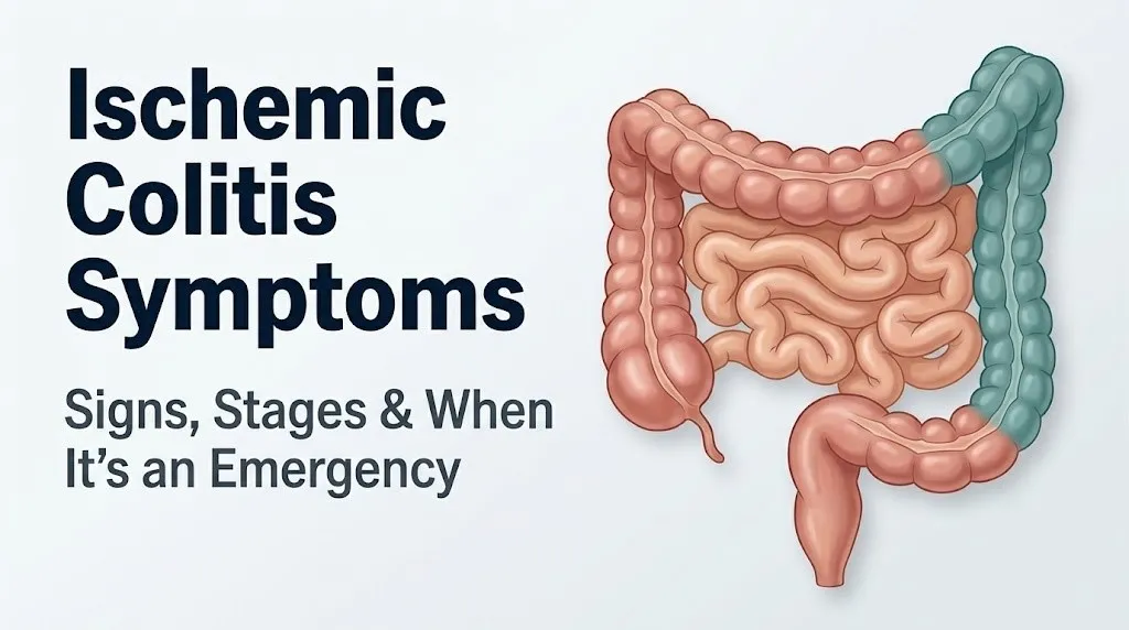
The colon receives its primary blood supply from three arterial systems. The superior mesenteric artery (SMA) supplies the right colon and transverse colon. The inferior mesenteric artery (IMA) supplies the descending colon, sigmoid colon, and upper rectum. The internal iliac arteries supply the lower rectum. Where these arterial territories meet, a zone of anatomically limited collateral blood supply exists. These zones, the splenic flexure (junction of transverse and descending colon) and the rectosigmoid junction, are called watershed areas.
Think of the colon’s blood supply like a city’s electrical grid. The high-traffic zones in the middle have the most redundancy. But the outskirts, where two power companies meet but neither fully covers the territory, lose power first when the main lines are stressed. The splenic flexure is the colon’s version of that boundary zone. When systemic blood pressure drops or an artery narrows, this is where the colon goes dark first.
When blood flow falls below the metabolic threshold of the colonic mucosal epithelium, ischemic injury begins in minutes to hours. The mucosa is the innermost and most metabolically active layer of the colon wall. Mucosal cells begin to die. Capillary walls break down. Fluid leaks into the bowel lumen. In most cases, ischemia is transient and limited to the mucosa. In severe cases, ischemia penetrates the muscularis propria and the serosa, producing transmural necrosis, a state called gangrenous ischemic colitis.
The National Institute of Diabetes and Digestive and Kidney Diseases (NIDDK) notes that ischemic colitis most commonly affects the left colon, particularly the descending and sigmoid colon, in alignment with the anatomical vulnerability of the IMA watershed territory.
Ischemic Colitis Symptoms: The Full Clinical Picture
The hallmark ischemic colitis symptoms are sudden onset left-sided or left lower abdominal cramping pain, followed within 24 hours by diarrhea that contains blood or bright red rectal bleeding.
The classic triad recognized in clinical gastroenterology practice includes abdominal pain, hematochezia (rectal bleeding), and diarrhea occurring together in a patient over age 60 with vascular risk factors. That said, all three symptoms are not always present simultaneously, and approximately 20 percent of patients present with only two of the three, according to a review published in the American Journal of Gastroenterology.
| Symptom | Mechanism | Onset Timing |
|---|---|---|
| Left-sided cramping pain | Mucosal ischemia, bowel wall edema, smooth muscle spasm | Sudden, within hours |
| Bloody diarrhea or rectal bleeding | Capillary breakdown, epithelial sloughing | Within 24 hours of pain onset |
| Urgent need to defecate | Colonic irritation from ischemic mucosal injury | Concurrent with pain |
| Nausea and vomiting | Visceral pain response, autonomic nervous system activation | Early phase |
| Fever | Systemic inflammatory response, bacterial translocation from ischemic mucosa | Moderate to severe presentations |
| Abdominal tenderness on palpation | Peritoneal irritation from bowel wall inflammation | Correlates with severity |
| Abdominal distension | Bowel dysmotility from ischemic injury to enteric nervous system | Later presentation or severe cases |
Atypical presentations occur in approximately 15 percent of patients, particularly those who are immunocompromised, on high-dose corticosteroids, or have diabetic neuropathy affecting visceral sensation. These individuals may have significant colonic ischemia with minimal or no pain, presenting instead with altered bowel habits or unexplained rectal bleeding alone.
Key Takeaway: The classic ischemic colitis symptom triad is sudden left-sided abdominal pain, bloody diarrhea, and an urgent need to defecate, but up to 20 percent of patients present without all three symptoms simultaneously.
Ischemic Colitis Abdominal Pain and Cramping
Ischemic colitis abdominal pain is characteristically sudden in onset, cramp-like in quality, and located in the left lower or left lateral abdomen, corresponding to the anatomical position of the descending colon and sigmoid colon.
The pain is generated at two levels. First, at the mucosal level, ischemic injury triggers the release of prostaglandins and substance P from damaged epithelial cells. These inflammatory mediators activate pain receptors (nociceptors) in the bowel wall. Second, ischemia triggers contraction of the colonic smooth muscle as the enteric nervous system responds to injury, producing the cramping character of the pain rather than a steady ache. This smooth muscle spasm is similar in mechanism to the cramping produced by irritable bowel syndrome but is far more acute and intense.
The pain of ischemic colitis is typically rated as moderate to severe by affected patients. It is described as a squeezing, cramping, or pressure-like sensation rather than the sharp stabbing pain associated with peritonitis. When the pain shifts from cramping to constant and severe, with a rigid or “boardlike” abdomen on palpation, this indicates disease progression toward transmural ischemia or perforation.
Patients with diabetic autonomic neuropathy or significant pre-existing visceral nerve damage may experience reduced pain intensity despite severe colonic ischemia. This population requires careful clinical evaluation even when pain is mild, as pain level alone does not reliably predict disease severity in this group.
Pain that is present but mild and self-resolves within 24 to 48 hours without worsening, rectal bleeding that stops, and the absence of fever are features associated with transient ischemic colitis, the least severe form. Pain that intensifies over hours, spreads across the abdomen, or is accompanied by fever and hemodynamic changes requires same-day emergency evaluation.
Blood in Stool With Ischemic Colitis
Rectal bleeding in ischemic colitis occurs because mucosal ischemia causes breakdown of the epithelial lining and rupture of the capillaries beneath it, releasing blood into the colon lumen that is passed during defecation.
The bleeding is classified as hematochezia: bright red or maroon blood visible in or mixed with stool. This is distinct from melena (black tarry stool), which indicates upper gastrointestinal bleeding. In ischemic colitis, the bleeding source is the left colon, close enough to the rectum that blood does not undergo the prolonged intestinal transit needed to produce the dark tarry appearance of upper GI bleeding.
The volume of bleeding in ischemic colitis is typically modest. Most patients pass blood-streaked stool or small amounts of frank blood mixed with diarrhea. Heavy rectal bleeding with large clots is less common in ischemic colitis than in lower GI arteriovenous malformations or diverticular bleeding, though it can occur in more severe presentations.
Bright red blood passed in the first 24 hours after sudden left-sided pain onset is one of the most clinically useful distinguishing features of ischemic colitis. A 2018 review in Gastroenterology noted that the combination of abdominal pain preceding rectal bleeding by 12 to 24 hours has a positive predictive value that strongly suggests colonic ischemia rather than infectious colitis, where diarrhea and bleeding often begin simultaneously.
Patients taking anticoagulant medications (warfarin, direct oral anticoagulants such as apixaban or rivaroxaban) may experience heavier or more prolonged rectal bleeding from the same degree of mucosal injury. Any rectal bleeding in a patient on anticoagulation warrants same-day evaluation by a primary care physician or emergency medicine physician, as the volume can escalate rapidly.
Ischemic Colitis Diarrhea and Urgency
The diarrhea of ischemic colitis is loose to watery, often mixed with blood, and accompanied by a sudden and intense urgency that is difficult or impossible to defer.
The urgency occurs because ischemic injury disrupts the normal neural coordination between the enteric nervous system (the network of neurons embedded in the bowel wall) and the internal anal sphincter. The colon loses its ability to store and modulate stool output. Contents move toward the rectum rapidly and trigger a strong defecation reflex that the affected person cannot comfortably suppress.
Unlike the diarrhea of viral gastroenteritis, which is typically watery and non-bloody in the first 24 to 48 hours, ischemic colitis diarrhea contains visible blood or blood-stained mucus from the onset or within hours of pain beginning. This distinction matters clinically: blood mixed in from the start of diarrhea, combined with left-sided abdominal pain, points toward colonic disease rather than a small intestinal or secretory cause.
The frequency of bowel movements varies. Most patients with mild to moderate ischemic colitis pass three to eight loose or bloody stools in the acute phase. Patients with more extensive colonic involvement may have higher frequency with progressively worsening urgency over 24 to 48 hours.
Older adults and patients with pre-existing fecal incontinence from prior anorectal surgery or pelvic floor dysfunction may lose continence entirely during an ischemic colitis episode. This is not a marker of disease severity by itself but can cause significant distress and should be addressed when taking the patient history, as it may affect the reported symptom timeline.
Key Takeaway: Ischemic colitis diarrhea contains blood from early in the episode and is paired with severe urgency, distinguishing it from most viral gastroenteritis where blood in stool is absent or a late feature.
Nausea, Vomiting, and Fever With Ischemic Colitis
Nausea and vomiting in ischemic colitis are reflexive responses driven by the autonomic nervous system’s reaction to acute visceral pain and intestinal wall ischemia, not by primary involvement of the stomach or small intestine.
When the colonic wall is ischemic, afferent pain signals travel through the splanchnic nerves to the spinal cord and then to the vomiting center in the medulla oblongata. This triggers the nausea and vomiting reflex as a non-specific response to severe abdominal visceral injury. Nausea is present in approximately 30 to 40 percent of ischemic colitis presentations, and vomiting is less common, occurring in roughly 15 to 25 percent, based on clinical series data from the American Journal of Gastroenterology.
Fever is a more ominous feature. Low-grade fever (37.8 to 38.5 degrees Celsius, or 100 to 101.3 degrees Fahrenheit) in ischemic colitis reflects systemic inflammation driven by mucosal injury and cytokine release, particularly TNF-alpha and interleukin-6 from ischemic intestinal epithelial cells. This level of fever can accompany moderate ischemic colitis and does not by itself indicate gangrenous disease.
High fever above 38.5 degrees Celsius (101.3 degrees Fahrenheit), particularly when accompanied by chills, rigors, and worsening abdominal pain, strongly suggests bacterial translocation: bacteria from the colon lumen crossing the damaged mucosal barrier into the bloodstream. This mechanism is the pathway to sepsis in severe ischemic colitis and gangrenous colitis. Any fever above 38.5 degrees Celsius in a patient with ischemic colitis symptoms warrants emergency evaluation, not watchful waiting.
Patients who are immunocompromised, including those receiving chemotherapy, long-term corticosteroid therapy, or biologic medications for autoimmune conditions, may not mount a fever even in the presence of serious ischemic colitis with bacterial translocation. In these patients, the absence of fever should not be taken as reassurance of a mild presentation.
Ischemic Colitis Risk Factors
Ischemic colitis risk factors operate through one of two main physiological pathways: reduced arterial blood flow to the colon (vascular insufficiency) or a hypercoagulable state that promotes clot formation in the mesenteric vessels.
The most well-established risk factors, according to the American College of Gastroenterology’s clinical guidelines on colonic ischemia, include:
- Atherosclerosis affecting the inferior mesenteric artery or its branches (most common underlying mechanism in older adults)
- Congestive heart failure and cardiac arrhythmias, particularly atrial fibrillation, which reduce cardiac output and mesenteric perfusion
- Chronic kidney disease, which accelerates vascular disease and alters platelet function
- Hypertension and diabetes mellitus, both of which contribute to small vessel disease in the mesenteric vasculature
- Obesity and hyperlipidemia, contributing to accelerated atherosclerosis
- Prior aortic or abdominal surgery, particularly aortic aneurysm repair, during which the inferior mesenteric artery may be sacrificed or temporarily clamped
- Systemic hypotension from any cause: septic shock, hemorrhage, cardiac failure, or aggressive diuresis
- Chronic use of stimulant laxatives (bisacodyl, senna in high doses), which cause colonic dysmotility and may alter mucosal perfusion
- NSAIDs (non-steroidal anti-inflammatory drugs), which inhibit prostaglandin synthesis and reduce mucosal protective mechanisms
- Heavy cocaine or methamphetamine use, which causes intense mesenteric vasoconstriction through sympathomimetic mechanisms
A 2021 analysis published in JAMA Internal Medicine found that patients with three or more cardiovascular risk factors (hypertension, diabetes, dyslipidemia, smoking) had a significantly higher rate of ischemic colitis hospitalization and recurrence compared to matched controls without cardiovascular comorbidity.
Key Takeaway: Atherosclerosis of the inferior mesenteric artery combined with any systemic cause of low blood pressure is the highest-risk combination for ischemic colitis in adults over 60.
Ischemic Colitis in Older Adults
Older adults account for the majority of ischemic colitis cases, with studies from the NIDDK indicating that the condition is most prevalent in adults aged 60 and above, and that age-related changes in mesenteric vascular anatomy are the primary physiological reason.
As humans age, the mesenteric arteries undergo progressive atherosclerotic narrowing: a buildup of cholesterol-laden plaques within the arterial walls that stiffens the vessels, reduces their internal diameter, and limits their capacity to increase flow in response to demand. The inferior mesenteric artery, already the smaller and lower-pressure of the two main mesenteric arteries, is particularly vulnerable to flow limitation from even moderate atherosclerotic disease. A vessel that is 50 percent narrowed under baseline conditions may become functionally insufficient during any episode of systemic hypotension.
Collateral blood vessel development also becomes less effective with age. In younger people, the mesenteric vascular bed can recruit alternative routes of blood supply (marginal artery of Drummond, arc of Riolan) when a primary vessel is compromised. These collateral pathways narrow and stiffen with age, reducing the colon’s ability to compensate for flow reduction.
Older adults with ischemic colitis frequently present in the context of a precipitating cardiovascular event. Common triggers include:
- Acute dehydration from vomiting or fever
- Cardiac arrhythmia onset (particularly atrial fibrillation with rapid ventricular response)
- Diuretic-induced volume depletion
- Post-procedural hypotension following cardiac catheterization or surgery
- Initiation of vasoconstrictive medications
Older adults who are taking beta-blockers or ACE inhibitors for cardiovascular conditions may have blunted heart rate response and may not mount the expected tachycardia during hemodynamic compromise, making the clinical picture of early shock less obvious. A primary care physician or emergency medicine physician evaluating an older adult with ischemic colitis symptoms should not rely on heart rate alone as a marker of hemodynamic stability.
Ischemic Colitis in Young Women and Medication Triggers
Ischemic colitis in adults under 50 is less common than in older adults, but when it occurs in younger women, oral contraceptive use is the most frequently identified contributing factor, making it a distinct clinical subgroup with a different risk profile and typically a better prognosis.
Combined oral contraceptives containing estrogen and progestin increase the coagulability of blood through several mechanisms: estrogen elevates levels of clotting factors II, VII, VIII, and X, suppresses protein S (a natural anticoagulant), and promotes platelet aggregation. In the mesenteric venous system, these pro-thrombotic changes can lead to small vessel thrombosis, reducing venous outflow from the colon and causing a form of ischemia driven by venous congestion rather than arterial insufficiency. This mechanism produces a slightly different clinical presentation than arterial ischemic colitis: symptoms may develop more gradually over 24 to 72 hours, and the pain is sometimes less acute in onset.
Research published in the American Journal of Gastroenterology has found that younger women with ischemic colitis who use oral contraceptives represent approximately 10 to 20 percent of ischemic colitis cases in patients under 50, with higher risk in women who smoke simultaneously (smoking compounds the pro-thrombotic effect of estrogen).
Other medication triggers that affect any age group include:
- Vasoconstrictive medications: triptans (sumatriptan, rizatriptan) used for migraine, pseudoephedrine in decongestants, ergotamine derivatives
- Immunomodulatory agents: interferon, some chemotherapy agents
- Constipation-causing drugs: opioids, anticholinergics, and calcium channel blockers that slow colonic transit and increase intraluminal pressure
- Alosetron (a 5-HT3 antagonist for irritable bowel syndrome with diarrhea): the FDA issued a black box warning for this drug specifically because of its association with ischemic colitis
Young women who present with ischemic colitis symptoms should be asked directly about oral contraceptive use, migraine medications, decongestant use, and smoking history. A board-certified gastroenterologist should evaluate these patients even when symptoms resolve, to determine whether medication cessation and risk factor modification are indicated.
Transient vs. Chronic vs. Gangrenous Ischemic Colitis
Ischemic colitis exists on a clinical spectrum with three distinct stages, each defined by the depth of bowel wall injury, the symptom pattern, and the treatment required.
The American College of Gastroenterology classifies ischemic colitis along this spectrum:
| Stage | Depth of Injury | Typical Symptoms | Duration | Treatment |
|---|---|---|---|---|
| Transient (mild) | Mucosa only | Mild to moderate pain, modest rectal bleeding, self-limiting diarrhea | Resolves within 1 to 2 weeks | Supportive: IV fluids, bowel rest, monitoring |
| Chronic (stricturing) | Mucosa and submucosa, fibrotic healing | Recurrent abdominal pain, incomplete bowel obstruction symptoms, constipation alternating with diarrhea | Weeks to months of symptoms, persistent structural change | Gastroenterology evaluation, possible endoscopic dilation or surgery |
| Gangrenous (transmural) | Full bowel wall thickness | Severe constant pain, peritoneal signs, fever above 38.5 C, hemodynamic instability, bloody stool | Rapid progression over hours | Emergency surgical resection |
Transient ischemic colitis accounts for approximately 80 to 85 percent of all cases. Symptoms improve spontaneously within 24 to 72 hours with supportive care. Colonoscopic findings typically show segmental mucosal edema, erythema, and petechial hemorrhage without ulceration or necrosis.
Chronic ischemic colitis occurs when repeated or sustained episodes of ischemia cause submucosal fibrosis and progressive stricture formation. The colon narrows at the site of prior ischemia (most commonly the splenic flexure or sigmoid colon), and the patient develops symptoms of partial bowel obstruction: abdominal distension, colicky pain, and altered bowel habits.
Gangrenous ischemic colitis is the most severe and life-threatening form. Ischemia penetrates through the muscularis propria and serosa, causing full-thickness bowel necrosis. This stage carries a mortality rate of 40 to 80 percent without emergency surgical intervention, according to data reviewed in Annals of Internal Medicine. It requires immediate surgical consultation and bowel resection.
Ischemic Colitis Complications and Stricture Formation
The primary complications of ischemic colitis, beyond the acute ischemic episode itself, are colonic stricture formation, recurrent ischemia, sepsis from bacterial translocation, and in the most severe cases, bowel perforation with peritonitis.
Colonic stricture formation occurs when the healing process after ischemic mucosal injury involves excessive submucosal fibrosis. As scar tissue replaces the injured bowel wall layers, the lumen narrows. This complication typically becomes apparent four to six weeks after the acute ischemic episode, when a patient who appeared to recover fully begins experiencing progressive abdominal distension, constipation, bloating, and colicky pain after meals. The physical mechanism is straightforward: stool that previously passed easily through a normal colon diameter now encounters a narrowed segment and cannot transit freely.
A 2019 study published in the American Journal of Gastroenterology found that stricture formation occurred in approximately 10 to 20 percent of patients hospitalized with ischemic colitis, with the sigmoid colon and descending colon as the most common sites.
Bacterial translocation occurs when the damaged mucosal barrier allows colonic bacteria (Gram-negative enteric organisms such as Escherichia coli and Klebsiella species) to cross from the colon lumen into the bloodstream. The clinical result is bacteremia and, in severe cases, Gram-negative septic shock. This complication is more common in patients with extensive colonic involvement, high-grade transmural ischemia, or co-existing immunosuppression.
Bowel perforation, while the least common complication, carries the highest mortality. Perforation allows fecal contents to enter the peritoneal cavity, producing bacterial peritonitis, rapid hemodynamic deterioration, and septic shock. Clinically, this presents as sudden severe worsening of abdominal pain that becomes diffuse and constant, accompanied by involuntary guarding, rebound tenderness, and fever. This is a surgical emergency with no acceptable delay in treatment.
Key Takeaway: Stricture formation appearing four to six weeks after an ischemic colitis episode is a recognized complication that warrants follow-up colonoscopy with a gastroenterologist to assess the need for endoscopic or surgical intervention.
How Ischemic Colitis Is Diagnosed
Ischemic colitis is confirmed through a combination of clinical presentation, cross-sectional imaging, and colonoscopy with mucosal biopsy, in that order of urgency.
The diagnostic sequence typically follows these steps:
- Initial clinical assessment: History of sudden left-sided abdominal pain, rectal bleeding, and diarrhea in a patient with known vascular risk factors. A physical examination assesses tenderness location, peritoneal signs (guarding, rebound tenderness), and hemodynamic stability.
- Blood tests: A complete blood count (CBC) checks for leukocytosis (elevated white blood cell count), which correlates with inflammatory severity. A serum lactate level is ordered: elevated lactate (above 2 mmol/L) suggests tissue-level ischemia and is a marker of disease severity. C-reactive protein (CRP) and erythrocyte sedimentation rate (ESR) assess systemic inflammation. A metabolic panel checks for metabolic acidosis, which accompanies severe ischemia.
- CT scan of the abdomen and pelvis with IV contrast: This is the recommended first-line imaging study. According to the American College of Gastroenterology, CT findings in ischemic colitis include segmental bowel wall thickening, mucosal enhancement, pericolonic fat stranding, and, in severe cases, intramural gas (pneumatosis intestinalis), which is an emergency-level finding indicating transmural necrosis.
- Colonoscopy with mucosal biopsy: Performed after the patient is hemodynamically stable and perforation has been excluded on imaging. Colonoscopy reveals the characteristic pattern of segmental colonic involvement, with abrupt transitions between affected and unaffected mucosa. Mucosal biopsy shows ischemic changes: ghost cells, lamina propria hemorrhage, and hemosiderin-laden macrophages.
- Stool culture and Clostridioides difficile testing: Ordered to exclude infectious colitis as the cause of rectal bleeding and diarrhea.
Patients with suspected gangrenous ischemic colitis should not undergo colonoscopy until surgical evaluation has occurred, as bowel distension during the procedure increases perforation risk.
Ischemic Colitis vs. Ulcerative Colitis Symptoms
Ischemic colitis and ulcerative colitis share several symptoms, including rectal bleeding, diarrhea, and left-sided abdominal pain, but the clinical context, age of onset, imaging pattern, and colonoscopic appearance allow experienced clinicians to distinguish them in most cases.
| Feature | Ischemic Colitis | Ulcerative Colitis |
|---|---|---|
| Typical age of onset | Over 60 (but can occur at any age) | 15 to 40 years (bimodal: second peak at 60 to 80) |
| Speed of onset | Hours (sudden) | Days to weeks (gradual flare) |
| Location of colonic involvement | Segmental, left colon, splenic flexure; spares rectum in most cases | Continuous from rectum extending proximally |
| Rectal sparing | Common | Rare (rectum almost always involved) |
| Risk factor context | Cardiovascular disease, medications, hypotension | Family history, autoimmune history, prior IBD diagnosis |
| Colonoscopic pattern | Segmental, abrupt transition zones, normal mucosa between affected areas | Continuous, diffuse inflammation from rectum upward |
| Response to treatment | Typically resolves within days to weeks with supportive care | Requires ongoing anti-inflammatory therapy (mesalamine, corticosteroids, biologics) |
| Recurrence pattern | May recur with vascular events; not inherently a chronic relapsing condition | Chronic relapsing-remitting disease with lifetime management |
The most clinically useful distinguishing feature on colonoscopy is rectal involvement. Ulcerative colitis almost always involves the rectum. Ischemic colitis, because the rectum receives a dual blood supply from both the inferior mesenteric artery and the internal iliac arteries, typically spares the rectum even when the sigmoid and descending colon are severely affected.
A board-certified gastroenterologist should evaluate any patient with rectal bleeding and left-sided abdominal pain who does not have a clear prior diagnosis, regardless of age, to confirm the diagnosis through colonoscopy and biopsy rather than relying on clinical pattern alone.
Key Takeaway: Rectal sparing on colonoscopy strongly favors ischemic colitis over ulcerative colitis, because the rectum has a dual arterial supply that protects it from ischemic injury.
Ischemic Colitis Recurrence and Long-Term Outlook
Most patients with transient ischemic colitis recover fully within one to two weeks, but approximately 5 to 20 percent will experience a recurrent episode, and the long-term outlook is influenced primarily by the severity of underlying cardiovascular disease.
Recurrence is not driven by the colitis itself recurring in the same location. Rather, it reflects the persistence of the underlying vascular disease that made the initial episode possible. A patient whose ischemic colitis was triggered by a hypertensive episode with cardiac arrhythmia is at ongoing risk for future ischemic events involving the colon if the arrhythmia and blood pressure are not adequately managed.
The Cleveland Clinic reports that patients who recover from ischemic colitis should undergo follow-up colonoscopy approximately four to six weeks after the acute episode, to assess mucosal healing, confirm the absence of an underlying colonic malignancy (which can impair local blood flow and predispose to ischemia), and identify early stricture formation.
Long-term management after ischemic colitis focuses on aggressive cardiovascular risk reduction:
- Blood pressure control to target levels recommended by the treating cardiologist or primary care physician
- Antiplatelet therapy for patients with atherosclerotic vascular disease, as directed by a cardiologist
- Medication review and discontinuation of agents that contributed to the episode (oral contraceptives, vasoconstrictors, high-dose stimulant laxatives) with guidance from the prescribing physician
- Hydration management, particularly in older adults at risk for dehydration-induced hypoperfusion
- Evaluation for hypercoagulable states in younger patients without clear vascular risk factors, through a hematologist or vascular medicine specialist
Patients with chronic ischemic colitis complicated by symptomatic stricture may require endoscopic balloon dilation or, in cases of complete or near-complete obstruction, surgical resection of the strictured bowel segment. These decisions should be made by a multidisciplinary team including gastroenterology and colorectal surgery.
Emergency Symptoms of Ischemic Colitis
Certain symptoms associated with ischemic colitis require immediate emergency evaluation. Do not wait to see if these resolve on their own.
Call 911 or go to the nearest emergency room immediately if you experience:
- Abdominal pain that is constant, severe, and spreading across the entire abdomen rather than remaining localized to the left side (suggests transmural ischemia or bowel perforation)
- A rigid, boardlike abdomen that is painful to touch with any pressure, or abdomen that feels different than any previous pain episode (peritoneal signs suggesting peritonitis)
- Fever above 38.5 degrees Celsius (101.3 degrees Fahrenheit) combined with shaking chills and worsening abdominal pain (suggests bacterial translocation or sepsis)
- Rapid heart rate (over 100 beats per minute), dizziness, lightheadedness, or fainting combined with rectal bleeding (suggests hemodynamic instability from blood loss)
- Profuse or continuous rectal bleeding with large blood clots that does not slow or stop within 30 minutes
- Blood pressure that has dropped measurably from your normal baseline combined with any abdominal pain and rectal bleeding
- Sudden severe pain without any prior bowel symptoms (the symptom pattern of bowel infarction rather than gradual ischemia)
- Absent or significantly reduced bowel sounds combined with severe abdominal distension (suggests paralytic ileus from transmural necrosis)
These presentations can indicate gangrenous ischemic colitis, bowel perforation with peritonitis, or Gram-negative septic shock, all of which require emergency surgical and medical assessment, not an urgent care appointment or a phone call to your primary care office.
Frequently Asked Questions About Ischemic Colitis Symptoms
What does ischemic colitis pain feel like?
Ischemic colitis pain feels like a sudden, cramping or squeezing pressure concentrated in the left lower or left lateral abdomen, corresponding to the descending or sigmoid colon.
Most patients describe the onset as rapid, reaching moderate to severe intensity within one to three hours, often accompanied by an urgent need to use the bathroom.
Pain that shifts from cramping to constant and severe, or that spreads across the entire abdomen, is a warning sign of disease progression that warrants immediate emergency evaluation.
Can ischemic colitis go away on its own?
Transient ischemic colitis, the mildest form, resolves without surgery in approximately 80 to 85 percent of cases, typically within one to two weeks of the initial episode.
Recovery with supportive care, including intravenous fluids, bowel rest, and close monitoring, is the standard approach when symptoms are mild and there are no peritoneal signs or hemodynamic instability.
Any patient with ischemic colitis should be evaluated by a physician, even if symptoms improve, because colonoscopy is needed to confirm mucosal healing and exclude a complicating stricture or underlying malignancy.
How do you know if ischemic colitis is getting worse?
Ischemic colitis is getting worse if pain intensifies from cramping to constant and severe, spreads beyond the left side, or is accompanied by high fever and shaking chills.
Increasing heart rate, dropping blood pressure, inability to keep fluids down, and increasing blood in the stool are additional markers of clinical deterioration.
Any worsening of these symptoms over a period of hours rather than days should prompt immediate emergency room evaluation, not a wait-and-see approach.
What is the difference between ischemic colitis and ulcerative colitis?
Ischemic colitis is caused by reduced blood flow to the colon and typically presents suddenly in older adults with cardiovascular risk factors, while ulcerative colitis is a chronic autoimmune inflammatory condition that usually begins in younger adults.
The most reliable distinguishing feature on colonoscopy is rectal involvement: ulcerative colitis almost always begins in the rectum and extends upward continuously, while ischemic colitis typically spares the rectum.
Only a gastroenterologist performing colonoscopy with biopsy can confirm which condition is present; the two cannot be reliably differentiated through symptoms alone.
Can medications cause ischemic colitis?
Yes, several commonly used medications are associated with ischemic colitis through mechanisms of mesenteric vasoconstriction or increased blood coagulability.
Oral contraceptives, triptans used for migraine (such as sumatriptan), pseudoephedrine in decongestants, alosetron (prescribed for irritable bowel syndrome with diarrhea), stimulant laxatives, and NSAIDs are among the most consistently implicated agents in the clinical literature.
Any patient diagnosed with ischemic colitis should have a full medication review with their physician to identify contributing drugs and assess whether discontinuation is appropriate.
How long does ischemic colitis last?
Transient ischemic colitis typically resolves within one to two weeks with appropriate supportive care.
Chronic ischemic colitis with stricture formation can produce ongoing symptoms for weeks to months until the stricture is treated endoscopically or surgically.
Gangrenous ischemic colitis is a surgical emergency with no defined recovery timeline at home; it requires immediate hospitalization, surgery, and intensive care management.
Ischemic colitis can appear identical to several other gastrointestinal conditions in its early hours. The combination of sudden left-sided pain, blood in the stool, and urgent diarrhea is a pattern that demands same-day evaluation, not a position on a wait-list for a routine colonoscopy. The physiological logic is clear: a colon that is starved of oxygen for hours becomes a colon that risks permanent structural damage within days.
If you or someone you care for has experienced the symptom triad described throughout this article, contact a primary care physician immediately for a same-day assessment or go directly to an emergency department if pain is severe, fever is present, or rectal bleeding is significant. Tell the evaluating physician the exact sequence: when the pain started, when the blood appeared in the stool, and whether pain has changed character since onset.
You now have a clinically accurate understanding of what ischemic colitis symptoms are, why they occur at the level of named arteries and named bowel wall layers, and exactly which symptoms mean the situation has crossed into emergency territory.