Vocal Nodules Symptoms: What Your Voice Is Telling You
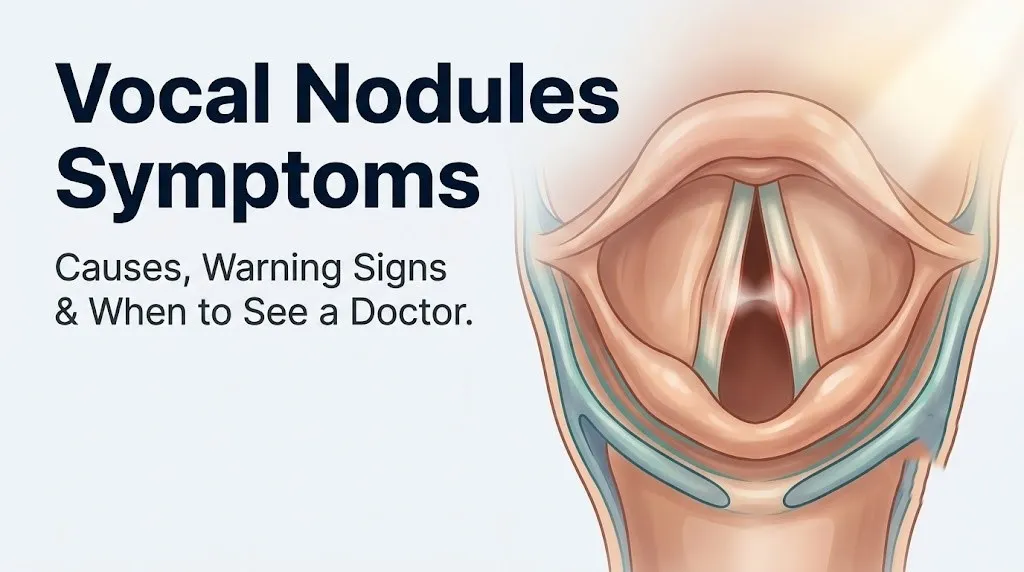
Vocal nodules symptoms develop when benign fibrous growths form on the vocal folds and disrupt the precise mechanics of normal voice production. The result is a recognizable cluster of changes: persistent hoarseness, a breathy or doubled quality to the voice, accelerating vocal fatigue, and a constant urge to clear the throat.
According to the National Institute on Deafness and Other Communication Disorders (NIDCD), voice disorders affect roughly 7.5 million people in the United States, and vocal fold nodules are among the most common benign laryngeal lesions in adults and children who use their voice heavily. Most people who develop nodules spend weeks misattributing the symptoms to allergies, a lingering cold, or simple overuse before a diagnosis is made.
This article covers every clinically recognized symptom of vocal nodules with the physiological explanation for why each one occurs, the difference between early and established nodule symptoms, how nodule symptoms compare to polyp and cyst symptoms, and the specific clinical thresholds that separate “monitor at home” from “call your ENT today.”
Vocal Nodules Symptoms: The Core Clinical Picture
Vocal nodules symptoms are produced by bilateral fibrous masses on the vocal folds that interfere with the smooth oscillation and complete closure needed for normal voice production.
The five primary symptoms, in order of clinical frequency, are:
- Hoarseness or roughness of voice: The most consistent presenting symptom, reported by the large majority of patients with confirmed nodules.
- Breathiness or air-wasting quality to the voice: Caused by incomplete glottal closure at the site of the nodule mass.
- Vocal fatigue: The voice deteriorates with use and requires increasing effort to sustain normal volume and clarity.
- Throat clearing or the sensation of something in the throat: A secondary symptom driven by abnormal airflow and mucosal irritation.
- Loss of upper pitch range or register breaks: Particularly prominent in singers, where the transition between chest and head voice becomes unreliable.
Symptoms typically begin gradually rather than suddenly. A sudden complete voice loss is far more consistent with a vocal fold hemorrhage or an acute illness than with nodule formation, which develops over weeks to months of repetitive vocal fold trauma.
People with early-stage pre-nodular lesions often report symptoms that are intermittent, worse in the evening after a day of voice use, and improved after overnight rest. As nodules become more fibrotic and established, the voice changes persist even after adequate rest, and this progression is one of the key clinical clues that separates a resolving reactive lesion from a true established nodule.
| Symptom | Frequency in Nodule Patients | Stage at Which It Typically Appears |
|---|---|---|
| Hoarseness or roughness | Very common (reported in most confirmed cases) | Early and established stages |
| Breathiness | Common | Early stage onward |
| Vocal fatigue | Common | Early stage, worsens as nodules enlarge |
| Throat clearing urge | Common | Early and established stages |
| Diplophonia (double voice) | Less common | Established or larger nodules |
| Loss of upper pitch range | Common in singers | Early stage in high-demand voice users |
| Throat pain with voicing | Less common | Established stage, associated with compensatory tension |
What Symptoms of Vocal Nodules Actually Feel Like
The subjective experience of vocal nodules symptoms goes beyond what a voice sounds like to an outside listener. For the person experiencing them, the sensations are often described in specific, consistent ways across patient populations.
Patients frequently describe the voice as feeling “effortful,” as if they are pushing air through a narrowed space. This is physiologically accurate. Because the nodular mass prevents the vocal folds from meeting cleanly at the midpoint, the body increases subglottic air pressure and recruits compensatory tension in the supraglottic musculature to try to achieve the closure that the folds cannot produce naturally. The result is a voice that requires noticeably more physical work to produce.
The throat sensation is often described as a lump, a tickle, or a feeling that something is stuck just behind the larynx. This is called globus pharyngeus when it occurs without a structural cause, but in the context of nodules, it reflects actual mucosal irregularity and the turbulent airflow patterns the nodules create. It is not a mass in the traditional sense; it is a sensory response to altered phonation mechanics.
Many patients also report that the voice “cuts out,” particularly at higher pitches or at the end of sentences when breath support is diminishing. This reflects the nodule’s greatest disruption occurring at the point of maximum oscillatory amplitude, which is the midpoint of the membranous fold where nearly all nodules develop.
Quick Tip:
- If your voice symptoms are noticeably worse at the end of the day and better after a full night of silence, this pattern is consistent with an early reactive or pre-nodular lesion, which has a good response rate to voice therapy and behavioral modification.
- If symptoms persist equally throughout the day regardless of voice use, the lesion may be more established and fibrotic.
- Postmenopausal women may notice that symptoms are slightly more pronounced due to reduced vocal fold tissue hydration associated with declining estrogen levels, which affects the pliability of Reinke’s space.
Vocal Cord Nodules Symptoms vs. Normal Voice Strain
Normal voice strain and vocal cord nodules symptoms share enough features that many people, and even some clinicians, initially conflate them. The distinguishing factor is time.
Normal voice fatigue after an unusually heavy voice day, such as a concert, a long lecture, or a day of phone calls in a noisy environment, resolves after 24 to 48 hours of reduced voice use. Vocal cord nodules symptoms do not. A 2019 review published in the Journal of Voice confirmed that the hallmark clinical feature separating nodules from acute overuse strain is persistence: symptoms lasting more than two weeks without a clear resolving trend are clinically suspicious for a structural lesion.
Normal strain produces a roughness that is consistent across pitch levels. Nodule-related dysphonia tends to be worse at specific pitch ranges, particularly in the upper register, and produces the characteristic “breaks” or “flips” when transitioning between chest and head voice that normal strain rarely causes.
The effort component is another distinction. Normal strain feels like a tired muscle. Nodule-related phonation feels like pushing against resistance that does not yield with rest in the way a muscle would. This is because a fibrotic nodule does not rest away. It remains physically present on the fold surface regardless of voice use, and its mass continues to interrupt the mucosal wave even when the surrounding musculature has recovered.
Children, particularly boys aged 5 to 10 years who are high-volume voice users (frequent shouters, children with loud play styles), develop nodules at higher rates than adults in the same age range. In children, the pattern is often described by parents as a chronically husky voice that has been present “for months” and does not respond to typical respiratory illness treatment.
Key Takeaway: Vocal nodule symptoms that persist beyond two weeks without improvement after voice rest are a clinical signal that requires professional laryngoscopic evaluation, not continued self-monitoring.
Throat Nodules Symptoms You Should Not Ignore
Throat nodules symptoms become clinically significant when they move beyond simple hoarseness into a pattern that suggests either a worsening structural lesion or a more serious underlying condition.
The following symptoms, when present alongside a voice change, warrant expedited medical evaluation:
- Dysphagia (difficulty swallowing): Swallowing difficulty is not a typical feature of isolated vocal fold nodules. Its presence suggests a lesion that may extend beyond the vocal fold, a concurrent laryngeal or hypopharyngeal process, or a neurological cause.
- Odynophagia (pain with swallowing): Similarly atypical for benign nodules and should prompt faster evaluation.
- Stridor (audible high-pitched breathing noise at rest): Stridor indicates a compromised airway and requires same-day or emergency evaluation.
- Unexplained weight loss accompanying voice change: This combination raises the clinical suspicion for a malignant laryngeal lesion and requires prompt otolaryngology referral.
- A visible or palpable neck mass with voice change: Not a feature of simple vocal fold nodules and must be evaluated clinically.
- Hemoptysis (coughing up blood): Not associated with benign nodules. Requires urgent evaluation.
The American Academy of Otolaryngology and Head and Neck Surgery (AAO-HNS) clinical practice guideline on hoarseness states explicitly that any patient with hoarseness persisting beyond three months should be evaluated by direct laryngoscopy, and any clinician who notes hoarseness in a patient with a history of tobacco use, heavy alcohol use, or prior head and neck radiation should expedite that referral rather than observe.
Adults over age 60 with new-onset voice changes, particularly those with a smoking history, should not accept a self-diagnosis of “nodules” without direct visualization of the larynx. The symptom overlap between benign and malignant laryngeal lesions at the level of patient self-report is too high to allow clinical confidence without endoscopy.
What Are Vocal Nodules and How Do They Form
Vocal nodules are bilateral, symmetric, benign fibrous growths that develop at the midpoint of the membranous portion of the vocal folds, which are the structures within the larynx responsible for voice production.
Think of the vocal folds like two lips of tissue that open and close thousands of times per minute during speech. The midpoint of each fold bears the greatest mechanical stress during vibration because it is the point of maximum oscillatory amplitude during normal phonation. When this area is subjected to repetitive trauma through chronic vocal overuse, misuse, or abuse, an initial edematous reaction develops in Reinke’s space, the loose connective tissue layer in the superficial lamina propria that allows the vocal fold epithelium to slide freely over the underlying muscle. This is the pre-nodular stage.
If the vocal trauma continues, inflammatory mediators including interleukin-1 and fibronectin drive progressive collagen deposition in Reinke’s space. The soft, fluid-filled reactive lesion transitions into a firmer, fibrotic mass. This is the established nodule.
Because nodules form bilaterally at the same midpoint on each fold, they meet each other like two small bumps during phonation, preventing the folds from achieving a clean, flat contact. The gap created by this kissing-lesion pattern is the primary cause of incomplete glottal closure and the breathiness it produces.
According to the NIDCD, vocal nodules are most commonly caused by vocal overuse or misuse. Contributing factors include inadequate hydration (which reduces the protective mucus film over the vocal fold epithelium), untreated laryngopharyngeal reflux (LPR), and habitual voice behaviors such as throat clearing, whispering in forced ways, or speaking at pitches outside the natural comfortable range.
Hoarseness and Voice Changes From Vocal Nodules
Hoarseness is the most consistent and recognizable symptom of vocal nodules, but the character of that hoarseness is clinically specific and differs from the hoarseness produced by laryngitis or simple inflammation.
Hoarseness from vocal nodules has a rough, irregular quality that may come and go early in the disease course, but becomes increasingly persistent as the lesion matures. The sound is produced because the nodular masses prevent the vocal folds from vibrating in their normal smooth, wave-like pattern. Instead of a clean, periodic waveform, the vocal fold oscillation becomes irregular and aperiodic, which the ear perceives as roughness or harshness.
In some patients, a phenomenon called diplophonia develops: the voice sounds doubled or has two simultaneous pitches. This occurs when the nodules disrupt the symmetry of the two vocal folds so severely that they begin vibrating at slightly different frequencies. Diplophonia is more common with larger or asymmetric nodular masses and is a reliable clinical indicator that a vocal fold lesion has reached a stage requiring specialist evaluation.
Voice changes from nodules are typically worse at the extremes of the pitch range. Speaking at a conversational pitch may be relatively preserved, while attempting high notes (in singers), projecting the voice across a room, or sustaining vowel sounds reveals the greatest disruption. This pitch-dependent quality is one clinical feature that separates nodule-related hoarseness from the generalized hoarseness of laryngitis, where the voice is often uniformly affected across the pitch range.
Adults with untreated gastroesophageal reflux disease (GERD) or laryngopharyngeal reflux should be aware that acid and pepsin exposure to the posterior larynx can exacerbate vocal fold inflammation and slow the resolution of nodular lesions. A 2021 study published in the Annals of Otology Rhinology and Laryngology found that LPR was a comorbid finding in a substantial proportion of patients with benign vocal fold lesions, and that treatment of reflux improved voice therapy outcomes.
Breathy Voice and Vocal Quality Changes
A breathy voice is one of the most diagnostically informative vocal nodules symptoms, because it points directly to a specific anatomical problem: incomplete glottal closure.
During normal phonation, the vocal folds adduct completely, allowing subglottic air pressure to build and then be released in controlled, periodic bursts that produce a clear, resonant voice. When nodules occupy the midpoint of each fold, they prevent the folds from meeting flat against each other. The result is a persistent gap at the point of contact, which allows air to escape continuously throughout phonation rather than in controlled periodic bursts. This turbulent, non-phonatory airflow is what the listener perceives as breathiness or an “airy” quality.
The clinical term for this insufficient closure is incomplete glottal closure, and it has a practical consequence beyond voice quality: the person has to use more air to produce the same loudness. This increased air demand means the breath runs out faster during speech, shortening phrase length and creating the need to pause more frequently to breathe. Many patients notice this as “running out of air” before reaching the end of a sentence, which is a direct consequence of the air-wasting effect of the nodule-created glottal gap.
In singers, breathiness from nodules often appears first in the upper passaggio (the transition zone between chest and head voice), where the demands on complete glottal closure are highest. A classical singer who notices a sudden “leaky” quality in the C5 to E5 range above the staff, with no new illness to explain it, should consider this pattern a clinical indicator of a lesion affecting the mid-membranous fold.
The breathy quality from nodules differs from the breathiness of vocal fold paralysis, in which one fold is immobile and the gap is lateral and persistent rather than confined to the mid-fold contact point. Stroboscopic laryngoscopy can distinguish these patterns clearly.
Key Takeaway: Breathiness in the voice caused by vocal nodules reflects a specific gap at the mid-fold contact point; this is mechanistically different from breathiness caused by vocal fold paralysis and requires laryngoscopy to confirm.
Vocal Fatigue and the Effort to Speak
Vocal fatigue is among the most disabling and least understood vocal nodules symptoms for patients who have never experienced a laryngeal disorder before. It is not simply a tired feeling in the throat. It is a specific deterioration of voice quality and function that occurs with extended voice use and worsens progressively throughout a speaking day.
The mechanism involves two overlapping processes. First, the nodular masses prevent efficient phonation, so the body must generate more subglottic air pressure and more muscular effort to produce the same vocal output. Second, the supraglottic musculature, including the false vocal folds, the aryepiglottic folds, and the intrinsic laryngeal muscles above the glottis, begins to recruit compensatorily to help achieve the closure the nodule-disrupted folds cannot produce on their own. This compensatory pattern is called supraglottic hyperfunction or muscle tension dysphonia when it becomes habitual, and it adds a significant layer of muscular fatigue on top of the phonatory dysfunction.
The result is a voice that may sound relatively acceptable in the morning but degrades noticeably by midday and becomes strained, low, or inconsistent by the end of the day. This temporal pattern, morning clarity with afternoon and evening deterioration, is one of the most characteristic and clinically informative features of nodule-related vocal fatigue.
According to a research review in the Journal of Voice, professional voice users with vocal fold lesions consistently report that vocal fatigue is the symptom most disruptive to their occupational functioning, rating it as more impactful than hoarseness in terms of daily limitation. Teachers, attorneys, clergy, and call center workers who must maintain voice quality across a full working day often describe the fatigue as a fundamental occupational impairment.
People with pre-existing cervical musculoskeletal tension, including those who work at a desk with forward head posture, often experience amplified laryngeal muscle fatigue because the extrinsic laryngeal muscles (particularly the sternohyoid and omohyoid) are already under tension from postural strain.
Throat Discomfort and Compulsive Throat Clearing
Throat discomfort and the persistent urge to clear the throat are secondary vocal nodules symptoms that many patients find more immediately bothersome than the voice changes themselves.
The sensation is often described as a tickle, a thick feeling, or a sense that mucus is sitting on or near the vocal folds. This perception is mechanistically accurate in two ways. First, the turbulent airflow created by the nodule-related glottal gap causes abnormal air-mucus interactions at the vocal fold surface, which can stimulate sensory nerve endings in the laryngeal epithelium and produce a tickling or foreign-body sensation. Second, if laryngopharyngeal reflux is contributing to the lesion, the posterior laryngeal mucosa may be directly irritated by acid or pepsin, adding a genuine mucous inflammatory response.
The problem is that throat clearing is itself a form of vocal trauma. Each forceful throat clear generates a sharp, high-velocity collision between the two vocal folds, similar in mechanical impact to a very brief cough. For someone with already-irritated or nodular vocal folds, habitual throat clearing becomes a cycle: the nodule creates the sensation that triggers the clearing, and the clearing worsens the mucosal inflammation that perpetuates the nodule.
ASHA (American Speech-Language-Hearing Association) identifies voluntary throat-clearing reduction as one of the first behavioral modifications taught in voice therapy for vocal fold lesions, specifically because of this compounding cycle of mucosal trauma.
Swallowing saliva is a clinically recommended substitute for throat clearing. It activates the same laryngeal muscle patterns but without the high-velocity fold collision. Sipping water immediately after sensing the urge to clear is another effective substitution strategy taught in voice therapy programs.
People with seasonal allergies or post-nasal drip are at higher risk for this symptom because the additional mucous drainage onto the posterior larynx compounds the sensation that drives the clearing behavior.
Key Takeaway: Throat clearing feels productive but worsens vocal fold inflammation with each repetition; replacing the habit with a sip of water or a deliberate swallow is one of the most effective immediate self-management steps for vocal nodule symptoms.
Vocal Polyps Symptoms
Vocal polyps symptoms share some features with nodule symptoms but have a distinct clinical profile that reflects their different underlying pathology.
Vocal fold polyps are typically unilateral (occurring on one fold only), whereas nodules are bilateral. A polyp is usually a single, well-demarcated, fluid-filled or fibrovascular lesion that arises from Reinke’s space following a single acute vocal fold hemorrhage or severe acute overuse event. This origin story matters to symptoms: because a polyp is unilateral and often larger than a nodule at the time of diagnosis, its effects on glottal closure are more pronounced and tend to produce more severe hoarseness from an earlier stage.
Key vocal polyps symptoms include:
- Moderate to severe hoarseness that is often immediately noticeable rather than gradual.
- A pronounced breathy or strained voice quality that may have appeared after a single event (a shouting episode, a long concert, a period of forced whispering).
- Diplophonia (double-pitch voice), which is more common with polyps than with nodules due to the unilateral asymmetry.
- A sensation of fullness or mass in the throat on the affected side, sometimes described as one-sided.
- Variable response to voice rest: A hemorrhagic polyp may be somewhat more responsive to strict voice rest in its early stage than a fibrotic nodule, particularly if the hemorrhagic component resolves.
Polyps can be pedunculated (on a stalk) or sessile (flat-based). A pedunculated polyp can occasionally move during phonation, causing intermittent voice obstruction and dramatic pitch breaks that are less typical of nodule behavior.
Throat Polyps Symptoms
Throat polyps symptoms is a colloquial term that typically refers to the patient experience of a vocal fold polyp rather than a distinct anatomical category. The term is commonly used by patients who have been told they have a “polyp in the throat” and are searching for what to expect.
From the patient’s perspective, the experience of a vocal fold polyp differs from a nodule in several practical ways:
- Onset is often linked to a specific memorable event: a period of intense voice use, screaming at a sporting event, or a bad respiratory infection involving severe coughing. Patients often say “it started the day after.”
- The voice change may be more abrupt and more severe from the outset than the gradual worsening typical of nodule formation.
- The sensation of something present in the throat is often more localized and described as one-sided.
- Complete loss of certain pitch registers (particularly the upper register in classical singers) can occur relatively quickly with a polyp compared to the gradual register loss of nodules.
According to information from the Mayo Clinic, vocal fold polyps can vary substantially in size, from a few millimeters to over a centimeter in some cases, and larger lesions can produce more severe airway sensation symptoms including a sense of breathing effort that nodules rarely cause.
Throat clearing with a polyp tends to be less effective as a short-term symptom reliever than with nodules, because the polyp’s mass does not move in the way that a mucous deposit would, and the sensation of foreign presence persists regardless of clearing behavior.
An untreated, persistent polyp does not resolve with voice rest and behavioral modification as readily as a pre-nodular reactive lesion. Most established polyps require either surgical microlaryngoscopy with excision or, in some cases, office-based laser or steroid injection treatment, followed by post-procedural voice therapy.
Vocal Nodules vs. Vocal Polyps Symptoms Compared
Distinguishing vocal nodules vs. vocal polyps symptoms matters clinically because the two conditions have different natural histories, different treatment timelines, and different expectations for non-surgical management.
| Feature | Vocal Nodules | Vocal Polyps |
|---|---|---|
| Location | Bilateral, mid-membranous fold | Typically unilateral, variable location |
| Onset | Gradual, over weeks to months | Often acute, following a single vocal event |
| Voice quality | Rough, breathy, fatigued | Severe hoarseness, strained or breathy |
| Diplophonia | Less common (bilateral symmetry) | More common (unilateral asymmetry) |
| Throat sensation | Bilateral tickle or fullness | Often one-sided foreign body sensation |
| Response to voice rest | Pre-nodular lesions may improve; fibrotic nodules do not fully resolve | Variable; hemorrhagic polyps may partially resolve |
| Typical patient | Heavy voice user over extended period | Any voice user after acute vocal event |
| Surgical frequency | Reserved for lesions unresponsive to therapy | More frequently requires surgery |
| Stroboscopic appearance | Small symmetric masses disrupting mucosal wave bilaterally | Single discrete lesion, may be pedunculated |
The patient experience of both conditions at the symptom level is close enough that self-diagnosis between the two is not clinically reliable. Both require direct laryngoscopy, ideally with videostroboscopy, for accurate diagnosis. The distinction matters because the treatment pathway diverges: early-stage nodules often respond well to voice therapy alone, while polyps frequently do not.
Key Takeaway: Vocal nodules and vocal polyps produce similar symptoms at the patient level, but their different laterality, onset patterns, and response to conservative therapy mean that only videostroboscopic laryngoscopy can reliably distinguish them.
Vocal Nodule Symptoms in Singers and Professional Voice Users
Singers and professional voice users experience vocal nodule symptoms in ways that are both earlier and more disruptive than in average speakers, because the demands they place on their vocal folds expose subclinical lesions that would remain unnoticed in someone with a lighter voice load.
A classical singer with a developing nodule often notices the problem first in the upper passaggio, the transition zone between chest and head voice. This area of the voice requires the most precise glottal closure and the most flexible mucosal wave, and a small nodular mass at the mid-membranous fold disrupts both. The result is a passaggio that suddenly feels inconsistent, sticky, or unreliable, where it was previously smooth. The singer may describe this as “the flip is gone” or “the voice breaks before it used to.”
Pop and rock singers, who rely more on chest-dominant technique and belt voice, may notice breathiness first in sustained high chest-voice notes. The air-wasting effect of incomplete glottal closure becomes intolerable at high intensities, where the gap creates audible air escape that recording equipment and live sound engineers detect even before the singer does.
Professional speakers, including teachers, clergy, attorneys, and voice-over artists, typically notice vocal fatigue as the first and most functionally impactful symptom. The ability to sustain voice quality across a full teaching day or a two-hour argument in court degrades before the voice quality itself becomes obviously abnormal to a casual listener.
According to research published in Laryngoscope, professional voice users are significantly more likely to delay seeking evaluation for vocal symptoms because they attribute changes to normal performance variation. This delay means that by the time a singer or teacher presents to an otolaryngologist, lesions are often more established than in a non-professional voice user with equivalent symptom duration.
Female singers and female professional voice users are at approximately twice the risk of developing vocal fold nodules compared to their male counterparts, a difference attributable to shorter vocal fold length (typically 12.5 to 17 mm in adult women versus 17 to 23 mm in adult men), higher fundamental speaking frequency (requiring faster and more frequent fold oscillation), and speaking pattern norms that often involve longer daily voice-use hours.
Nodules in Throat Symptoms in Adults and Population Variation
Nodules in throat symptoms do not present identically across all adult populations. Biological sex, age, hormonal status, and occupational voice load all modify the symptom picture in clinically meaningful ways.
In adult women: Adult women develop vocal fold nodules more frequently than adult men, with some clinical series reporting a female-to-male ratio of approximately 2:1 in adults. Women also tend to present with symptoms earlier in the lesion’s development, possibly because their higher fundamental frequency means the mid-fold contact point is stressed more frequently per unit of time. Perimenstrual voice changes (increased breathiness or reduced range in the days before menstruation, when progesterone and estrogen levels shift and vocal fold tissue becomes slightly more edematous) can amplify nodule-related symptoms and lead to misattribution. Postmenopausal women may experience greater vocal fold tissue dryness due to declining estrogen levels, which reduces the protective mucous film over the epithelium and can worsen both the underlying lesion and the perceived symptoms.
In adult men: Male vocal fold nodules are less common but not rare, particularly among men who are heavy occupational voice users. Men with nodules tend to present at a more advanced lesion stage because the lower fundamental frequency and longer vocal folds provide somewhat more tolerance before symptoms become functionally limiting.
In older adults: New-onset dysphonia in adults over 60 should always prompt laryngoscopic evaluation rather than a clinical assumption of nodules, because the differential diagnosis in this age group includes laryngeal malignancy and vocal fold paresis or paralysis, both of which can present with similar patient-reported symptoms.
In pediatric patients: Children, particularly boys aged 5 to 10, develop vocal nodules at a higher rate than girls of the same age, reversing the adult pattern. Pediatric nodules are almost universally managed with voice therapy and behavioral modification; surgical intervention is rarely required and generally deferred until the larynx has reached adult proportions.
| Population | Relative Risk | Distinguishing Symptom Feature |
|---|---|---|
| Adult women (reproductive age) | Higher than adult men | Symptoms may vary with menstrual cycle |
| Adult women (postmenopausal) | Moderate | Voice dryness amplifies breathiness |
| Adult men (occupational voice users) | Moderate | Later symptom presentation, more established lesions |
| Boys aged 5 to 10 | Higher than girls same age | Chronic husky voice; often misattributed to repeated colds |
| Adults over 60 (any sex) | Variable; malignancy risk elevated | Requires faster laryngoscopic evaluation |
| Professional singers | High | Symptoms appear earlier in lesion development |
Vocal Cord Nodule Warning Signs and Red Flags
Certain presentations alongside or beyond the typical vocal nodules symptoms require faster evaluation than the standard “wait three weeks then see your doctor” guidance.
Seek evaluation sooner than three weeks if any of the following are present:
- Hoarseness accompanied by difficulty swallowing solid foods or liquids (dysphagia)
- Pain when swallowing (odynophagia) in combination with voice change
- A visible or palpable lump in the neck alongside a voice change
- Unexplained weight loss of more than five pounds over one to two months in combination with voice change
- Any history of tobacco use or heavy alcohol consumption in a person with new or worsening hoarseness
- History of head and neck radiation and new voice changes
- Blood in sputum (hemoptysis) in any form, regardless of voice symptoms
- Voice changes following a period of intubation (general anesthesia with a breathing tube), which can cause contact injury, granulomas, or intubation trauma distinct from typical nodules
These features move the differential diagnosis away from benign nodules and toward conditions including laryngeal squamous cell carcinoma, vocal fold paralysis, laryngeal papillomatosis (caused by human papillomavirus), or other structural lesions requiring biopsy or imaging.
The AAO-HNS hoarseness clinical practice guideline recommends that clinicians use laryngoscopy rather than empirical treatment in any patient with hoarseness where there is a risk factor for serious laryngeal pathology. Waiting three weeks is appropriate for a healthy 30-year-old teacher with gradual onset hoarseness and no other symptoms. It is not appropriate for a 58-year-old former smoker with the same complaint.
Patients who have been diagnosed with a vocal fold lesion and notice a sudden, sharp worsening of symptoms, particularly the development of a voice that drops to near-aphonia, should contact their otolaryngologist promptly. A sudden worsening can indicate a vocal fold hemorrhage (bleeding into the fold), which requires voice rest and may require more urgent assessment.
Key Takeaway: The “wait three weeks” rule for hoarseness applies to healthy adults with no other risk factors; tobacco use, alcohol use, neck mass, dysphagia, or unexplained weight loss alongside voice change require expedited laryngoscopy, not watchful waiting.
How Vocal Nodules Are Diagnosed and When to Seek Care
Diagnosing vocal nodules requires direct visualization of the vocal folds. No combination of symptom duration, voice quality description, or physical examination of the neck and throat from the outside can confirm the presence of nodules or distinguish them reliably from polyps, cysts, granulomas, or early laryngeal malignancy.
The diagnostic pathway typically unfolds as follows:
- Primary care physician evaluation: The patient presents with hoarseness lasting more than two to three weeks. The physician takes a voice history (onset, duration, pattern, voice use, occupational demands, smoking history, reflux symptoms) and examines the oral cavity and external larynx. If no obvious cause is found, referral to otolaryngology is appropriate.
- Otolaryngologist (ENT) examination: The ENT performs a flexible laryngoscopy, passing a thin flexible endoscope through the nose and down the throat to visualize the larynx directly. This provides a real-time view of the vocal folds at rest and during phonation. The physician can see nodular masses, asymmetry, incomplete closure, and abnormal mucosal changes.
- Videostroboscopy: For a complete picture of how the vocal folds are actually vibrating, the gold standard is videostroboscopy, which uses a strobe light synchronized to the vocal fundamental frequency to create a slow-motion visualization of the mucosal wave. This reveals whether mucosal wave propagation is disrupted, whether closure is incomplete, and whether the lesion is soft (edematous, pre-nodular) or stiff (fibrotic, established). This distinction directly guides treatment decisions.
- Voice assessment by a speech-language pathologist: A qualified voice pathologist may administer the Voice Handicap Index (VHI), a validated self-report instrument that quantifies the functional, physical, and emotional impact of the voice disorder. This baseline measurement guides therapy goals and documents outcomes.
- Additional imaging or biopsy if indicated: If the stroboscopic appearance is atypical, if the patient has risk factors for malignancy, or if a lesion is unresponsive to therapy, a biopsy under general anesthesia (microlaryngoscopy) may be required. CT imaging of the neck and chest is ordered if there is concern for regional spread or a submucosal process.
According to the AAO-HNS clinical practice guideline on hoarseness (updated 2018), the recommended evaluation point for persistent hoarseness is three weeks in adults without risk factors and immediately (same-visit laryngoscopy or expedited referral) in adults with tobacco or alcohol use history, neck mass, or associated dysphagia.
For a professional voice user (singer, teacher, actor, attorney), the AAO-HNS and ASHA both recommend earlier referral, ideally at two weeks, because the functional consequences of delay are greater and early diagnosis improves both therapy outcomes and career impact.
Emergency Symptoms: When to Call 911 or Go to the ER
Certain symptoms associated with vocal cord disease require immediate emergency evaluation. Do not wait to see if these resolve on their own.
Call 911 or go to the nearest emergency room immediately if you experience:
- Stridor (a high-pitched, noisy sound during breathing at rest): This indicates a narrowed airway and is a medical emergency regardless of the suspected cause.
- Severe difficulty breathing that is worsening rapidly or came on suddenly, in the context of known vocal cord disease or not.
- Complete sudden voice loss accompanied by difficulty breathing or swallowing: This pattern is not consistent with simple vocal nodules and suggests an acute airway or neurological event.
- Severe throat swelling that makes swallowing or speaking feel obstructed, particularly if it came on suddenly or after a new food, medication, or allergen exposure (which may indicate angioedema).
- Coughing up blood in any quantity alongside a voice change.
These presentations can indicate acute airway compromise, angioedema, a large hemorrhagic vocal fold lesion, epiglottitis, or a rapidly progressing laryngeal process. None of these are appropriate for an urgent care appointment or a scheduled ENT visit. They require emergency medical assessment.
Routine vocal nodule symptoms such as hoarseness, breathiness, and vocal fatigue are not 911 emergencies. However, they do require timely medical evaluation rather than indefinite self-monitoring.
Frequently Asked Questions About Vocal Nodules Symptoms
What are the first signs of vocal nodules?
The first signs of vocal nodules are typically a gradual change in voice quality, most often increased roughness or breathiness, that is worse at the end of the day and after extended voice use.
Vocal fatigue that was not present before, combined with a reduced upper pitch range, is among the earliest functional indicators in professional voice users.
These early symptoms often precede the development of a fully established fibrotic nodule, which means early intervention at this stage has the best response rate.
Can vocal nodules go away on their own?
Early-stage, pre-nodular reactive lesions can resolve with strict voice rest, adequate hydration, and elimination of the vocal behaviors that caused them.
Fully established, fibrotic nodules rarely resolve completely without voice therapy or surgical intervention, because the collagen deposits in Reinke’s space do not reabsorb on their own.
A speech-language pathologist specializing in voice disorders can determine which stage the lesion is at and whether conservative management is a realistic primary treatment.
How long does hoarseness from vocal nodules last?
Hoarseness from vocal nodules typically persists for as long as the underlying lesion is present, which may be months if untreated.
Pre-nodular reactive lesions may improve within two to four weeks of strict voice rest and behavioral modification.
Established fibrotic nodules produce hoarseness that persists indefinitely without treatment, which is why the AAO-HNS recommends laryngoscopic evaluation at the three-week mark for unexplained hoarseness in adults.
What does it feel like to have vocal nodules?
Most people with vocal nodules describe the experience as the voice feeling effortful, as if pushing air through a resistance that did not previously exist.
The throat often has a persistent tickling or foreign-body sensation, and the voice may “break” or “cut out” at specific pitches, particularly in the upper range.
Vocal fatigue is typically described as the voice becoming noticeably worse and requiring more physical effort to produce as the day progresses.
Can vocal nodules cause pain in the throat?
Vocal nodules themselves do not typically cause significant pain, because the vocal fold epithelium has limited pain fiber innervation.
Some patients experience a dull ache or tension sensation in the laryngeal area and anterior neck, which reflects the compensatory muscle tension generated by supraglottic hyperfunction rather than pain from the nodule itself.
True pain with swallowing or speaking is not a typical feature of isolated vocal nodules and warrants evaluation to rule out other laryngeal conditions.
When should I see a doctor for vocal nodule symptoms?
You should see a primary care physician and request otolaryngology referral if voice changes have persisted for more than two to three weeks without a clear explanation such as a current respiratory illness.
See your doctor sooner, within one week, if your hoarseness is accompanied by difficulty swallowing, a neck lump, unexplained weight loss, or you have a history of tobacco or heavy alcohol use.
A speech-language pathologist specializing in voice disorders is appropriate to involve early, as voice therapy is a first-line treatment for many vocal fold lesions and can be initiated while laryngoscopy is being arranged.
Closing
Vocal nodules symptoms follow a recognizable pattern: a voice that gradually becomes rougher, breathier, and more effortful to produce, with the worst symptoms arriving at the end of a heavy voice-use day. Understanding the mechanism behind each symptom, specifically the disruption of mucosal wave propagation and the compensatory muscle tension that follows, helps explain why these symptoms behave the way they do and why throat clearing and forced whispering make things worse rather than better.
If your voice has been consistently different for more than two weeks without a clear cause, the right next step is a referral to an otolaryngologist for direct laryngoscopy. Do not self-diagnose between nodules, polyps, and cysts. Do not assume hoarseness with any of the red flag features described above is a benign lesion without visualization of the vocal folds.
Your voice is a finely calibrated instrument. Getting accurate answers about what is happening to it requires the same directness.