Broken Jaw Symptoms: Signs, Pain, and When to Act (2026)
Broken jaw symptoms include jaw pain, facial swelling, a changed bite, difficulty opening the mouth, and numbness of the chin or lower lip, and any of these signs after face or jaw trauma should be treated as a potential medical emergency until a fracture is confirmed or excluded. A jaw fracture is not a bruise or a muscle strain: it is a break in the mandible, the only movable bone of the face, and the clinical consequences range from a manageable non-displaced crack to an immediately life-threatening airway obstruction.
According to the American Association of Oral and Maxillofacial Surgeons (AAOMS), mandibular fractures are among the most frequent facial skeletal injuries seen in trauma settings, with motor vehicle accidents, falls, sports injuries, and interpersonal violence representing the four primary mechanisms. Research published in the International Journal of Oral and Maxillofacial Surgery has documented that male patients account for approximately 75 to 80 percent of mandibular fracture cases, with peak incidence in the 20 to 40 year age range.
This guide covers every clinically recognized symptom of a jaw fracture, the exact anatomical mechanism producing each symptom, how different fracture locations produce different presentations, which signs require emergency evaluation right now, and how a fracture is confirmed and managed. The mechanistic depth here goes well beyond a standard symptom list.
Broken Jaw Symptoms Overview
Broken jaw symptoms are the clinical signs produced when one or more fracture lines disrupt the cortical and cancellous bone of the mandible, straining the surrounding periosteum, nerve supply, musculature, and soft tissue in predictable, anatomically explicable ways.
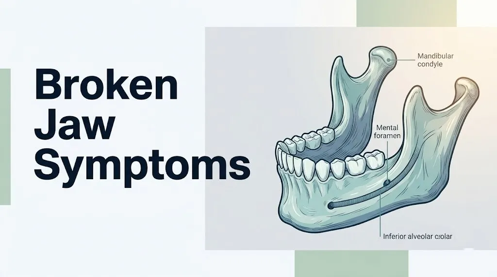
The mandible is a horseshoe-shaped bone consisting of a horizontal body, two vertical rami, two condylar processes (which form the temporomandibular joint with the skull base), two coronoid processes (which anchor the temporalis muscle), and the mental symphysis at the chin. Any of these regions can fracture independently or in combination. Multiple fracture sites are common: because the mandible forms a ring-like arch, a force applied at one point often transmits energy to a second, distant location, producing a second fracture line away from the point of impact.
The periosteum covering the mandible is densely supplied by branches of the inferior alveolar nerve (a branch of the mandibular division V3 of the trigeminal nerve, cranial nerve V). Fracture tears the periosteum, activates nociceptive nerve endings, triggers the release of substance P and prostaglandin E2 from damaged tissue, and initiates a fracture hematoma within minutes. This hematoma, combined with acute soft tissue edema from histamine-driven mast cell degranulation, produces the swelling and escalating pain pattern characteristic of jaw fractures.
| Fracture Site | Approximate Clinical Frequency | Key Symptom Produced |
|---|---|---|
| Condyle (including subcondylar) | 29 to 36% of mandibular fractures | Ear pain, limited opening, anterior open bite |
| Angle of mandible | 25 to 31% | Step deformity at molar region, trismus |
| Body of mandible | 15 to 20% | Numbness, step deformity, tooth pain |
| Symphysis and parasymphysis | 14 to 22% | Chin deformity, airway risk if bilateral |
| Ramus | 3 to 5% | Swelling over angle, limited opening |
| Coronoid process | Less than 1% | Limited opening, swelling below temporal region |
Children and adolescents with unfused condylar cartilage experience fracture patterns that differ meaningfully from adult presentations, a distinction covered in detail in a dedicated section below.
Jaw Pain and Swelling After a Fracture
Jaw pain after a fracture is produced by periosteal nociceptor activation and fracture hematoma pressure, and it typically presents as a deep, throbbing ache that intensifies dramatically with any jaw movement, including speaking, swallowing, or attempting to chew.
The periosteum of the mandible is the most pain-sensitive membrane covering bone in the craniofacial skeleton, because it is densely innervated by terminal branches of the inferior alveolar nerve. When a fracture tears the periosteum, pain signals travel through the inferior alveolar nerve to the trigeminal nucleus caudalis in the brainstem, where they are processed as intense, localized facial pain. The fracture hematoma that forms within the first minutes to hours increases intraosseous pressure, adding a second pain mechanism on top of the periosteal injury.
Swelling follows a predictable time course. Immediate soft tissue edema results from histamine release and vascular permeability changes at the fracture site. Over the following 12 to 24 hours, the inflammatory response amplifies swelling further as interleukin-6 and other acute-phase cytokines drive fluid into the extracellular space. Bruising, medically termed ecchymosis, typically appears at two specific locations: on the floor of the mouth (sublingual ecchymosis) and in the buccal mucosa (inner cheek lining). Sublingual ecchymosis visible in the mouth floor is considered a reliable clinical sign of mandibular fracture in trauma assessment.
Quick Tip:
- Palpate gently along the lower jaw from chin to ear: a painful, localized bony step or gap at the fracture line can often be felt through the skin.
- Sublingual bruising (visible bluish-purple discoloration on the floor of the mouth) visible within hours of injury is a specific clinical indicator of mandibular fracture.
- Patients taking anticoagulant medications (warfarin, rivaroxaban, apixaban) or antiplatelet agents (clopidogrel, aspirin) are at elevated risk for rapid hematoma expansion and should be evaluated in an emergency setting without delay.
Malocclusion and Bite Change as Broken Jaw Symptoms
A changed bite, the sensation that your upper and lower teeth no longer fit together correctly, is one of the most specific clinical indicators of a mandibular fracture and is produced by the mechanical displacement of fracture fragments under masticatory muscle pull.
Think of the mandible like a picture frame hanging on the wall. The masticatory muscles, including the masseter, medial pterygoid, and lateral pterygoid, attach to specific surfaces of the mandibular ramus and pull in specific directions. When a fracture line crosses the mandible, these muscle forces act on the separated fragments independently, pulling each fragment in the direction its attached muscle dictates. A fracture line running from lower front (mandibular symphysis) diagonally upward toward the molar region may be “favorable,” meaning muscle pull tends to hold fragments together, or “unfavorable,” meaning muscle pull separates them. Unfavorable fractures produce visible malocclusion.
The specific bite change pattern depends on fracture location. A condylar fracture causes loss of posterior vertical height on the fractured side, driving that side of the jaw upward and forward, and producing an anterior open bite: the front teeth cannot contact when the mouth is nearly closed because the back teeth on the fracture side are in premature contact. A displaced symphyseal fracture may shift the front teeth sideways, producing a midline deviation. A displaced angle fracture on one side may cause the back teeth on that side to touch prematurely while the opposite side is open.
The American Association of Oral and Maxillofacial Surgeons (AAOMS) identifies malocclusion as a primary clinical indicator guiding surgical management decisions: fractures that produce malocclusion generally require either closed reduction with intermaxillary fixation (wiring the jaws together) or open reduction with internal fixation plates to restore correct occlusal relationships.
People who wear complete dentures may not report bite change in the same way as dentate patients, because they have no natural teeth to perceive occlusal discrepancy. Clinicians examining edentulous patients must rely more heavily on skeletal asymmetry and range of motion to assess fracture displacement.
Broken Jaw Difficulty Opening the Mouth (Trismus)
Trismus in a jaw fracture is limited mouth opening caused by a combination of masticatory muscle spasm, pain inhibition of jaw-opening muscles, and mechanical blockage from displaced fracture fragments, and it reduces normal mouth opening (typically 40 to 50 millimeters between upper and lower central incisors) to as little as 5 to 15 millimeters in severe cases.
The jaw-closing muscles, including the masseter and medial pterygoid, respond to adjacent skeletal injury with reflex spasm through a protective withdrawal mechanism mediated by the trigeminal motor nucleus in the brainstem. This spasm is compounded by pain inhibition of the jaw-opening muscles: the digastric, mylohyoid, and geniohyoid muscles cannot generate sufficient force against a spasming antagonist group. The result is a jaw that the patient can barely open at all, despite a clear voluntary effort.
Displaced fracture fragments at the coronoid process or sigmoid notch can also physically block the path of the coronoid as it attempts to slide under the zygomatic arch during opening, creating a mechanical (rather than muscular) limitation.
To describe jaw opening limitation accurately to an emergency physician:
- Measure mouth opening by placing two fingers vertically between your upper and lower front teeth at maximum opening. Normal is three finger-widths. One finger-width or less suggests severe restriction.
- Note whether the jaw deviates to one side on opening: deviation toward the side of pain suggests condylar fracture on that side.
- Record whether jaw opening has worsened, stayed the same, or improved in the hours since the injury.
- Tell the clinician whether chewing, swallowing, or speaking has become progressively more difficult since the injury.
- Note any popping, grating, or complete loss of smooth jaw movement since the injury occurred.
Patients with pre-existing TMJ disorders or prior jaw surgery may have baseline limited opening that complicates accurate assessment of fracture-related restriction. This history should be disclosed immediately to the examining clinician.
Key Takeaway: A changed bite (malocclusion) after jaw trauma is one of the most specific clinical indicators of mandibular fracture, because it reflects actual displacement of fracture fragments driven by masticatory muscle forces on the separated bone segments.
Chin and Lip Numbness in Jaw Fracture
Numbness of the chin and lower lip after jaw trauma is a direct indicator of inferior alveolar nerve injury at the fracture site, and it is one of the most clinically useful signs for confirming a mandibular fracture without imaging.
The inferior alveolar nerve runs through the entire length of the mandibular body inside the inferior alveolar canal, a bony tunnel in the lower jaw. It enters the mandible at the mandibular foramen on the inner surface of the ramus and exits at the mental foramen on the outer surface of the body, just below the first and second premolars. At the mental foramen, it becomes the mental nerve, which supplies sensation to the skin of the chin, the lower lip, and the adjacent gingiva.
When a fracture line crosses the mandibular body or angle, it disrupts the inferior alveolar canal. The nerve inside can be compressed by displaced bone edges, stretched by fragment separation, or in severe cases, directly lacerated by a sharp bone fragment. Any of these injuries reduce or eliminate nerve conduction, producing the sensation that the chin and lower lip have been given a dental anesthetic that won’t wear off. This clinical finding is called Vincent’s sign, or more formally, hypoesthesia or anesthesia of the lower lip, and its presence is considered a reliable clinical predictor of mandibular fracture on the affected side.
According to research published in the Journal of Oral and Maxillofacial Surgery, chin and lip numbness is present in a clinically meaningful proportion of confirmed mandibular body and angle fractures, and its persistence after fracture healing can indicate incomplete nerve recovery requiring monitoring by the treating oral and maxillofacial surgeon.
- Numbness affecting both sides of the chin simultaneously suggests either a symphyseal fracture with bilateral mental nerve involvement or, rarely, a central nervous system lesion requiring immediate neurological evaluation.
- Patients with diabetes mellitus have pre-existing peripheral nerve vulnerability (diabetic peripheral neuropathy) and may experience more severe or prolonged inferior alveolar nerve symptoms after fracture.
- Numbness that worsens after initial evaluation, rather than remaining stable, may indicate progressive nerve compression from an expanding hematoma and warrants immediate reassessment.
Bleeding in the Mouth After Jaw Fracture
Oral bleeding after jaw trauma signals either laceration of the overlying gingival or mucosal tissue at a fracture line, or avulsion of a tooth from its socket, and its clinical significance depends on the source, volume, and rate of bleeding rather than its mere presence.
Mandibular fractures that pass through the alveolar process (the tooth-bearing portion of the jaw) are classified as compound fractures because they communicate with the oral environment through a wound in the gingival tissue. This communication is why some jaw fractures bleed visibly into the mouth even without any external skin wound. The mucosal tear over the fracture line is the entry point for oral bacteria, which is one reason compound mandibular fractures carry a risk of osteomyelitis (bone infection) if antibiotic prophylaxis is not initiated promptly.
Bleeding from within tooth sockets indicates tooth root fracture, avulsion (complete displacement of a tooth from its socket), or dentoalveolar fracture (fracture of the alveolar bone holding the tooth in place). A tooth that has been fully avulsed from its socket should not be placed back without professional guidance, because improper reimplantation of a fractured or contaminated tooth can introduce infection directly to the fracture hematoma.
Mayo Clinic identifies uncontrolled oral bleeding after jaw trauma as an indication for immediate emergency evaluation. Oral bleeding that cannot be controlled with direct pressure applied to the bleeding site for 10 continuous minutes, or that is associated with blood pooling in the throat, is an airway risk and constitutes an emergency.
Older adults with liver disease or those taking anticoagulant medications may experience disproportionate oral hemorrhage from what appears to be a minor laceration, because clotting factor synthesis or platelet function is impaired. This population should be evaluated in an emergency setting rather than waiting for a scheduled dental or surgical appointment.
Condylar Jaw Fracture Symptoms
Condylar jaw fracture symptoms are distinct from other mandibular fractures because the fracture site is at the very top of the jaw joint, producing symptoms that mimic TMJ disorder or ear injury rather than the more obvious jaw deformity seen in body or angle fractures.
The condyle is the rounded superior end of the mandibular ramus that articulates with the glenoid fossa of the temporal bone to form the temporomandibular joint. A fracture here, which accounts for roughly 29 to 36 percent of all mandibular fractures according to data in the Journal of Oral and Maxillofacial Surgery, disrupts the vertical height of the posterior mandible. When the condyle collapses or displaces medially (inward, pulled by the lateral pterygoid muscle attached to the condylar neck), the posterior vertical height on that side is lost.
The specific clinical picture of a condylar fracture:
- Preauricular pain (pain directly in front of the ear) worsened by jaw movement
- Jaw deviation toward the injured side on opening, because the intact opposite condyle translates forward normally while the fractured side cannot
- Anterior open bite: the back teeth contact prematurely on the fracture side, tipping the front teeth apart
- Limited mouth opening, usually less than 30 millimeters, from pain, muscle spasm, and mechanical disruption
- Swelling and point tenderness at the preauricular region, just anterior to the tragus of the ear
- In some cases, bleeding from the external ear canal if the fracture displaces into the bony ear canal wall (a finding called hemotympanum when blood collects behind the eardrum)
Bilateral condylar fractures after a midline chin impact (the classic “chin-first” fall or dashboard impact) produce an anterior open bite affecting the entire front section of the dentition, because posterior height is lost on both sides simultaneously.
Key Takeaway: Chin and lip numbness after jaw trauma indicates inferior alveolar nerve compression or injury at the fracture site, specifically through disruption of the inferior alveolar canal; its presence is a reliable clinical predictor of mandibular body or angle fracture on the affected side.
Symphyseal and Parasymphyseal Jaw Fracture Symptoms
Symphyseal and parasymphyseal jaw fracture symptoms are centered at the chin and front of the lower jaw, and they carry the most serious potential complication of any mandibular fracture location: posterior tongue displacement causing upper airway obstruction.
The mandibular symphysis is the midline vertical joint at the chin where the two halves of the mandible fuse during childhood. The parasymphyseal region extends laterally from the symphysis to the canine tooth socket on each side. Fractures in this region are common with direct chin trauma, sports impacts to the chin, and dashboard injuries. When a single parasymphyseal fracture occurs, the chin may deviate to one side, the lower front teeth may be displaced or avulsed, and a visible step deformity may be palpable at the chin.
The dangerous scenario is bilateral parasymphyseal fracture, which releases the entire anterior mandibular segment as a free-floating block. The genioglossus muscle, the primary tongue protrusion and airway maintenance muscle, attaches to the genial tubercles on the inner surface of the mandibular symphysis. When the symphysis becomes free of the rest of the mandible, the genioglossus loses its anterior skeletal anchor and can no longer hold the tongue base forward. The tongue falls posteriorly by gravity, particularly in a supine patient, and can completely obstruct the oropharynx.
This is the mechanism behind the most feared complication of jaw fractures: tongue-based airway obstruction. The patient may appear to be breathing but with increasing effort, stridor (high-pitched inspiratory sound), and worsening oxygen desaturation. In an unconscious or confused patient after facial trauma, this presentation can be rapidly fatal without immediate airway intervention.
Cleveland Clinic identifies bilateral jaw fractures as a primary surgical emergency requiring airway assessment before any other treatment. If you are with someone who has sustained significant chin trauma and is having difficulty breathing, call 911 immediately.
Broken Jaw Ear Pain and Referred Symptoms
Ear pain after a jaw fracture is not always a sign of ear injury: it often represents referred pain from the condylar fracture site transmitted through the auriculotemporal nerve, the same nerve responsible for TMJ-related ear pain.
The auriculotemporal nerve is a branch of the mandibular nerve (V3) that wraps around the neck of the mandibular condyle before ascending to supply the external ear canal and periauricular skin. A condylar fracture places mechanical disruption and fracture hematoma pressure directly adjacent to this nerve’s course. The resulting pain is perceived as deep, aching ear pain, frequently described as identical to the pain of an ear infection, even though the tympanic membrane and middle ear are undisturbed.
Additional referred symptoms from jaw fractures include:
- Headache radiating from the preauricular region into the temple, produced by temporalis muscle tension and trigeminal nerve sensitization at the fracture site
- Throat pain or difficulty swallowing, particularly with parasymphyseal fractures where displacement affects the floor of mouth musculature (mylohyoid and genioglossus)
- Tooth pain in the upper teeth, which can occur from trigeminal crossover: the injured mandibular nerve system sensitizes adjacent maxillary nerve (V2) pathways, producing referred pain perceived in upper dentition that is structurally intact
According to the U.S. National Library of Medicine, referred pain patterns from mandibular fractures are frequently the reason patients initially present to general practitioners or emergency departments describing an ear problem rather than a jaw injury, particularly with condylar fractures that produce minimal visible facial deformity.
Patients with pre-existing migraine disorder may experience a disproportionate headache response to jaw fracture due to pre-existing trigeminal sensitization, and their pain level may significantly underestimate the severity of the underlying fracture when used as a triage indicator.
Broken Jaw in Children
Broken jaw symptoms in children differ substantially from adult presentations because the pediatric mandible has unique structural properties that produce different fracture patterns and healing trajectories.
The most important anatomical difference is the condylar cartilage. In children younger than approximately 12 years, the condylar head is covered by a thick layer of growth cartilage that is resilient and less brittle than the cortical bone of the adult condyle. When a child sustains a force that would fracture an adult condyle, the pediatric condylar cartilage may produce a greenstick fracture: an incomplete break in which the bone bends and partially cracks, like a green wood branch, without fully separating. Greenstick condylar fractures in young children may produce surprisingly limited swelling and pain, leading to delayed diagnosis.
Research published in the Journal of Craniofacial Surgery has documented that children under age 5 sustain the majority of their mandibular fractures from falls, while older children and adolescents are more likely to sustain fractures from sporting collisions and, unfortunately, from child abuse. Any mandibular fracture in a child under age 5 without a plausible mechanism of injury should prompt evaluation for non-accidental trauma by the examining emergency medicine physician.
Key symptoms to watch for in a child after a jaw or chin impact:
- Refusal to eat, chew, or open the mouth widely
- Crying or flinching when the cheek or jaw area is gently touched
- Visible swelling at the preauricular region (in front of the ear) or at the chin
- A preference for soft or liquid foods for more than 24 hours after a fall
- Any complaint of ear pain after a chin impact
Children’s condylar fractures carry a specific long-term concern not present in adults: damage to the condylar growth center can impair subsequent mandibular growth, potentially causing facial asymmetry or restricted jaw opening years after the original injury. This makes follow-up with a pediatric oral and maxillofacial surgeon important even for fractures that appear clinically minor.
Key Takeaway: Bilateral parasymphyseal jaw fractures are the most immediately life-threatening jaw fracture pattern because they release the anterior mandibular segment, causing the genioglossus muscle to lose its anchor and allowing the tongue to fall back and obstruct the oropharynx.
Broken Jaw in Older Adults
Broken jaw symptoms in older adults may be less dramatic than those in younger patients despite the fracture being equally or more severe, because age-related physiological changes alter pain perception, bone quality, and healing capacity in ways that can mask clinical severity.
Osteoporosis reduces bone mineral density in the mandible just as it does in the vertebrae and hip. According to the National Institute of Dental and Craniofacial Research (NIDCR), age-related bone loss affects the mandibular body and alveolar process. An older adult with severe osteoporosis may sustain a pathological mandibular fracture from minimal trauma: biting on a hard food, a low-energy fall, or even prolonged chewing. The fracture force needed is far lower than for a healthy adult mandible, but the fracture displacement and comminution (shattering into multiple fragments) can be far greater because osteoporotic bone lacks the structural integrity to maintain a clean fracture line.
Edentulous older adults (those without natural teeth) who wear dentures present specific diagnostic challenges. The absence of teeth means clinicians cannot use occlusal change (bite shift) as a symptom indicator, and bone resorption in the edentulous mandible produces a much thinner, more fragile jaw. A fracture of an edentulous atrophic mandible is a specialized surgical problem that standard plating techniques may not adequately address.
Additional concerns specific to older adults with jaw fractures:
- Anticoagulant use (warfarin, apixaban, rivaroxaban, dabigatran) increases oral hemorrhage risk and complicates surgical management
- Cognitive impairment may prevent accurate symptom reporting, requiring clinicians to rely more heavily on clinical examination and imaging
- Reduced immune function and comorbid diabetes increase risk of postoperative infection and delayed bone healing
- Prior radiation therapy to the head and neck for cancer treatment creates osteoradionecrosis risk, where the mandible fails to heal normally after fracture or surgery
An older adult with unexplained jaw pain after minimal trauma should be evaluated by a primary care physician with imaging ordered promptly, specifically to exclude pathological fracture and, less commonly, to exclude metastatic disease to the mandible as a cause.
Jaw Fracture Symptoms by Severity
Jaw fracture symptoms vary from subtle and easily missed in a non-displaced crack to immediately life-threatening in a severely displaced bilateral fracture, and understanding this severity spectrum is the key to making the right decision about urgency of care.
| Severity Level | Clinical Features | Required Action |
|---|---|---|
| Mild (non-displaced, single-site) | Localized jaw tenderness, minimal swelling, no bite change, no numbness, mouth opens to at least 30mm | Urgent evaluation by emergency physician within hours; imaging required to confirm |
| Moderate (minimally displaced, single-site) | Moderate pain, visible swelling, mild bite change, possible lip numbness, mouth opening 20 to 30mm | Emergency department evaluation the same day; oral and maxillofacial surgeon consultation |
| Severe (displaced, multiple sites) | Significant malocclusion, step deformity, chin/lip numbness, oral bleeding, mouth opening less than 20mm | Immediate emergency department evaluation; expect imaging and surgical consultation |
| Emergency (bilateral, airway-involved) | Difficulty breathing, stridor, posterior tongue displacement, uncontrolled hemorrhage, unconsciousness | Call 911 immediately. Do not attempt to drive. Airway management is required. |
A non-displaced mandibular fracture can be deceptively mild in its immediate presentation. Pain may be moderate, swelling minimal, and mouth opening preserved. The absence of dramatic symptoms does not exclude fracture. Any jaw pain after face or chin trauma that makes chewing or wide mouth opening uncomfortable warrants imaging to exclude a hairline fracture, because untreated even non-displaced fractures can displace secondarily with normal function.
People with high pain tolerance, including trained athletes, military personnel, and individuals with chronic pain conditions that have already raised their pain threshold, may significantly underreport jaw fracture severity. Clinicians and first responders should not use pain level alone as the primary indicator of fracture severity in these groups.
Broken Jaw vs Dislocated Jaw
A broken jaw and a dislocated jaw are distinct injuries with overlapping symptoms but entirely different anatomical mechanisms, and distinguishing them correctly determines whether the treatment is emergency relocation maneuver or surgical repair.
A jaw dislocation (temporomandibular joint dislocation, medically termed mandibular condyle luxation) occurs when the condyle translates so far forward during wide mouth opening that it passes anterior to the articular eminence (the bony ridge at the front of the glenoid fossa) and cannot return. The result is a jaw that is stuck open. The patient is distressed, unable to close their mouth, and has visible anterior displacement of both condyles producing a characteristic appearance where the chin protrudes forward and down.
A jaw fracture, by contrast, does not necessarily leave the mouth stuck open. The mandible is broken but the condyle remains within the joint space (unless the condyle itself is the fracture site, in which case the two injuries overlap).
| Feature | Broken Jaw (Mandibular Fracture) | Dislocated Jaw (Condyle Luxation) |
|---|---|---|
| Mouth position | Usually can partially open and close | Stuck open, cannot close at all |
| Mechanism | High-energy impact, direct trauma | Wide mouth opening, yawning, dental procedure |
| Visible deformity | Possible step at fracture site, swelling | Bilateral preauricular depression, protruding chin |
| Imaging needed | Yes, always (plain film or CT) | Often clinical diagnosis; X-ray confirms |
| Treatment | Surgery or immobilization | Manual reduction under sedation |
| Recurrence risk | Low with healed fracture | High in hypermobile TMJ patients |
| Numbness (chin/lip) | Yes, if inferior alveolar nerve involved | No, nerve is not disrupted |
The overlap occurs specifically with condylar fractures: a forceful condylar fracture with medial or anterior displacement can produce a presentation that partially resembles dislocation, with limited opening and preauricular swelling. Imaging is required to differentiate the two.
Key Takeaway: Jaw fractures range from a hairline non-displaced crack (moderate pain, normal bite, urgent imaging needed) to a bilateral displaced fracture (airway emergency requiring immediate 911 activation); the severity spectrum determines urgency, but no suspected jaw fracture should wait more than a few hours for professional evaluation.
Broken Jaw Red Flag Symptoms
Red flag symptoms in jaw fractures signal either an immediately life-threatening complication, a co-existing injury to adjacent structures that changes management entirely, or a presentation severe enough that delay in evaluation will worsen outcome.
The following red flag symptoms require emergency evaluation without any delay:
- Difficulty breathing or noisy breathing (stridor) after jaw trauma: this indicates posterior tongue displacement from a bilateral jaw fracture obstructing the airway; this is a primary emergency
- Blood pooling in the throat or the sensation of blood running down the back of the throat: this indicates pharyngeal hemorrhage or posterior fracture hematoma threatening the airway
- Loss of consciousness, confusion, or amnesia after jaw trauma: these findings indicate concurrent traumatic brain injury requiring neurosurgical evaluation independent of the jaw fracture
- Vision changes, double vision, or facial asymmetry at rest after jaw trauma: these suggest concurrent orbital floor fracture, zygomaticomaxillary complex fracture, or skull base fracture extending beyond the mandible
- Numbness spreading beyond the chin and lower lip, particularly involving the cheek, upper lip, or the area around the nose: this suggests fractures involving the infraorbital nerve (V2 branch) or skull base nerve involvement rather than isolated mandibular injury
- A jaw that cannot be closed at all after trauma: this may indicate dislocation, condylar fracture into the glenoid fossa, or a severely displaced ramus fracture
- Teeth that can be freely wiggled or have been knocked out of their sockets: this indicates alveolar fracture with aspiration risk if loose teeth are swallowed or inhaled
- Rapidly expanding neck or facial swelling after jaw trauma: this suggests spreading hematoma or deep space infection with potential airway compromise
Research published in the Journal of Oral and Maxillofacial Surgery has identified concurrent cervical spine injury in a clinically meaningful proportion of patients with high-energy facial trauma. Any mechanism capable of fracturing the jaw is also capable of injuring the cervical spine; immobilization of the cervical spine is standard trauma protocol until injury is excluded by imaging.
Emergency Symptoms: When to Call 911 or Go to the ER
Certain symptoms associated with a broken jaw require immediate emergency evaluation. Do not wait to see if these resolve on their own.
Call 911 or go to the nearest emergency room immediately if you or someone with you experiences:
- Difficulty breathing, noisy breathing, or stridor after jaw trauma: posterior tongue displacement from a bilateral jaw fracture is blocking the airway and is a life-threatening emergency
- Blood running into the throat or blood-tinged vomit after jaw injury: pharyngeal hemorrhage can obstruct breathing and constitutes an airway emergency
- Loss of consciousness at any point after the injury, even briefly: concurrent traumatic brain injury must be assessed immediately
- Confusion, slurred speech, or inability to follow simple instructions after jaw trauma: these are signs of intracranial injury, not isolated jaw fracture
- Vision changes or double vision alongside jaw pain after facial trauma: this suggests co-existing orbital or skull base fracture
- A jaw that is completely stuck and cannot move at all after trauma: this requires immediate imaging and potential airway evaluation
- Loose or missing teeth with no memory of where they went: tooth aspiration into the airway is a medical emergency requiring imaging to locate the tooth
- Rapidly growing swelling in the neck or under the chin after jaw trauma: deep space hematoma can compress the airway within minutes to hours
Call 911 immediately if the injured person shows any difficulty breathing, loss of consciousness, or rapidly expanding neck or facial swelling. Do not attempt to drive an unconscious or confused trauma patient to the hospital. These presentations can indicate airway obstruction, traumatic brain injury, or major vascular hemorrhage and require emergency medical assessment and immediate airway management, not an urgent care visit or a call to a dental office.
How a Broken Jaw Is Diagnosed and Which Specialist to See
A broken jaw is definitively diagnosed through imaging, with clinical examination providing the initial suspicion and guiding which views are ordered, and the gold standard for fracture confirmation is computed tomography (CT) of the face with three-dimensional reconstruction.
The clinical examination by an emergency medicine physician or oral and maxillofacial surgeon includes: palpation of the entire mandible for step deformity, tenderness, or crepitus (a grating sensation indicating bone fragment motion), assessment of occlusion (bite relationship), measurement of maximum mouth opening, assessment of chin and lower lip sensation, examination of the oral cavity for sublingual ecchymosis and mucosal lacerations, and neurological screening for concurrent brain or cervical spine injury.
The imaging sequence for suspected mandibular fracture:
| Imaging Study | What It Shows | When It Is Ordered |
|---|---|---|
| Panoramic radiograph (OPG) | Full mandible in one image; good for body, angle, and ramus fractures | First-line outpatient or stable emergency evaluation |
| CT scan with 3D reconstruction | Full fracture geometry, fragment displacement, condylar detail | Standard of care in emergency settings; best for condylar, comminuted, or complex fractures |
| Cone-beam CT (CBCT) | High-resolution condylar detail with lower radiation than conventional CT | Outpatient specialist evaluation of condylar fractures |
| Plain film (PA and lateral oblique mandible views) | Limited; useful when CT is unavailable | Rarely used now that CT is widely accessible |
The specialist who manages jaw fractures is an oral and maxillofacial surgeon (OMFS), a physician-dentist with specialty training in facial skeletal trauma. In complex cases involving concurrent skull base or orbital fractures, a neurosurgeon or plastic and reconstructive surgeon may be involved in a multidisciplinary team.
To prepare for your emergency or specialist evaluation:
- Do not eat or drink anything after a suspected jaw fracture: you may require surgery, and a full stomach increases anesthetic risk.
- If wearing a denture, bring it to the appointment: the clinician will use it to assess your correct bite relationship.
- Disclose all medications, particularly anticoagulants, antiplatelet agents, and corticosteroids, which affect surgical planning and hemorrhage risk.
- Provide a precise account of the mechanism and timing of injury: the force direction helps the surgeon predict fracture patterns before imaging.
- Bring any dental records or prior jaw imaging if you have a history of jaw surgery or prior mandibular fractures.
Frequently Asked Questions About Broken Jaw Symptoms
Can you have a broken jaw without knowing it?
A non-displaced mandibular fracture can produce such mild symptoms that the injury is initially dismissed as a bruise or muscle strain, particularly after low-energy trauma.
The absence of severe pain, visible deformity, or obvious bite change does not exclude fracture, especially in people with high pain tolerance, alcohol intoxication at the time of injury, or condylar fractures that may not produce dramatic swelling.
Any jaw pain after face or chin trauma that persists beyond 24 to 48 hours, or that makes chewing uncomfortable, warrants imaging by an emergency physician or oral and maxillofacial surgeon to exclude a hairline fracture.
What does a broken jaw feel like compared to a bruised jaw?
A bruised jaw produces localized surface tenderness over the point of impact without any change in bite, numbness, or jaw movement limitation.
A broken jaw produces pain that worsens with jaw movement (not just pressure on the skin), is frequently associated with a changed bite, chin or lip numbness, limited mouth opening, or floor-of-mouth bruising visible inside the mouth.
The single most distinguishing feature is bite change: if your teeth don’t meet the way they did before the injury, treat this as a fracture until imaging proves otherwise.
How long does jaw fracture pain last?
Acute jaw fracture pain typically peaks in the first 48 to 72 hours and begins to lessen with appropriate immobilization or surgical fixation, though residual soreness in the masticatory muscles and fracture site can persist for two to six weeks.
Pain that escalates after the first week of treatment, or that is accompanied by swelling, fever, and an unpleasant taste in the mouth, may indicate early osteomyelitis (bone infection) and requires immediate reassessment by the treating oral and maxillofacial surgeon.
Persistent or worsening pain beyond the expected healing window is never normal and should prompt imaging to assess fracture healing and exclude complication.
Can a broken jaw heal on its own without surgery?
Some non-displaced mandibular fractures in favorable locations, particularly condylar fractures in children, can heal adequately with a soft diet, restricted jaw movement, and close specialist monitoring rather than surgery.
Displaced fractures, fractures producing malocclusion, bilateral fractures, and compound fractures communicating with the oral cavity generally require either closed reduction (intermaxillary fixation) or open reduction with internal fixation plates to heal correctly.
Whether surgery is needed is a decision made by an oral and maxillofacial surgeon after clinical examination and imaging; it should never be made by the patient based on pain level alone.
What is the first thing to do if you think your jaw is broken?
If breathing is normal and there are no signs of airway compromise, keep the jaw as still as possible and go to the nearest emergency room immediately.
Do not eat, drink, or take oral medications in case surgery is required. Support the jaw gently with your hand or a cloth wrap to limit movement during transport.
If there is any difficulty breathing, blood running into the throat, or loss of consciousness, call 911 immediately rather than attempting to drive to the hospital.
Which doctor should I see for a broken jaw?
A suspected jaw fracture requires initial evaluation by an emergency medicine physician in a hospital emergency department, who can order imaging and assess for airway compromise and concurrent injuries.
Confirmed jaw fractures are managed by an oral and maxillofacial surgeon (OMFS), who specializes in facial skeletal trauma and determines whether closed or open surgical reduction is needed.
Children with jaw fractures should be seen by a pediatric oral and maxillofacial surgeon when available, because condylar growth center damage requires specific expertise and long-term growth monitoring.
Closing
A suspected jaw fracture is not a “wait and see” injury. The bite change that appears after a chin impact, the chin numbness that develops hours after a fall, the ear pain after a sports collision to the jaw: these are not coincidences. They are predictable anatomical consequences of specific fracture patterns, and they point directly to which structures are injured and how urgently evaluation is needed.
If you or someone with you has jaw pain, a changed bite, or any difficulty breathing after facial trauma, go to an emergency department and tell the triage team you suspect a jaw fracture. Ask specifically for imaging and for an oral and maxillofacial surgery consultation. If breathing is compromised at any point, call 911 immediately.
The anatomy tells a precise story. Lip numbness means the inferior alveolar nerve is compromised. A changed bite means fracture fragments are displaced. Stridor after bilateral jaw trauma means the tongue has lost its anchor and the airway is at risk. Knowing what each symptom means is what gets the right care started without delay.