What Causes Bulging Eyes? Causes, Signs & When to Act
Bulging eyes, known clinically as proptosis or exophthalmos, occur when one or both eyes protrude forward beyond the normal position within the bony orbital socket, typically as the result of increased volume or pressure in the space behind the eye. The most common cause of bulging eyes in adults is thyroid eye disease, but the full list of causes ranges from autoimmune inflammation and bacterial infection to orbital tumors and life-threatening vascular emergencies.
According to the National Eye Institute, the bony orbit is a rigid, fixed-volume cavity with no room to expand when internal pressure rises. Any process that adds volume behind the eye, whether inflamed tissue, an abscess, a tumor, or an engorged blood vessel, pushes the globe forward. That forward displacement is what produces the visible protrusion most people recognize as bulging eyes.
This article covers 16 specific causes of proptosis with the anatomical mechanism behind each one, explains why unilateral (one-eye) bulging carries different clinical implications than bilateral bulging, identifies which populations face the highest risk, and tells you clearly which presentations require emergency evaluation.
What Causes Bulging Eyes?
Bulging eyes are caused by any process that increases the volume of tissue, fluid, or vascular content within the rigid bony orbit, forcing the globe (eyeball) to displace forward because the orbital walls cannot expand to accommodate the added pressure.
The bony orbit is bounded by four walls: the frontal bone superiorly, the ethmoid and lacrimal bones medially, the maxillary bone inferiorly, and the sphenoid bone posterolaterally. These walls form a rigid cone roughly the size and shape of a small pear. Inside that cone sits the globe, orbital fat, the extraocular muscles (medial rectus, inferior rectus, superior rectus, lateral rectus, superior oblique, and inferior oblique), the optic nerve, and the orbital blood vessels.
When any of these structures enlarges, or when a new mass, abscess, or fluid collection appears in this space, the only decompression pathway available is forward. The globe moves anteriorly (forward) beyond the orbital rim. Clinically, this is measured in millimeters using a Hertel exophthalmometer. A protrusion of more than 20mm in adults, or more than 2mm greater than the other eye in a unilateral case, is considered outside the normal range, though normal values vary by race and age.
The list of causes spans the full spectrum from chronic autoimmune conditions to acute life-threatening infections. According to the American Academy of Ophthalmology, thyroid eye disease accounts for approximately 60% of all adult proptosis cases, making it far and away the most common cause. But the remaining 40% includes conditions that carry significant vision and life-threatening risk, which is why every new presentation of proptosis warrants formal ophthalmologic evaluation.
Key causes at a glance:
- Thyroid eye disease (Graves’ ophthalmopathy)
- Orbital cellulitis (postseptal)
- Cavernous sinus thrombosis
- Orbital tumors (primary or metastatic)
- Idiopathic orbital inflammation (orbital pseudotumor)
- Carotid-cavernous fistula and vascular malformations
- Orbital lymphoma
- Rhabdomyosarcoma (in children)
- High myopia (axial elongation)
Proptosis Causes and What the Condition Means
Proptosis is the technical term for forward displacement of the globe, and understanding what it means anatomically helps explain why the underlying cause matters so much for treatment and urgency.
The term comes from the Greek “pro” (forward) and “ptosis” (falling). In clinical practice, ophthalmologists use it interchangeably with exophthalmos, though some specialists reserve “exophthalmos” specifically for proptosis caused by thyroid disease. The distinction is largely semantic. What matters clinically is the direction of displacement, the rate of onset, whether one or both eyes are affected, and whether the eye can be gently pushed back into the socket (reducible) or resists with firm pressure (non-reducible, which suggests a space-occupying mass).
Think of the orbit like a rigid cup with a soft marble placed inside. The marble represents the globe. If you add anything else into that cup, the marble must rise. Slowly added pressure from a growing tumor produces gradual displacement over weeks to months. Rapid pressure from an expanding abscess produces displacement over hours to days, with pain and systemic illness.
| Feature | Clinical Implication |
|---|---|
| Bilateral, gradual onset | Systemic cause likely (thyroid disease, lymphoma) |
| Unilateral, rapid onset | Orbital infection, tumor, or vascular lesion likely |
| Reducible on gentle pressure | Less likely to be a solid mass |
| Non-reducible, firm | Strongly suggests space-occupying lesion; urgent evaluation |
| Pulsatile (beating with pulse) | Vascular cause likely (carotid-cavernous fistula) |
| Associated with fever and pain | Orbital cellulitis or cavernous sinus thrombosis until proven otherwise |
The Cleveland Clinic notes that race-based differences in normal exophthalmometry values are well-documented, with Black individuals having higher normal values than white individuals on average. This means the 20mm threshold used as a general cutoff requires clinical context, not blanket application.
Thyroid Eye Disease and Bulging Eyes
Thyroid eye disease (TED), also called Graves’ ophthalmopathy, is the single most common cause of bulging eyes in adults, accounting for roughly 60% of proptosis cases referred to ophthalmologists.
The mechanism is specific and worth understanding. In Graves’ disease, the immune system produces TSH receptor antibodies (TRAb). These antibodies bind not only to TSH receptors on the thyroid gland but also to TSH receptors and insulin-like growth factor 1 receptors (IGF-1R) expressed on orbital fibroblasts, the connective tissue cells that populate the retrobulbar space. When activated by TRAb, these fibroblasts proliferate and begin producing large amounts of glycosaminoglycans (GAGs), particularly hyaluronic acid. GAGs are water-attracting molecules. They draw fluid into the orbital fat and the extraocular muscles, causing both fat expansion and muscle thickening.
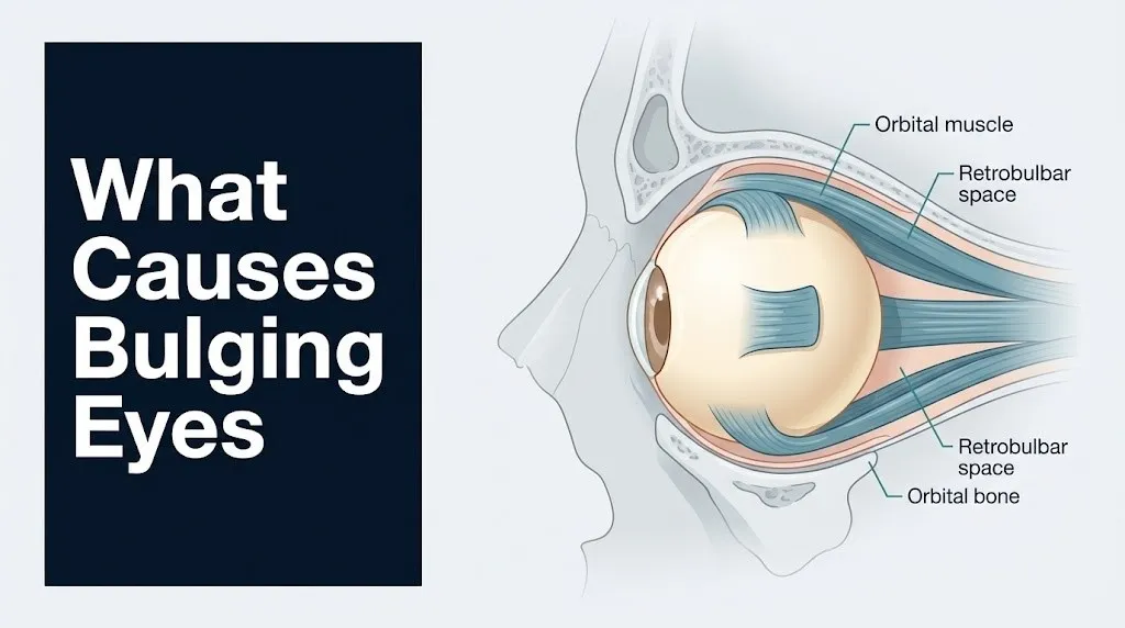
The result is a dramatic increase in retrobulbar volume. The orbital fat swells. The extraocular muscles enlarge, sometimes to several times their normal diameter. Because the bony orbit cannot stretch, the globe is pushed forward. A 2019 study published in JAMA Ophthalmology confirmed that the degree of proptosis in thyroid eye disease correlates directly with the degree of extraocular muscle and fat volume expansion on MRI.
Thyroid eye disease does not always coincide with abnormal thyroid hormone levels. Approximately 5% of patients with TED have normal thyroid function tests at the time of orbital diagnosis, according to the American Thyroid Association. This means a normal TSH level does not rule out thyroid eye disease as a cause of proptosis.
Clinical features of thyroid eye disease:
- Bilateral proptosis in the majority of cases (though often asymmetric)
- Eyelid retraction, producing a wide-eyed stare
- Periorbital puffiness, particularly on waking
- Dryness, grittiness, and redness from incomplete eyelid closure (lagophthalmos)
- Double vision (diplopia) from extraocular muscle restriction
- Optic nerve compression in severe cases, causing vision loss
Orbital Cellulitis as a Cause of Bulging Eyes
Orbital cellulitis is a bacterial infection of the tissues within the orbital septum (the fibrous barrier that separates the preseptal eyelid tissues from the deeper orbital contents), and it is one of the most time-sensitive causes of bulging eyes.
The distinction between preseptal cellulitis (infection anterior to the septum, which is less dangerous) and postseptal orbital cellulitis (infection posterior to the septum, which is the true emergency) is clinically critical. Postseptal orbital cellulitis produces proptosis. Preseptal cellulitis does not. Both cause periorbital swelling and redness, which is why misdiagnosis can occur without imaging.
The most common route of infection is direct spread from ethmoid sinusitis. The ethmoid sinuses lie immediately medial to the orbital wall, separated only by the thin lamina papyracea (a paper-thin bony plate that is literally named for its resemblance to paper). When infection erodes through or tracks around this barrier, bacteria enter the orbital fat, causing rapid cellulitis and sometimes a subperiosteal abscess (a pus collection between the orbital wall and the periorbita). The most common organisms are Staphylococcus aureus, Streptococcus pneumoniae, and, particularly in children, non-typeable Haemophilus influenzae.
Untreated or inadequately treated orbital cellulitis can progress to cavernous sinus thrombosis, meningitis, intracranial abscess, and vision loss from optic nerve compression. A 2021 review published in the British Journal of Ophthalmology found that subperiosteal abscess formation occurred in approximately 15 to 20% of pediatric orbital cellulitis cases.
Orbital cellulitis warning signs requiring same-day emergency evaluation:
- Proptosis developing over hours to 1 to 2 days
- Periorbital erythema and warmth
- Fever (temperature above 38.5°C / 101.3°F)
- Restricted, painful eye movement
- Vision change or diplopia
- Signs of meningeal irritation (stiff neck, severe headache, altered mental status)
Children under 9 years old develop orbital cellulitis more frequently than adults, primarily because their sinuses are still developing and the lamina papyracea is thinner.
Key Takeaway: Thyroid eye disease causes bilateral proptosis through TSH receptor antibody activation of orbital fibroblasts and GAG deposition in retrobulbar fat, while orbital cellulitis causes typically unilateral, rapidly progressive, painful proptosis from bacterial spread through the ethmoid sinus wall.
Cavernous Sinus Thrombosis and Eye Bulging
Cavernous sinus thrombosis (CST) is a rare but life-threatening cause of proptosis produced by septic clot formation in the cavernous sinus, a large venous channel located at the base of the skull just behind the orbit.
The cavernous sinus is a paired dural venous sinus that receives blood from the superior ophthalmic vein, which drains the orbit. When infection in the face, sinuses, or orbit spreads to the cavernous sinus (most commonly from severe sinusitis, dental infections, or orbital cellulitis), it triggers thrombosis of the sinus. The clot obstructs venous outflow from the orbit, causing rapid venous congestion, orbital edema, and proptosis, typically bilateral and rapidly evolving.
Cranial nerves III (oculomotor), IV (trochlear), V1 and V2 branches of the trigeminal nerve, and VI (abducens nerve) all pass through or adjacent to the cavernous sinus. This explains the classic neurological findings: multiple cranial nerve palsies, ophthalmoplegia (inability to move the eye in multiple directions), ptosis, and facial numbness. These neurological signs alongside proptosis, fever, and toxic appearance are the red flag constellation for cavernous sinus thrombosis.
The National Institutes of Health categorizes CST as a neurovascular emergency with mortality rates ranging from 20 to 30% even with modern antibiotic therapy, reflecting how rapidly this condition can progress to septicemia and intracranial spread.
Treatment requires urgent hospital admission, high-dose intravenous antibiotics, and neurosurgical or neurovascular consultation. There is no appropriate outpatient pathway for suspected CST.
Signs distinguishing CST from orbital cellulitis:
| Feature | Orbital Cellulitis | Cavernous Sinus Thrombosis |
|---|---|---|
| Proptosis laterality | Typically unilateral | Rapidly bilateral |
| Eye movement restriction | Limited, one eye | Multiple cranial nerves, both eyes |
| Neurological signs | Usually absent | Ptosis, facial numbness, altered consciousness |
| Progression | Days | Hours |
| Systemic toxicity | Moderate | Severe |
Orbital Tumors and Proptosis
Orbital tumors produce proptosis through direct mass effect: as the tumor occupies space within the rigid bony orbit, the globe is displaced forward, usually slowly and progressively over weeks to months.
The type of tumor determines both the speed of displacement and the associated clinical features. Primary orbital tumors arise from structures within the orbit itself. These include cavernous venous malformations (formerly called cavernous hemangiomas), which are the most common benign orbital tumors in adults, and optic nerve gliomas and optic nerve sheath meningiomas, which produce axial (straight-forward) proptosis along the optic nerve axis without much deviation of the globe to one side.
Secondary orbital tumors arise from structures adjacent to the orbit. Lacrimal gland tumors are particularly important. The lacrimal gland sits in the superolateral (upper outer) corner of the orbit. A tumor there displaces the globe inferomedially (down and toward the nose), which is a fairly specific clinical sign suggesting lacrimal gland pathology.
Metastatic tumors can also reach the orbit through hematogenous (bloodstream) spread. Breast cancer, lung cancer, and prostate cancer are the primary cancers most likely to metastasize to the orbit in adults. Breast cancer orbital metastases can actually cause enophthalmos (backward displacement of the globe) rather than proptosis, due to scirrhous (fibrotic) tumor contraction in the orbital fat.
According to research published in JAMA Ophthalmology, orbital tumors account for approximately 17 to 22% of proptosis cases referred to tertiary orbital centers.
Common orbital tumors by type and patient age:
| Tumor Type | Most Common Age Group | Proptosis Pattern |
|---|---|---|
| Cavernous venous malformation | Adults 20 to 60 | Gradual, axial, painless |
| Rhabdomyosarcoma | Children under 10 | Rapid, unilateral, painful |
| Optic nerve glioma | Children and young adults | Axial, associated with vision loss |
| Orbital lymphoma | Adults over 50 | Gradual, can be bilateral |
| Metastatic carcinoma | Adults with known primary cancer | Variable, often rapid |
| Lacrimal gland carcinoma | Adults 40 to 70 | Inferonmedial globe displacement |
Idiopathic Orbital Inflammation (Orbital Pseudotumor)
Idiopathic orbital inflammation (IOI), commonly called orbital pseudotumor, is a nonspecific inflammatory condition of the orbital soft tissues with no identifiable cause, and it is one of the more frequent causes of painful proptosis in adults.
The name reflects its history: before modern imaging, these lesions were biopsied and found to be inflammatory, not neoplastic, hence “pseudotumor.” IOI can involve any part of the orbital soft tissue: the orbital fat, the extraocular muscles, the lacrimal gland, or the optic nerve sheath. When it primarily affects the extraocular muscles, it resembles thyroid eye disease on imaging and requires careful distinction, typically by the pattern of muscle involvement. In thyroid eye disease, the muscle bellies are enlarged but the tendons are spared. In IOI myositis, both the bellies and the tendinous insertions of the extraocular muscles are involved.
Pain is a defining feature. IOI typically presents with acute or subacute orbital pain, proptosis, restricted eye movement, and conjunctival redness. The pain is often severe, deep within the orbit, and worsens with eye movement.
A subset of IOI is now recognized as IgG4-related orbital disease, in which elevated serum IgG4 antibodies and characteristic tissue infiltration by IgG4-positive plasma cells drive the inflammatory process. This distinction matters because IgG4-related disease may require different management and may involve other organ systems (salivary glands, pancreas, kidneys) simultaneously.
IOI typically responds to systemic corticosteroid therapy (noted here for context, not as a recommendation; management is directed by a board-certified ophthalmologist or neuro-ophthalmologist). Approximately 60 to 70% of patients show dramatic improvement within days of starting treatment, according to reviews in the British Journal of Ophthalmology.
Key Takeaway: Orbital tumors produce slow, progressive proptosis through mass effect within the fixed orbital volume, while idiopathic orbital inflammation causes rapid, painful proptosis and is distinguished from thyroid eye disease by tendon involvement on MRI.
Vascular Causes of Proptosis
Vascular abnormalities in or near the orbit produce proptosis through abnormal blood flow, either by creating high-pressure arterial input into venous spaces, or by obstructing venous outflow and causing orbital venous congestion.
The most clinically distinctive vascular cause is carotid-cavernous fistula (CCF): an abnormal connection between the carotid artery (or its branches) and the cavernous sinus, which is normally a low-pressure venous structure. When arterial blood is redirected at high pressure into the cavernous sinus, it backs up through the superior ophthalmic vein into the orbital venous system, causing engorgement of the orbital vasculature, chemosis (swelling of the conjunctival lining), proptosis, and a characteristic pulsating sensation behind the eye.
The clinical hallmark of CCF is pulsatile proptosis: the globe literally pulses forward and back in synchrony with the heartbeat. Patients and clinicians may also hear a bruit (a rushing, machine-like sound) over the orbit or temple when using a stethoscope or even, in some cases, with the naked ear. This combination of pulsatile proptosis and audible bruit is essentially pathognomonic for CCF.
CCF can be traumatic (caused by head injury, usually from a basal skull fracture that tears arterial vessels into the cavernous sinus) or spontaneous (arising from rupture of a cavernous sinus aneurysm or dural arteriovenous fistula). Spontaneous CCF is more common in postmenopausal women and individuals with connective tissue disorders.
Orbital varices (abnormal dilated venous structures within the orbit) are another vascular cause. These produce intermittent proptosis that worsens when the patient bends forward, coughs, or performs the Valsalva maneuver (bearing down), because increased venous pressure engorges the varix and pushes the globe forward. This position-dependent, intermittent pattern is a strong clinical clue.
According to the American Academy of Ophthalmology, vascular causes account for a small but diagnostically important proportion of proptosis referrals and often require specialized vascular imaging (CT angiography or digital subtraction angiography) for confirmation.
One Eye Bulging Causes (Unilateral Proptosis)
Unilateral proptosis (one eye bulging) carries distinct diagnostic implications compared to bilateral bulging and should prompt a more urgent search for a local orbital cause.
When only one eye protrudes, the clinical probability shifts strongly toward a cause that is anatomically local to that orbit. A space-occupying lesion (tumor, abscess, or vascular malformation) in the right orbit, for example, will displace the right globe forward without significantly affecting the left. By contrast, bilateral proptosis more often reflects a systemic process, typically thyroid eye disease or lymphoproliferative disease, that affects both orbits symmetrically (though often asymmetrically in degree).
The difference matters practically. A patient with bilateral, gradually progressive proptosis and a known history of Graves’ disease can reasonably pursue an outpatient ophthalmology evaluation scheduled within days to weeks (depending on whether vision is affected). A patient with new, unilateral proptosis and no prior orbital history needs to be seen urgently, because the list of causes includes orbital cellulitis, a rapidly growing tumor, and carotid-cavernous fistula.
One nuance: thyroid eye disease can present as unilateral proptosis in roughly 5 to 10% of cases, according to clinical series reviewed in JAMA Ophthalmology. This is called “unilateral Graves’ orbitopathy” and occurs when one orbit is far more severely affected than the other. Without a high index of suspicion and thyroid function testing, these cases can initially be misattributed to a unilateral mass lesion.
Causes of one eye bulging, ranked by clinical frequency in adults:
| Cause | Relative Frequency | Speed of Onset |
|---|---|---|
| Thyroid eye disease (asymmetric) | Most common overall | Weeks to months |
| Orbital cellulitis | Common, urgent | Hours to days |
| Orbital tumor (benign or malignant) | Moderately common | Weeks to months |
| Idiopathic orbital inflammation | Moderately common | Days to weeks |
| Carotid-cavernous fistula | Less common | Hours to days (traumatic) or gradual (spontaneous) |
| Subperiosteal or orbital abscess | Less common | Hours to days |
| Orbital varix | Rare | Intermittent |
Bilateral Proptosis and Systemic Disease
When both eyes bulge simultaneously, even asymmetrically, the cause is more likely systemic than local, and thyroid eye disease remains the leading explanation.
Bilateral proptosis is the presentation pattern that most strongly points a clinician toward thyroid eye disease. Both orbits contain TSH receptors and IGF-1R on their fibroblasts, so when TRAb circulates systemically, both orbits respond. One often responds more severely than the other, which is why thyroid eye disease can sometimes look unilateral. But when an imaging study (CT or MRI of the orbits) is done, bilateral extraocular muscle enlargement and bilateral orbital fat expansion are usually visible even in apparently unilateral cases.
Beyond thyroid eye disease, bilateral proptosis can reflect bilateral orbital involvement from lymphoproliferative disease (orbital lymphoma or leukemic orbital infiltration), systemic granulomatous disease (sarcoidosis involving the orbit), or metastatic disease to both orbits simultaneously (though bilateral orbital metastases are uncommon).
High intracranial pressure from any cause can theoretically contribute to bilateral periorbital changes, but true bilateral proptosis from elevated ICP alone is unusual and would be accompanied by papilledema (swelling of the optic discs) and other neurological signs.
According to the Mayo Clinic, bilateral proptosis that develops gradually over months, particularly in a woman aged 30 to 60 with any thyroid-related symptoms or history, should prompt TRAb testing and thyroid function evaluation even if no thyroid diagnosis has been previously established.
The rare but important condition Wegener’s granulomatosis (now formally called granulomatosis with polyangiitis, or GPA) can produce bilateral orbital involvement through granulomatous tissue deposition in the orbital fat. This is typically accompanied by sinus disease, respiratory symptoms, and positive ANCA (antineutrophil cytoplasmic antibody) serology.
Key Takeaway: Unilateral proptosis demands urgent evaluation to rule out infection, tumor, or vascular lesion, while bilateral proptosis most commonly indicates thyroid eye disease but can reflect systemic lymphoma, sarcoidosis, or granulomatosis with polyangiitis.
What Causes Bulging Eyes in Children
In children, the cause of proptosis differs substantially from the adult spectrum, and the most alarming pediatric cause, rhabdomyosarcoma, is also one of the most rapidly progressive orbital emergencies in pediatric medicine.
Rhabdomyosarcoma (RMS) is the most common primary malignant orbital tumor in children, accounting for approximately 35 to 40% of pediatric orbital malignancies, according to data compiled by the National Cancer Institute. It typically presents in children under 10 years old, with a peak incidence between 5 and 8 years. The onset is strikingly rapid, often producing visible, painful proptosis over a period of days to 1 to 2 weeks. The speed of onset is what makes RMS a medical emergency: delays in diagnosis worsen prognosis significantly. Any child with new, rapidly progressive unilateral proptosis without fever or signs of infection should be evaluated by a board-certified ophthalmologist and pediatric oncologist urgently.
Capillary hemangioma is the most common benign orbital tumor in infants and young children. Unlike the adult cavernous venous malformation, capillary hemangioma typically presents in the first few weeks to months of life, grows rapidly through the first year, and then involutes (shrinks) spontaneously over several years. Parents may notice an increasing protrusion of one eye in an infant that seems to enlarge with crying (because crying increases venous pressure, engorging the lesion).
Orbital cellulitis is also more common in children than adults, particularly in the 2 to 10 year age group, because ethmoid sinuses are more prone to infection in this developmental window and the lamina papyracea is thinner.
Optic nerve glioma in children is strongly associated with neurofibromatosis type 1 (NF1). Any child with known NF1, café-au-lait spots, or Lisch nodules who develops visual changes or axial proptosis should be screened by ophthalmology and neurology for optic pathway glioma.
Age-specific orbital causes in children:
| Age Group | Most Likely Cause | Clinical Clue |
|---|---|---|
| Infants (0 to 12 months) | Capillary hemangioma | Worsens with crying; soft, compressible |
| Toddlers 1 to 4 years | Rhabdomyosarcoma, dermoid cyst | Rapid onset; dermoid is firm, non-pulsatile |
| School age 5 to 10 years | Rhabdomyosarcoma, orbital cellulitis | Rapid onset pain and fever suggest cellulitis |
| Adolescents | Orbital cellulitis, Graves’ disease beginning | Fever, sinus history; or thyroid symptoms |
Causes of Bulging Eyes in Older Adults
In adults over 50, the differential diagnosis for proptosis shifts, and orbital lymphoma moves from a rare consideration to one of the more common diagnoses seen in tertiary orbital disease centers.
Orbital lymphoma most commonly represents a localized form of mucosa-associated lymphoid tissue (MALT) lymphoma, a low-grade B-cell lymphoma that arises from lymphoid tissue in the orbit and conjunctiva. It presents as slowly progressive, painless proptosis, often with a “salmon-colored” subconjunctival lesion visible to the examining clinician. Unlike orbital cellulitis or RMS, orbital lymphoma causes no pain and no fever. The gradual, painless quality of the presentation can delay diagnosis because patients may attribute the subtle change in eye prominence to aging or weight changes.
According to a review published in the British Journal of Ophthalmology, orbital lymphoma accounts for approximately 10 to 15% of orbital tumors in adults and the median age at diagnosis is 65 years. Systemic lymphoma evaluation is always required at diagnosis, because a proportion of patients with apparently localized orbital lymphoma will have or develop systemic disease.
Thyroid eye disease remains possible in older adults, but its onset in patients over 65 is less common than in middle-aged adults and may present more atypically with less dramatic lid retraction and more pronounced extraocular muscle restriction.
Metastatic disease to the orbit becomes a progressively more likely cause of proptosis as patients age, particularly in individuals with a history of breast, lung, prostate, or kidney cancer. Orbital metastases can grow rapidly, cause diplopia, or cause the paradoxical enophthalmos from scirrhous metastases described earlier.
Older adults with unexplained new proptosis should be evaluated by a board-certified ophthalmologist and, depending on imaging findings, referred to an orbital oncology specialist or hematologist-oncologist for systemic workup.
Key Takeaway: In children, rapidly progressive unilateral proptosis without fever should be assumed to be rhabdomyosarcoma until imaging and biopsy prove otherwise; in adults over 50, painless, gradual proptosis requires evaluation for orbital lymphoma and metastatic disease.
Bulging Eyes Causes in Females vs. Males
Thyroid eye disease is substantially more common in women than men, but male patients with this condition tend to experience more severe orbital involvement, and this sex-based difference has direct clinical implications.
The American Thyroid Association reports that Graves’ disease (the most common cause of hyperthyroidism and the underlying systemic disease in thyroid eye disease) affects women approximately 5 to 7 times more often than men. This reflects the broader female predominance of autoimmune thyroid conditions, which is driven in part by estrogen’s modulatory effects on the immune system, particularly its upregulation of B-cell activity and autoantibody production. Women are therefore far more likely than men to present to an ophthalmologist with bilateral, gradually progressive proptosis associated with thyroid dysfunction.
However, when thyroid eye disease does develop in men, the clinical severity is on average worse. Men with TED have higher rates of optic nerve compression (known as dysthyroid optic neuropathy), more extensive extraocular muscle involvement, and less favorable treatment outcomes in some series. A 2020 study published in JAMA Ophthalmology found that male sex was independently associated with more severe thyroid eye disease at presentation, even after controlling for duration of disease and thyroid hormone levels.
For other causes of proptosis, the sex distribution is more balanced. Orbital cellulitis, orbital tumors, and idiopathic orbital inflammation do not show strong sex predilection in adult populations.
One notable exception: carotid-cavernous fistula of the spontaneous (non-traumatic) type is more common in postmenopausal women, likely related to increased vascular fragility from reduced estrogen-mediated effects on vessel wall integrity. Women who develop pulsatile proptosis in the fifth decade or later without a history of head trauma should be evaluated for spontaneous CCF.
Risk Factors for Developing Proptosis
Several factors increase the likelihood of developing proptosis, and most of these act by increasing the probability of one specific underlying cause rather than proptosis in general.
Autoimmune thyroid disease is the dominant risk factor for proptosis in the general adult population. Having Graves’ disease places a person at lifetime risk for thyroid eye disease, though only approximately 25 to 50% of patients with Graves’ disease develop clinically significant orbital involvement.
Tobacco smoking is the most important modifiable risk factor for thyroid eye disease severity. Research published in the New England Journal of Medicine has confirmed that smokers with Graves’ disease have significantly higher rates of thyroid eye disease, more severe proptosis, and worse responses to orbital treatment compared to non-smokers. The mechanism is multifactorial: nicotine and tobacco combustion products activate orbital fibroblasts, increase TRAb levels, and promote orbital fat expansion. Smoking cessation is the single most impactful lifestyle modification for a patient with Graves’ disease and eye involvement.
Prior or current sinusitis is the primary risk factor for orbital cellulitis. Chronic or recurrent ethmoid sinusitis, dental infections in the upper jaw, and dacryocystitis (tear duct infections) all provide potential routes of bacterial spread into the orbital space.
Age and sex act as risk modifiers for specific causes as described in previous sections.
Pre-existing orbital abnormalities, including large orbital varices, sinus tumors adjacent to the orbit, or a history of prior orbital surgery, increase the risk that pathological processes will affect orbital volume and produce proptosis.
Key modifiable and non-modifiable risk factors:
- Autoimmune thyroid disease (Graves’ disease): strongest overall risk factor for adult proptosis
- Active tobacco smoking: dramatically worsens thyroid eye disease severity
- Chronic sinusitis, particularly ethmoid sinusitis: primary risk factor for orbital cellulitis
- Female sex: higher Graves’ disease incidence; however, male sex correlates with worse TED severity
- Age under 10: highest risk for rhabdomyosarcoma
- Age over 50: higher risk for orbital lymphoma and metastatic disease
- History of systemic cancer (breast, lung, prostate, kidney): orbital metastasis risk
- Neurofibromatosis type 1 (NF1): optic pathway glioma risk in children
- IgG4-related systemic disease: bilateral lacrimal gland enlargement and orbital inflammation
- Head trauma or basal skull fracture: traumatic carotid-cavernous fistula risk
Key Takeaway: Smoking is the most impactful modifiable risk factor for proptosis because it dramatically worsens thyroid eye disease, which accounts for 60% of all adult proptosis cases, and this effect is dose-dependent and partially reversible with cessation.
How Proptosis Is Diagnosed
The diagnosis of proptosis and its underlying cause requires a structured clinical evaluation that combines physical examination, measurement, and targeted imaging, and in many cases laboratory testing to identify systemic contributors.
The clinical examination starts with simple observation: does one or both eyes appear to protrude? Is the protrusion axial (straight forward, suggesting a retrobulbar mass along the optic nerve axis) or deviated to one side (suggesting a lesion in a specific orbital quadrant)? Can the eye be gently pushed backward into the socket (reducibility suggests no large solid mass)? Is there pulsation (vascular cause)?
Hertel exophthalmometry is the standard method for measuring proptosis objectively. A calibrated instrument measures the distance from the lateral orbital rim to the most anterior surface of the cornea in millimeters. Values above 20mm in adults (or greater than 2mm asymmetry between the two eyes) are generally considered outside the normal range, though the reference values vary by race: the American Academy of Ophthalmology recognizes higher normal values in Black patients compared to white patients.
CT scanning of the orbit is the first-line imaging study in most cases of new or unclear proptosis. It provides excellent bony orbital detail and can identify abscess formation, bony destruction by tumor, and the characteristic muscle enlargement pattern of thyroid eye disease rapidly and without the need for sedation in most adults. MRI of the orbit provides superior soft tissue characterization and is preferred for evaluating optic nerve lesions, orbital apex involvement, and distinguishing tumor types.
Thyroid function tests (TSH, free T4, free T3) and TSH receptor antibody (TRAb) levels are ordered in most adult patients with bilateral or unexplained proptosis, because thyroid eye disease is the most common cause and can be present even with normal thyroid hormone levels.
Diagnostic pathway for new proptosis:
- Visual acuity and visual field testing to assess optic nerve function
- Exophthalmometry to quantify the degree of protrusion
- Assessment of eye movement restriction and presence of diplopia
- Orbital CT scan (contrast-enhanced if infection or vascular lesion suspected)
- MRI orbit with and without contrast if CT is non-diagnostic or optic nerve involvement is suspected
- Thyroid function panel and TRAb antibodies for all adult bilateral cases and most unilateral cases without clear infectious cause
- CBC with differential if infection or lymphoma is suspected
- Tissue biopsy if imaging suggests mass lesion without a clear systemic diagnosis
- Referral to board-certified ophthalmologist or neuro-ophthalmologist for specialist evaluation and management planning
When Bulging Eyes Require Emergency Evaluation
Certain presentations of bulging eyes are medical emergencies that require evaluation at an emergency room or urgent call to a medical provider the same day, not a scheduled outpatient appointment.
The key clinical signals that indicate an emergency are: rapid onset (hours to days rather than weeks to months), presence of fever, pain on eye movement, restricted eye movement, vision change or loss, altered consciousness, severe headache, or a pulsating sensation in the eye. Any one of these in combination with proptosis raises the possibility of orbital cellulitis, cavernous sinus thrombosis, rapidly expanding orbital abscess, or acute vision loss from optic nerve compression, all of which can cause permanent blindness or death without rapid intervention.
Optic neuropathy from thyroid eye disease, while developing over a slower timeline than acute infection, can cause irreversible vision loss if not recognized and treated. Any patient with known thyroid eye disease who notices dimming of vision, loss of color saturation (colors appearing less vivid), or a new visual field defect needs to contact their ophthalmologist the same day, not at the next scheduled appointment.
The table below provides a clear framework for urgency assessment:
| Presentation Pattern | Likely Cause | Action Required |
|---|---|---|
| Gradual bilateral, no pain, known thyroid disease | Thyroid eye disease | Ophthalmology appointment within 1 to 2 weeks |
| Gradual unilateral, no pain, no systemic history | Orbital tumor or IOI | Ophthalmology appointment within days |
| Rapid unilateral, fever, periorbital redness | Orbital cellulitis | Emergency room same day |
| Rapid bilateral, fever, cranial nerve signs | Cavernous sinus thrombosis | Call 911 immediately |
| Pulsatile proptosis, audible bruit | Carotid-cavernous fistula | Emergency room same day |
| Any proptosis with acute vision change | Optic nerve compression | Emergency room same day |
| Child with rapid unilateral proptosis, no fever | Rhabdomyosarcoma | Ophthalmology within 24 to 48 hours |
Emergency Symptoms: When to Call 911 or Go to the ER
Certain presentations of bulging eyes require immediate emergency evaluation. Do not wait to see if these resolve on their own.
Call 911 or go to the nearest emergency room immediately if you experience:
- Sudden onset of proptosis (eye protrusion developing within hours) in one or both eyes
- Proptosis accompanied by fever above 38.5°C (101.3°F) and periorbital redness or swelling
- Any vision loss, sudden blurring, or sudden change in color perception alongside eye protrusion
- Severe pain behind or around the eye, particularly pain that worsens with eye movement
- Inability to move the eye in one or more directions combined with proptosis
- Severe headache, stiff neck, altered consciousness, or confusion alongside eye protrusion (possible cavernous sinus thrombosis or meningitis)
- A pulsing or throbbing sensation within the eye accompanied by proptosis and a rushing sound (possible carotid-cavernous fistula)
- Proptosis in a child under 10 that has developed or worsened within days without fever or signs of infection
These presentations can indicate orbital cellulitis with abscess formation, cavernous sinus thrombosis, acute optic nerve compression, or a rapidly expanding orbital malignancy, and they require emergency medical assessment, not an urgent care appointment or a phone call.
Frequently Asked Questions About Bulging Eyes
What is the most common cause of bulging eyes?
The most common cause of bulging eyes in adults is thyroid eye disease (Graves’ ophthalmopathy), which accounts for approximately 60% of adult proptosis cases according to the American Academy of Ophthalmology.
This condition occurs when TSH receptor antibodies activate orbital fibroblasts to produce glycosaminoglycans (GAGs) in the retrobulbar fat and extraocular muscles, increasing orbital volume within the fixed bony socket.
Adults with Graves’ disease who notice any change in eye prominence should be evaluated by a board-certified ophthalmologist, even if thyroid hormone levels are currently within normal range.
Can one eye bulging be more serious than both eyes bulging?
Yes, unilateral (one-eye) proptosis carries a higher immediate concern for a localized serious cause such as orbital cellulitis, tumor, or vascular lesion than bilateral proptosis, which more commonly reflects a systemic condition like thyroid eye disease.
A new, unexplained unilateral proptosis should be evaluated urgently by a board-certified ophthalmologist, with imaging ordered to rule out a space-occupying lesion or infection.
The speed of onset is equally important: rapid unilateral proptosis with pain and fever is an emergency, while slow unilateral proptosis without systemic signs still requires prompt but non-emergency evaluation.
What does it mean if a child suddenly has one bulging eye?
Sudden or rapidly progressive unilateral proptosis in a child under 10 years old without fever should raise immediate concern for rhabdomyosarcoma, the most common primary orbital malignancy in children.
If fever and periorbital redness are present, orbital cellulitis is more likely and requires same-day emergency evaluation.
Any child with new unilateral proptosis developing over days should be seen by a board-certified ophthalmologist within 24 to 48 hours for imaging and specialist assessment.
Can stress or anxiety cause eyes to bulge?
Stress and anxiety do not directly cause anatomical proptosis (true forward displacement of the globe within the orbit).
Stress can worsen autoimmune conditions including Graves’ disease, which is a leading cause of proptosis, and psychological stress has been reported as a trigger for Graves’ disease flares in some patients, but this is an indirect association rather than a direct mechanical cause.
If eyes appear more prominent during periods of high stress, an evaluation by a physician to check thyroid function and rule out thyroid eye disease is appropriate.
What specialist do I see for bulging eyes?
A board-certified ophthalmologist is the appropriate first specialist for any new or progressive proptosis.
Depending on the underlying cause identified, the ophthalmologist may co-manage the patient with an endocrinologist (for thyroid eye disease), a neuro-ophthalmologist (for optic nerve involvement or cavernous sinus lesions), an oculoplastic surgeon (for orbital decompression or tumor resection), or a hematologist-oncologist (for orbital lymphoma).
For acute presentations with fever, vision loss, or neurological signs, the first contact should be an emergency medicine physician, not an outpatient specialist.
How much does an eye need to protrude to be considered proptosis?
Proptosis is generally defined as globe protrusion greater than 20mm from the lateral orbital rim as measured by a Hertel exophthalmometer, or greater than 2mm of asymmetry between the two eyes.
The Cleveland Clinic notes that normal exophthalmometry values vary by race, with Black individuals having slightly higher normal values on average than white individuals.
A measurement within these ranges does not rule out early or mild thyroid eye disease if asymmetry is present, eyelid retraction exists, or there is a change from a prior baseline measurement.
Putting This Together: What to Do Next
Bulging eyes are not a single disease. They are a physical sign produced by a wide range of processes, some manageable and slowly progressive, others capable of destroying vision or threatening life within hours. The clinical priority is always the same: determine the cause and the urgency of intervention.
If you or someone you care for has noticed new or worsening eye protrusion, the first step is an objective assessment of the speed and pattern of onset. Gradual, bilateral, painless bulging in an adult with known or suspected thyroid disease requires a scheduled ophthalmology evaluation within days to weeks. Rapid, painful, one-sided bulging with fever requires an emergency room visit today, not tomorrow.
The one fact worth carrying from this article: no degree of proptosis is safe to observe without medical evaluation. A board-certified ophthalmologist can measure, image, and differentiate the cause with precision that no symptom checker or article can replicate. Make that appointment, and if any of the emergency signals described here are present, go to the nearest emergency room without delay.