All Feline Leukemia Symptoms and Warning Signs in 2026
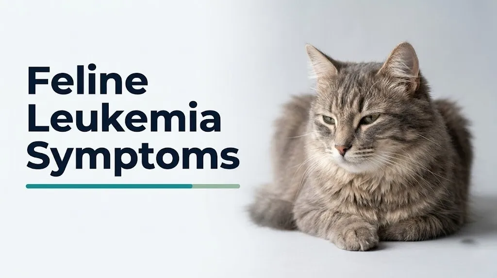
Feline leukemia symptoms range from subtle early changes like mild weight loss and reduced energy to life-threatening emergencies like open-mouth breathing and seizures, and knowing the full clinical picture of feline leukemia virus (FeLV) infection is the most important thing you can do for a cat that has tested positive or is showing concerning signs. This disease does not follow a single predictable path, and the symptoms depend entirely on which body system the virus has attacked most severely.
The American Association of Feline Practitioners (AAFP) identifies FeLV as one of the most consequential infectious diseases in domestic cats, estimating that approximately 2 to 3% of cats in the United States are FeLV-positive, with prevalence rising to 13% or higher in cats that are ill, have outdoor access, or live in multi-cat environments. The virus is entirely preventable through vaccination, yet it remains a leading cause of illness and death in cats worldwide.
This article covers every clinically recognized feline leukemia symptom, explains the specific virological and immunological mechanism behind each one, distinguishes early from late-stage disease, and gives you an unambiguous framework for deciding when your FeLV-positive cat needs a scheduled appointment versus an emergency veterinary hospital visit tonight.
Feline Leukemia Symptoms: What You Are Actually Observing
Feline leukemia symptoms are the observable behavioral and physical changes produced when FeLV, a gammaretrovirus, damages the cat’s immune system, suppresses bone marrow function, and in some cats drives the malignant transformation of lymphoid cells into lymphoma or leukemia.
FeLV does not produce a single fixed set of symptoms. The virus creates conditions for disease by destroying the cat’s immune defenses rather than causing direct tissue damage in most cases. Think of FeLV like a power outage in a security system: the damage is not from the outage itself but from everything that gets in when the security goes down. Secondary bacterial, viral, and fungal infections exploit the immunosuppressed state; cancers that the normal immune system would suppress grow unchecked; and organs that depend on normal blood cell production begin to fail.
The specific symptoms a cat displays depend on which secondary problem has emerged, whether FeLV-associated lymphoma is developing, and whether the virus is actively replicating (progressive infection) or suppressed in a latent state (regressive infection).
Symptom urgency overview:
| Observable Change | Urgency Level | Action |
|---|---|---|
| Mild weight loss, slight appetite decrease | Monitor closely | Vet appointment within 48 hours |
| Recurring infections, mild lethargy | Moderate concern | Vet appointment same or next day |
| Pale gums, significant weight loss, persistent vomiting | High concern | Same-day veterinary evaluation |
| Open-mouth breathing, collapse, seizures | Emergency | Go to emergency vet immediately |
Kittens under 16 weeks of age that test FeLV-positive are at dramatically higher risk for rapid symptom progression than adult cats, because their thymus and T-lymphocyte system have not fully matured. An FeLV-positive kitten showing any symptom other than mild lethargy warrants same-day veterinary evaluation rather than a wait-and-see approach.
What Is Feline Leukemia Virus and How Does It Affect Cats
Feline leukemia virus is a gammaretrovirus that infects domestic cats by integrating its genetic material permanently into the cat’s own chromosomes, giving it the ability to persist for the cat’s lifetime and to dysregulate the genes that control cell growth and immune function.
The infection process begins when FeLV enters the body through mucosal surfaces, most often through the mouth and nasal passages after contact with an infected cat’s saliva. The virus attaches to receptor proteins on susceptible cells, particularly lymphocytes and bone marrow progenitor cells, and uses its reverse transcriptase enzyme to convert its RNA genome into DNA. That DNA then inserts into the cat’s chromosomes as a provirus.
Once integrated, the provirus does two things that produce the observable disease. First, it uses the cat’s own cellular machinery to replicate new virus particles, which spread to infect additional cells throughout the body. Second, when the provirus integrates near a proto-oncogene (particularly c-myc) in a lymphoid cell, it dysregulates that gene’s expression through a process called insertional mutagenesis. The proto-oncogene, which normally controls when cells divide and when they die, becomes constitutively active. The result is a clone of lymphoid cells that divides without stopping, which is the biological definition of lymphoma or leukemia.
According to the Merck Veterinary Manual, FeLV is classified into four subgroups (A, B, C, and T) based on envelope protein differences, with subgroup A being the primary infectious form transmitted between cats. Subgroup C specifically impairs erythroid colony-forming units in the bone marrow, producing the severe non-regenerative anemia that is one of the most clinically devastating FeLV-associated conditions.
Adult cats with competent immune systems have a roughly 30 to 40% chance of suppressing active viral replication and entering a regressive infection state, according to AAFP 2020 testing guidelines. Cats that fail this immune suppression enter progressive infection, where the virus replicates continuously and the risk of clinical disease is high.
Early Feline Leukemia Symptoms in Cats
Early feline leukemia symptoms in cats are often so mild and non-specific that they are indistinguishable from common, self-limiting illnesses, which is precisely why the disease is so often advanced by the time a definitive diagnosis is made.
The very first detectable change in many FeLV-positive cats is a reduction in energy and enthusiasm. A cat that previously played readily, greeted family members at the door, or patrolled the house actively may begin sleeping more, engaging less with toys, and showing reduced interest in its environment. Owners frequently attribute this early lethargy to “getting older” or “having a lazy day,” even when the change is subtle but persistent over days to weeks.
A second early sign is intermittent mild fever. FeLV infection triggers cytokine release from activated immune cells, including tumor necrosis factor-alpha (TNF-alpha) and interleukins, which act on the hypothalamic thermoregulatory center to raise the body’s set point temperature. Cats with FeLV-related fever may feel warm to the touch on the ears, paws, and abdomen, appear slightly less comfortable than usual, and eat slightly less. This pattern recurs periodically even between periods of apparent normalcy.
Recurring mild respiratory symptoms, particularly episodic sneezing, mild nasal discharge, or eye discharge, can be early signs that FeLV-mediated immunosuppression is allowing latent feline herpesvirus-1 (FHV-1) or calicivirus infections to reactivate. These secondary infections cycle in and out because the immune system cannot maintain the suppression it normally achieves.
Quick Tip:
- Track your FeLV-positive cat’s weight weekly using a digital kitchen scale and a consistent technique (weigh yourself holding the cat, then subtract your own weight)
- Note the frequency and duration of any sneezing, nasal discharge, or eye discharge episodes in a simple log
- Any new symptom that persists for more than 3 to 5 days in an FeLV-positive cat warrants a call to a licensed veterinarian, not just observation
Cats that are FeLV-positive and also living with a feline immunodeficiency virus (FIV) co-infection tend to show earlier and more severe early symptoms because the combined retroviral load on the immune system is greater than either virus alone.
Weight Loss and Appetite Loss as Feline Leukemia Symptoms
Weight loss and appetite loss (anorexia) are among the most consistently observed feline leukemia symptoms across all stages of disease, and they develop through multiple simultaneous physiological mechanisms rather than a single cause.
The primary driver of appetite loss in FeLV-infected cats is the systemic inflammatory state produced by chronic viral replication and immune activation. Elevated circulating levels of TNF-alpha and interleukin-1 (IL-1) act directly on hypothalamic appetite centers, suppressing the normal signals that drive hunger. This is the same cytokine-mediated anorexia mechanism observed in any severe systemic infection or cancer, and it produces appetite suppression that does not improve simply by offering more palatable food.
Weight loss in FeLV cats has an additional mechanism beyond reduced caloric intake. When FeLV-associated lymphoma develops in the gastrointestinal tract (alimentary lymphoma), the tumor infiltrates the wall of the small intestine, disrupting normal mucosal absorption. Nutrients from food pass through an intestinal wall that cannot absorb them normally, producing protein-losing enteropathy and progressive cachexia, the extreme muscle and fat wasting characteristic of cancer-related weight loss. The cat eats something but loses weight anyway.
Observable weight loss patterns to report to your veterinarian:
- Visible prominence of the spine, hip bones, or shoulder blades where they were previously covered by muscle
- Feeling of reduced muscle mass when lifting the cat compared to weeks prior
- Consistent food avoidance or eating only a few bites before walking away
- Weight loss of more than 10% of normal body weight over 1 to 2 months
Senior FeLV-positive cats over 10 years of age are more likely to develop alimentary lymphoma than younger FeLV-positive cats, and in senior cats, weight loss from lymphoma can be particularly rapid because age-related muscle mass decline is already occurring in parallel.
Lethargy and Weakness as Feline Leukemia Symptoms
Lethargy and physical weakness in FeLV-positive cats develop through three converging mechanisms that worsen each other as the disease progresses: immune system exhaustion, anemia-related reduced oxygen delivery, and the direct energy-depleting effect of fighting chronic infection or cancer.
The immune system’s continuous mobilization against FeLV replication and secondary infections requires enormous metabolic resources. Activated lymphocytes, neutrophils, and macrophages consume glucose and amino acids at accelerated rates. The cat diverts energy from normal physical activity and behavioral engagement toward immune defense. The result is a cat that spends an increasing proportion of its time resting, loses interest in play, and responds to stimuli with less speed and enthusiasm than before.
When non-regenerative anemia develops from FeLV’s direct infection of bone marrow erythroid precursor cells, the reduced red blood cell mass means less oxygen is delivered to skeletal muscles per heartbeat. Muscles that cannot receive adequate oxygen fatigue rapidly even with minimal exertion. A cat with a hematocrit below 20% may be unable to play for more than a few seconds before stopping, and may breathe faster than normal even at rest as the cardiovascular system attempts to compensate by increasing heart rate.
The distinction between lethargy that warrants monitoring and lethargy that warrants immediate evaluation is practical:
| Lethargy Pattern | Clinical Interpretation | Action |
|---|---|---|
| Slightly less active than usual, still eating and drinking | Mild, monitor | Vet appointment within 48 hours |
| Reluctant to move, eating minimally, warm to touch | Moderate, likely systemic disease | Same-day veterinary evaluation |
| Will not rise from lying position, not eating or drinking | Severe, possible crisis | Emergency vet immediately |
| Cannot stand, unresponsive to handling | Emergency, collapse | Go to emergency vet immediately |
Male cats that are intact and outdoor-roaming may have acquired FeLV through bite wounds from fighting, meaning the history of fighting behavior combined with new lethargy is a clinically useful combination to report to a licensed veterinarian at the first appointment.
Key Takeaway: In an FeLV-positive cat, lethargy that progresses from “sleeping more than usual” to “won’t leave one spot” over 24 to 48 hours is not a wait-and-see situation; it warrants same-day evaluation because it may indicate acute decompensation from anemia, infection, or lymphoma.
Pale Gums and Anemia as Feline Leukemia Symptoms in Cats
Pale, white, or yellowish gum color in an FeLV-positive cat is one of the most clinically specific observable signs that something is seriously wrong, because the color of the mucous membranes directly reflects the cat’s blood oxygen-carrying capacity and overall circulatory status.
Normal cat gums are pink and moist. When a cat’s hematocrit (the percentage of blood volume occupied by red blood cells) drops significantly from the normal range of 25 to 45%, the gums lose their pink color because there are fewer red blood cells per unit of blood to provide the characteristic color. Pale gums in an FeLV-positive cat indicate non-regenerative anemia from bone marrow suppression.
FeLV-C subgroup specifically targets and infects the erythroid colony-forming units (precursor cells that develop into red blood cells) in the bone marrow. By directly infecting these precursor cells, FeLV-C impairs their maturation and replication, producing a severe, progressive non-regenerative anemia. “Non-regenerative” means the bone marrow is not compensating by producing new red blood cells at an increased rate, as it would in response to blood loss or immune-mediated hemolysis. The anemia simply worsens over time without effective intervention.
Yellowish gum color (icterus or jaundice) is a distinct and separate finding from paleness. If the gums appear yellow in addition to pale, this indicates that bilirubin is accumulating in the tissues, which can occur from liver disease, hemolytic disease, or biliary tract obstruction. FeLV-positive cats with jaundice should be evaluated the same day.
To check your cat’s gum color at home:
- Gently lift the upper lip to expose the gum tissue above the upper canine teeth.
- Observe the color in bright natural light or indoor lighting (flash photography on a phone camera works).
- Press one finger gently on the gum, release, and count the seconds until color returns (normal is less than 2 seconds, the capillary refill time).
- Compare gum color to the inside of your own lower lip for reference.
- Report any pale, white, grey, blue, or yellow gum color to a veterinarian immediately.
Cats receiving chemotherapy for FeLV-associated lymphoma may develop treatment-related anemia in addition to FeLV-related anemia. A board-certified veterinary oncologist should monitor complete blood counts throughout chemotherapy in FeLV-positive cats.
Respiratory Symptoms of Feline Leukemia
Respiratory symptoms in FeLV-positive cats arise from two entirely different mechanisms, and distinguishing between them determines the urgency level completely.
The first mechanism is secondary opportunistic respiratory infection. FeLV-mediated immunosuppression, particularly the depletion of CD4+ T-helper lymphocytes and impaired neutrophil function, allows pathogens that a healthy cat’s immune system would suppress to establish active infections. Latent feline herpesvirus-1 reactivates and causes rhinosinusitis. Feline calicivirus produces oral ulcers and upper respiratory tract inflammation. Bordetella bronchiseptica and Mycoplasma spp. cause bacterial bronchopneumonia. The observable symptoms include sneezing, nasal discharge (clear, yellow, or green), eye discharge (conjunctivitis), labored nasal breathing, and in more severe pneumonia cases, an audible wet or crackling sound when breathing.
The second mechanism is mediastinal lymphoma. When FeLV drives the malignant transformation of thymocytes (T-cells in the thymus) in the anterior mediastinum, the resulting tumor mass compresses the lungs, heart, and major thoracic vessels. As the mass grows, it physically reduces the space available for lung expansion. FeLV can also cause pleural effusion: fluid accumulation in the space between the lung surface and the chest wall, further compressing lung tissue. Both the mass and the effusion produce the same alarming symptom: dyspnea, or difficult breathing.
According to the Cornell Feline Health Center, open-mouth breathing in a cat is always abnormal. Cats do not pant like dogs. Open-mouth breathing in a cat indicates severe respiratory distress from any cause and requires immediate emergency veterinary evaluation.
Dyspnea from pleural effusion or a mediastinal mass is the single most common respiratory emergency in young FeLV-positive cats. A cat that is breathing with visible abdominal effort, extending its neck forward, sitting in a hunched posture with elbows pointed outward, or breathing with its mouth open must be taken to a 24-hour emergency veterinary hospital immediately.
Young male cats between 1 and 3 years of age with outdoor access are disproportionately represented among cats that develop mediastinal lymphoma from FeLV, making respiratory symptoms in this specific population a particularly urgent finding.
Lymphoma and Enlarged Lymph Nodes as Feline Leukemia Symptoms
Lymphoma associated with FeLV is the most common malignant disease in cats, and the enlarged lymph nodes or visible masses it produces are among the most diagnostically important feline leukemia symptoms an owner can observe.
The mechanism is specific. When FeLV’s proviral DNA integrates into a feline lymphoid cell near the c-myc proto-oncogene, it places c-myc under the continuous transcriptional influence of the viral promoter, the long terminal repeat (LTR). C-myc normally drives cell proliferation when stimulated by growth factors, then turns off. With the viral LTR driving its expression, c-myc remains continuously active, and the cell never stops dividing. A single transformed lymphoid cell produces a growing clone of identical malignant lymphocytes, which is lymphoma.
Owners can sometimes observe the physical consequences of this process. The peripheral lymph nodes, including the submandibular nodes under the jaw, the prescapular nodes in front of the shoulder blades, the axillary nodes in the armpits, and the popliteal nodes behind the knee, may become visibly or palpably enlarged. A healthy lymph node in a cat is small, soft, and moves slightly when pressed. An enlarged, firm, immobile, or significantly larger-than-normal lymph node in an FeLV-positive cat is clinically suspicious for lymphoma until proven otherwise.
Mediastinal lymphoma produces a visible bulging of the chest region or, more commonly, produces the respiratory symptoms described in the preceding section because of its anatomical location in the chest cavity.
According to published research in the Journal of Veterinary Internal Medicine, the two predominant FeLV-associated lymphoma types differ by age: mediastinal (thymic) lymphoma occurs predominantly in cats under 3 years of age, while alimentary (gastrointestinal) lymphoma occurs more often in older FeLV-positive cats, reflecting the different target cell populations that FeLV preferentially infects at different life stages.
Any palpable lump, swelling, or firm mass in an FeLV-positive cat, anywhere on the body surface, warrants evaluation by a licensed veterinarian within 24 to 48 hours for fine-needle aspirate cytology to determine whether the mass contains malignant lymphoid cells.
Key Takeaway: Lymphoma in FeLV-positive cats strikes young cats in the chest (mediastinal lymphoma producing breathing problems) and older cats in the gut (alimentary lymphoma producing weight loss and vomiting), and a licensed veterinarian who knows your cat’s FeLV status should evaluate any new lump, swelling, or mass within 48 hours.
Neurological Symptoms of Feline Leukemia
Neurological symptoms in FeLV-positive cats are among the least recognized by owners but among the most clinically important, because they indicate that either the central nervous system has been directly infected by FeLV, or a lymphoma is physically compressing neural structures.
FeLV can infect glial cells (the supportive cells of the central nervous system) and produce a condition called FeLV-associated neuropathy. The virus disrupts normal neural signaling by altering myelin production and axonal function in peripheral nerves and spinal cord tracts. The observable result is a cat that walks with an abnormal, wobbly gait (ataxia), that appears to have weakness or dragging of one or more limbs (paresis), or that holds its head tilted to one side (head tilt).
Behavioral changes are a neurological symptom that owners frequently misattribute to aging or personality changes. An FeLV-positive cat that becomes uncharacteristically aggressive, confused, disoriented in familiar spaces, or vociferous at unusual times may be experiencing FeLV-related central nervous system involvement. Vocalization in cats, particularly at night, combined with apparent confusion, is a specific behavioral indicator of neurological disease.
Seizures represent the most severe neurological manifestation of FeLV infection. They can arise from direct FeLV central nervous system invasion, from lymphoma compressing or infiltrating brain tissue, or from severe metabolic disturbances such as the hypocalcemia that can accompany certain mediastinal masses. A seizure in any cat is a veterinary emergency. A seizure in an FeLV-positive cat is a double emergency because the range of serious underlying causes is broad and all of them require immediate assessment.
Observable neurological signs to watch for:
- Wobbly or uncoordinated gait (ataxia)
- Weakness or dragging of one or more legs
- Head tilt to one side
- Rapid, involuntary eye movements (nystagmus)
- Sudden behavioral change: aggression, confusion, disorientation
- Nighttime vocalization or apparent hallucination behavior
- Seizure: loss of consciousness, muscle convulsions, paddling of limbs, loss of bladder or bowel control
Older FeLV-positive cats with systemic hypertension from FeLV-associated kidney disease can develop hypertensive retinopathy and sudden blindness, which may initially appear as a behavioral change or disorientation rather than an obvious neurological sign.
Kidney Disease and Mouth Sores as Feline Leukemia Symptoms
Kidney disease and oral lesions are two anatomically unrelated but physiologically connected feline leukemia symptoms that both arise from the immune dysregulation FeLV produces rather than from direct viral infection of kidney or oral tissue.
FeLV-associated immune complex glomerulonephritis develops when the immune system produces antibodies against FeLV antigens. These antigen-antibody complexes circulate in the bloodstream and deposit in the glomerular basement membrane of the renal glomeruli, triggering complement activation and local inflammation that damages the filtration surface. Over time, progressive glomerular damage reduces the kidney’s filtration capacity, producing renal proteinuria, eventual azotemia, and clinical kidney disease. The observable symptoms of FeLV-associated kidney disease include increased thirst, increased urination, weight loss, reduced appetite, and vomiting, a presentation that overlaps substantially with primary chronic kidney disease and requires blood and urine testing to differentiate.
Mouth sores in FeLV-positive cats (stomatitis and gingivitis) develop because the immunosuppressed cat cannot control the normal oral bacteria and secondary viral pathogens that healthy cats keep in balance. The oral cavity becomes inflamed at the gum line, the palate, and in severe cases throughout the entire oral mucosa. Owners observe a cat that is reluctant to eat hard food, drops food from its mouth while chewing, has noticeably bad breath, pawing at its mouth, drooling, or visible redness or ulceration of the gums visible when the mouth is opened.
According to the Journal of Feline Medicine and Surgery, severe lymphocytic-plasmacytic stomatitis, a painful and difficult-to-manage condition producing oral lesions, occurs at higher rates in cats that test positive for FeLV or FIV compared to retrovirus-negative cats, underscoring the role of immune dysfunction in perpetuating oral inflammatory disease.
Senior FeLV-positive cats are at compounded risk for oral disease because both FeLV-mediated immunosuppression and age-related decline in immune surveillance contribute simultaneously to the failure to control oral pathogens.
Progressive Versus Regressive Feline Leukemia Infection and Symptoms
Whether a cat develops observable feline leukemia symptoms depends critically on which of the three recognized FeLV infection outcomes occurs after exposure, and understanding this distinction is the single most important clinical concept for owners of FeLV-positive cats.
Progressive infection means the cat’s immune system failed to suppress viral replication after exposure. The virus replicates continuously in bone marrow, salivary glands, and other tissues. The FeLV p27 antigen is detectable in the blood by ELISA testing. This cat sheds live virus in saliva and other secretions and can infect other cats. Progressive infection carries a high risk of developing FeLV-associated disease within months to years.
Regressive infection means the cat’s immune system mounted a sufficient response to suppress active viral replication, but the proviral DNA is permanently integrated in bone marrow cells. The ELISA antigen test is negative (no circulating viral proteins), but PCR testing for proviral DNA in bone marrow or blood is positive. This cat does not shed live virus under normal circumstances and is not a transmission risk to other cats in its current state. Regressive infection does not typically produce clinical symptoms. However, the provirus can reactivate to progressive infection under significant immunosuppressive stress, such as high-dose corticosteroid therapy, co-infection with another serious illness, or extreme physiological stress.
Focal or atypical infection is a less common outcome where FeLV is detected in specific tissues (such as mammary tissue or the eye) but not consistently in blood or saliva.
The 2020 AAFP FeLV/FIV Testing Guidelines state that a positive FeLV ELISA test should be confirmed with IFA testing before a diagnosis of progressive infection is established, because ELISA false positives can occur. A positive ELISA with a negative IFA warrants PCR testing to determine whether regressive infection is present.
Cats in regressive infection may show no symptoms at all for their entire lives. The decision to treat or isolate these cats requires veterinary guidance based on individual circumstances.
Key Takeaway: A negative FeLV ELISA test does not rule out regressive infection detectable only by PCR for proviral DNA in bone marrow. If your cat was exposed to an FeLV-positive cat and tested negative on ELISA, ask your veterinarian whether PCR testing is appropriate to confirm the result.
End-Stage Feline Leukemia Symptoms
End-stage feline leukemia symptoms represent the point at which the cat’s remaining immune function and organ reserve are insufficient to sustain quality of life, and they require an honest clinical conversation with a licensed veterinarian about the cat’s experience and the appropriate path forward.
End-stage disease in FeLV-positive cats typically involves the convergence of multiple failing systems. Bone marrow has been so severely suppressed that the hematocrit is critically low, white blood cell counts are inadequate to mount any immune response, and platelets may be insufficient to prevent spontaneous bleeding. Lymphoma may be widespread. Kidneys may be failing. The cat’s body, in short, can no longer sustain itself.
End-stage signs that indicate severe, advanced disease:
- Complete and persistent refusal to eat or drink over more than 48 hours
- Body weight loss so severe that ribs, spine, and hip bones are visible and prominent
- Gums that are white, grey, or have a faint yellowish tinge
- Extreme weakness with inability to rise or maintain a normal resting posture
- Open-mouth breathing or breathing with visibly increased abdominal effort
- Severe oral ulceration making eating painful or impossible
- Neurological signs: seizures, stupor, or loss of responsiveness to familiar voices or touch
- Spontaneous bleeding from the nose, mouth, or visible under the skin as purple bruising spots (petechiae)
- Body temperature below normal (hypothermia) indicating circulatory failure
The timing of end-stage disease varies. Some FeLV-positive cats remain healthy with good quality of life for years with appropriate management. Others decline rapidly over weeks to months. A board-certified veterinary oncologist (Diplomate ACVIM-Oncology) can provide a prognosis based on the cat’s specific IRIS lymphoma stage, hematocrit, complete blood count, and overall condition.
Cats with FeLV-associated lymphoma that have achieved remission with chemotherapy and then relapse typically show the end-stage signs above within weeks of relapse.
What Causes Feline Leukemia and How FeLV Spreads
Feline leukemia symptoms are caused by feline leukemia virus, a gammaretrovirus transmitted between cats primarily through mucosal contact with infected bodily secretions, and understanding transmission routes is the foundation of preventing new infections in multi-cat households.
The primary transmission route is contact with infected saliva. Cats that groom each other, share food and water bowls, or engage in play biting exchange saliva in ways that allow FeLV to reach the nasal passages, oropharynx, and conjunctiva of a susceptible cat. Bite wounds from fighting are a particularly efficient transmission route because the virus is directly inoculated into tissue with disrupted mucosal barriers.
FeLV transmission routes by efficiency:
| Route | Efficiency | Common Context |
|---|---|---|
| Mutual grooming and shared bowls | High | Multi-cat households, cat colonies |
| Bite wounds from fighting | High | Outdoor cats, intact males, feral cats |
| Vertical transmission (queen to kittens) | High | In utero via placenta, nursing via milk |
| Shared litter boxes | Moderate | Virus detectable in urine and feces |
| Casual environmental contact | Low | Virus survives only hours outside the host |
| Human to human feline transmission | None | FeLV does not infect humans |
Vertical transmission from an FeLV-positive queen to her kittens occurs both in utero through the placenta and postnatally through nursing. The kittens receive a high viral inoculation at a stage when their immune systems are most vulnerable, producing progressive infection in most affected kittens. The AAFP recommends testing all breeding queens for FeLV before breeding and removing FeLV-positive queens from breeding programs.
FeLV does not infect humans, other domestic species, or wildlife. It is strictly a feline retrovirus. Family members handling an FeLV-positive cat have no risk of acquiring FeLV infection. This is an important clarification for worried owners, and it is supported by decades of epidemiological evidence and the American Veterinary Medical Association’s (AVMA) public health guidance on zoonotic risks from companion animal diseases.
How a Veterinarian Diagnoses Feline Leukemia Symptoms
A veterinarian diagnoses feline leukemia virus infection and its associated clinical presentations through a structured sequence of tests, beginning with simple in-clinic antigen screening and advancing to confirmatory and staging procedures as needed.
The standard diagnostic sequence:
- FeLV p27 ELISA test (point-of-care): A blood or saliva sample detects the FeLV p27 core antigen produced by actively replicating virus. This is the standard first-line screening test, available at most veterinary clinics within minutes. A positive result indicates circulating FeLV antigen. A negative result does not rule out regressive infection.
- Immunofluorescence assay (IFA): A blood smear is sent to a reference laboratory. The IFA detects FeLV antigen within circulating white blood cells and platelets, confirming that the virus has spread to bone marrow and is in active progressive infection. A positive IFA after a positive ELISA confirms progressive infection. A negative IFA after a positive ELISA suggests early infection or a false-positive ELISA, warranting retesting in 60 days or PCR testing.
- PCR for FeLV proviral DNA: The most sensitive test available. PCR detects proviral DNA in bone marrow or blood regardless of whether the virus is actively replicating, making it the only test that identifies regressive infection. Cats in regressive infection test ELISA-negative and IFA-negative but PCR-positive.
- Complete blood count (CBC): Quantifies red blood cells (hematocrit), white blood cells (neutrophil count for infection risk), and platelets (bleeding risk). Non-regenerative anemia, leukopenia, and thrombocytopenia are common in progressive FeLV disease.
- Serum chemistry panel: Assesses kidney function (BUN, creatinine), liver values, protein levels, and electrolytes. Identifies FeLV-associated kidney disease from immune complex glomerulonephritis.
- Thoracic radiography: Identifies mediastinal mass, pleural effusion, or pulmonary infiltrates in cats with respiratory symptoms.
- Abdominal ultrasound and fine-needle aspirate: Assesses lymph node and organ size; cytology of aspirated lymph nodes identifies malignant lymphoid cells in lymphoma.
A board-certified veterinary oncologist (Diplomate ACVIM-Oncology) should be involved in the management of any FeLV-positive cat with confirmed lymphoma for staging and treatment planning.
Which Cats Are at Highest Risk for Feline Leukemia
Feline leukemia virus does not affect all cats equally, and several specific biological and lifestyle factors determine which cats are most likely to be exposed, most likely to develop progressive infection after exposure, and most likely to develop severe clinical disease rapidly.
Age is the most powerful biological determinant of infection outcome. Kittens under 16 weeks of age have an immature thymus and T-lymphocyte system that cannot mount the adaptive immune response needed to achieve regressive infection. Studies indicate that nearly all kittens exposed to FeLV during this developmental window develop progressive infection, compared to approximately 30 to 40% of immunocompetent adult cats. Every week of additional immune maturation before first exposure improves the chance of a regressive outcome.
Lifestyle and environment are the most powerful exposure determinants:
- Outdoor cats and cats with outdoor access encounter FeLV-positive cats through social grooming and fighting
- Multi-cat households where one cat is FeLV-positive create ongoing daily exposure for other cats through shared food bowls, grooming, and litter boxes
- Shelter and rescue cats have higher prevalence rates (up to 13% in at-risk populations per AAFP data) than privately owned indoor cats
- Intact male cats that roam and fight have higher exposure from bite wound inoculation
- Kittens born to FeLV-positive queens are exposed at the highest viral dose at the most immunologically vulnerable period
According to the American Association of Feline Practitioners (AAFP), FeLV vaccination is highly effective and is recommended for all cats with any outdoor access or potential exposure to FeLV-positive cats. Vaccination does not protect 100% of cats, but two appropriately timed doses produce protective immunity in the majority of vaccinated cats.
Purebred cats from closed breeding colonies have theoretically reduced genetic immune diversity compared to outbred domestic cats, though no specific breed has been confirmed to have statistically higher FeLV susceptibility in peer-reviewed veterinary literature.
When to Take Your Cat to the Emergency Vet for Feline Leukemia Symptoms
Knowing exactly which feline leukemia symptoms require a 2 a.m. emergency hospital drive versus a morning phone call to your regular veterinarian is the most practically urgent information in this article.
Symptoms that allow monitoring with a scheduled appointment within 24 to 48 hours:
- Mild, gradual weight loss over several weeks without other concerning signs
- Slightly reduced appetite but still eating and drinking
- Occasional sneezing or mild nasal discharge without breathing difficulty
Symptoms that require same-day evaluation by a licensed veterinarian:
- Persistent complete appetite loss for more than 48 hours
- Visible pale gum color when the mouth is lifted
- Persistent vomiting or diarrhea for more than 24 hours
- Any new palpable lump or swelling
- Gradual increase in breathing rate at rest
Symptoms that require immediate emergency veterinary hospital evaluation tonight:
- Open-mouth breathing at any time for any duration
- Breathing with visible abdominal effort or neck extended forward
- Collapse or inability to stand
- Seizure of any duration
- Gums that are white, grey, or blue
- Spontaneous bleeding from nose, mouth, or visible skin bruising
- Extreme, sudden deterioration in condition over hours
Take your cat to the nearest 24-hour emergency veterinary hospital immediately if any of the emergency symptoms above are present. Do not wait to see if the cat improves. Do not call your regular vet first if the clinic is not open. Open-mouth breathing in a cat indicates severe respiratory distress that can progress to respiratory failure within minutes to hours.
Emergency Signs: When to Take Your Cat to the Vet Immediately
Certain feline leukemia symptoms indicate a life-threatening emergency. Do not monitor these at home or wait for a regular veterinary clinic to open.
Take your cat to a 24-hour emergency veterinary hospital immediately if you observe:
- Open-mouth breathing or panting: Cats never normally breathe with an open mouth. This indicates severe respiratory distress from pleural effusion or a mediastinal mass compressing the lungs. This can become fatal within hours without treatment.
- Complete collapse or inability to stand: Indicates severe anemia, circulatory failure, or neurological crisis requiring immediate stabilization.
- Seizures or muscle convulsions: Indicate FeLV-associated neurological involvement or severe metabolic crisis. Any seizure in an FeLV-positive cat is an emergency.
- White, grey, or blue gum color: Severe pallor or cyanosis indicates critically low red blood cell count or inadequate circulatory oxygen delivery.
- Spontaneous bleeding: Visible petechiae (pinpoint red-purple spots) under the skin, bleeding from the nose or mouth without trauma, or blood in urine indicates thrombocytopenia from bone marrow failure.
- Sudden complete behavioral collapse: A cat that was alert and is now completely unresponsive or stuporous is in neurological or circulatory crisis.
These presentations indicate severe FeLV-associated disease including acute respiratory failure, uremic crisis, neurological involvement, or complete circulatory decompensation. They require emergency stabilization at a 24-hour veterinary hospital, not a phone consultation or a wait-and-see approach.
Frequently Asked Questions About Feline Leukemia Symptoms
How do I know if my cat has feline leukemia?
The only reliable way to know if your cat has feline leukemia is through veterinary testing, specifically the FeLV p27 antigen ELISA test performed on a blood or saliva sample.
Observable symptoms including weight loss, lethargy, pale gums, recurring infections, and respiratory distress are consistent with FeLV but also occur in many other feline diseases, making symptom observation alone insufficient to confirm or rule out the diagnosis.
Any cat showing persistent symptoms or with known exposure to an FeLV-positive cat should be tested by a licensed veterinarian, with positive ELISA results confirmed by immunofluorescence assay (IFA) and, if needed, PCR for proviral DNA.
Can a cat with feline leukemia live a normal life?
Some cats with FeLV, particularly those in regressive infection who test antigen-negative on ELISA, can live normal or near-normal lives with no observable symptoms for years or even their entire lives.
Cats with progressive FeLV infection that develops into lymphoma, severe anemia, or significant immunosuppression will have a markedly shortened lifespan and require active veterinary management to maintain quality of life.
A licensed veterinarian can assess which infection category your cat falls into through testing and help you understand what that means for your cat’s specific prognosis and management needs.
How long can a cat live with feline leukemia?
Survival time for FeLV-positive cats varies widely based on infection category and disease status; cats with regressive infection may live normal lifespans, while cats with progressive infection and clinical disease have a median survival of approximately 2 to 3 years from diagnosis, according to observational data in veterinary internal medicine literature.
Cats with FeLV-associated lymphoma treated with combination chemotherapy can achieve remission periods of months to over a year in some cases, depending on lymphoma type, stage, and response to treatment.
Individual prognosis should be discussed with a licensed veterinarian or board-certified veterinary oncologist based on the cat’s specific test results, overall condition, and disease manifestations.
What does feline leukemia do to a cat’s immune system?
FeLV progressively depletes CD4+ T-helper lymphocytes, impairs neutrophil phagocytic function, and disrupts both cell-mediated and humoral immune responses, leaving the cat unable to control secondary infections or suppress developing malignancies.
The virus integrates into immune cell precursors in the bone marrow, disrupting the production of lymphocytes, neutrophils, and other immune cells from their earliest developmental stage.
The observable result is a cat that develops recurring infections that are difficult to clear, fails to respond normally to routine vaccination, and is susceptible to opportunistic pathogens that would cause minimal disease in an immunocompetent cat.
Is feline leukemia contagious to other cats or to humans?
Feline leukemia virus is contagious to other cats through direct contact with infected saliva, nasal secretions, and other bodily fluids, meaning FeLV-positive cats in progressive infection should be isolated from FeLV-negative cats in the household.
FeLV does not infect humans, dogs, or other domestic species; it is strictly a feline retrovirus with no documented zoonotic transmission, and this is supported by decades of epidemiological research and confirmed by the American Veterinary Medical Association.
All cats sharing a household with a known FeLV-positive cat should be tested by a licensed veterinarian and vaccinated against FeLV if they are not already protected.
What are the final signs of feline leukemia in a cat?
The final signs of feline leukemia in a cat include complete refusal to eat or drink, extreme muscle wasting with visible prominence of bones, white or yellowish gum color, inability to rise or maintain normal posture, open-mouth breathing, and in some cases seizures or complete loss of responsiveness.
These signs indicate that the remaining organ reserve and immune function are insufficient to sustain quality of life, and a frank conversation with a licensed veterinarian about the cat’s experience and the appropriateness of continued treatment versus palliative care is warranted.
A board-certified veterinary oncologist or the cat’s primary veterinarian can assess whether any further intervention would benefit the cat or whether comfort-focused care is the most appropriate path.
What to Do Right Now
If your cat has tested positive for FeLV or is showing the symptoms described in this article, the single most useful immediate step is an honest assessment of where on the urgency scale your cat currently sits. Mild weight loss and a slightly reduced appetite over two weeks: call a licensed veterinarian today, schedule an appointment within 48 hours, and specifically ask for a CBC, serum chemistry panel, and chest radiographs if respiratory symptoms are present.
Any open-mouth breathing, white gums, seizure, or collapse: go to a 24-hour emergency veterinary hospital immediately without waiting for your regular clinic to open.
For FeLV-positive cats without current symptoms, the most protective step is a baseline diagnostic workup including CBC, serum chemistry, and urinalysis every six months, so that changes from your cat’s individual normal can be detected early rather than discovered at a crisis stage. The disease this article describes is serious, but an FeLV-positive cat that is well-monitored, kept indoors, isolated from FeLV-negative housemates, and evaluated promptly at every new symptom gives both the cat and its owner the best possible position.