Low Alkaline Phosphatase Symptoms: Complete Guide 2026
Low alkaline phosphatase symptoms include bone pain, stress fractures, premature tooth loss, muscle weakness, fatigue, and in severe cases, seizures and respiratory failure, all produced by the body’s inability to properly mineralize bone and regulate key metabolic substrates. The specific symptoms you experience depend on how low your ALP level is, which underlying condition is causing the deficiency, and which organ system is most affected.
Low ALP is not simply a number on a lab report. According to the National Institute of Arthritis and Musculoskeletal and Skin Diseases (NIAMS), alkaline phosphatase plays a direct physiological role in bone formation, liver function, and intestinal absorption. When ALP activity falls below the functional threshold, the consequences are visible in bones, teeth, muscles, and the nervous system, sometimes years before a diagnosis is made.
This article covers the full symptom profile associated with low alkaline phosphatase, explains the specific biochemical mechanism behind each symptom, addresses the most medically serious condition linked to chronically low ALP (hypophosphatasia), and tells you precisely when and to whom you should bring these findings.
What Is Alkaline Phosphatase and Why Does It Matter
Alkaline phosphatase (ALP) is a zinc-dependent enzyme found on the surface of multiple cell types throughout the body, where it cleaves phosphate groups from a range of molecules to regulate bone mineralization, liver bile flow, and intestinal phosphate absorption.
ALP is not a single enzyme but a family of four isoenzymes, each produced by a different tissue. The liver isoenzyme is expressed on the hepatic canalicular membrane, the specialized surface of liver cells that faces the bile ducts. The bone isoenzyme is produced by osteoblasts, the cells responsible for building and mineralizing new bone tissue. The placental isoenzyme (sometimes called the Regan isoenzyme) is produced only during pregnancy. The intestinal isoenzyme is expressed on the brush border epithelium of the small intestine.
In a standard blood test, the reported ALP value reflects the combined activity of all circulating isoenzymes. The liver and bone isoenzymes contribute the majority of the total ALP reading in healthy, non-pregnant adults. Children and adolescents have substantially higher ALP than adults because their rapidly growing bone produces large amounts of the bone isoenzyme during skeletal development. This physiological difference matters enormously: a total ALP reading that would be “normal” for an adult can represent a genuinely low result in a growing child, and vice versa.
ALP requires zinc as a structural cofactor embedded within its active site. Without adequate zinc, the enzyme cannot maintain its three-dimensional shape and loses catalytic activity, which is why zinc deficiency is one of the dietary causes of measurably low ALP in blood tests.
The National Institutes of Health states that ALP is one of the most frequently measured enzymes in clinical laboratory medicine, appearing on the standard comprehensive metabolic panel (CMP) ordered during routine physical examinations and hospital admissions worldwide.
Low Alkaline Phosphatase Symptoms: The Full Clinical Picture
Low alkaline phosphatase symptoms span multiple body systems, ranging from mild fatigue and bone aching to severe fractures, seizures, and respiratory failure, depending on the degree of enzyme deficiency and the age at which deficiency develops.
The symptoms arise because ALP performs specific biochemical work that no other enzyme fully replaces. When ALP activity is inadequate, three natural substrates accumulate in the blood and tissues: inorganic pyrophosphate (PPi), pyridoxal-5-phosphate (PLP), and phosphoethanolamine (PEA). Each accumulated substrate produces a distinct set of clinical problems. PPi blocks hydroxyapatite crystal formation in bone. PLP cannot cross cell membranes without being dephosphorylated first, leading to intracellular vitamin B6 deficiency that affects the nervous system. PEA accumulation contributes to impaired cellular membrane function across multiple tissues.
The clinical picture of low ALP differs substantially between mild deficiency and severe deficiency. Mild, intermittent low ALP (from temporary causes like zinc deficiency, hypothyroidism, or recent cardiac surgery) may produce no symptoms at all, or only subtle fatigue and mild musculoskeletal aching. Chronically low ALP from genetic causes, particularly hypophosphatasia (HPP), produces a progressive, multi-system symptom burden that worsens without treatment.
Individual symptom burden also varies by sex and age. According to a review published in the Journal of Bone and Mineral Research, women with HPP tend to present with stress fractures of the metatarsal bones and femoral shaft more frequently than men, possibly reflecting the additional demand of pregnancy and lactation on bone mineral reserves in an already-compromised mineralization system. Older adults with low ALP may have reduced bone density as their primary finding, without the dramatic early-onset fracture pattern seen in younger patients with the same enzyme deficiency.
| Symptom Category | Specific Symptoms | Underlying Mechanism |
|---|---|---|
| Musculoskeletal | Bone pain, stress fractures, joint pain, skeletal deformity | PPi accumulation inhibits hydroxyapatite mineralization in osteoid |
| Dental | Premature tooth loss before age 5, absent cementum, poor enamel | Cementum production requires ALP; absent cementum detaches tooth roots |
| Neurological | Seizures (in infants), B6-related neuropathy, craniosynostosis | PLP accumulation; intracellular B6 deficiency; skull suture hypomineralization |
| Muscular | Proximal muscle weakness, difficulty walking, exercise intolerance | Impaired phosphate metabolism affecting muscle energy production |
| Systemic | Fatigue, chronic pain, reduced quality of life | Multi-system substrate accumulation and mineralization failure |
| Renal | Nephrocalcinosis (calcium deposits in kidneys) | Calcium and phosphate dysregulation from abnormal mineral metabolism |
Bone Pain and Stress Fractures From Low Alkaline Phosphatase
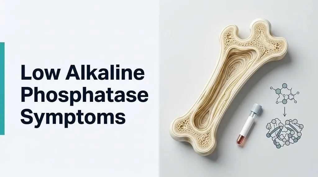
Bone pain and recurrent stress fractures are the most clinically prominent low alkaline phosphatase symptoms in adolescents and adults, produced by the failure of the osteoid matrix to mineralize properly when ALP activity is insufficient.
To understand why, think of bone as a construction project with two separate crews. The first crew (osteoblasts) lays down the protein scaffold, called osteoid, made primarily of type I collagen. The second crew deposits mineral crystals (hydroxyapatite, a calcium-phosphate compound) into that scaffold, hardening it into rigid bone. ALP’s role is to destroy PPi, which is a natural byproduct of collagen synthesis that would otherwise block hydroxyapatite from being deposited. When ALP is deficient, PPi accumulates at the mineralization front, and the second crew’s work stops. The protein scaffold is laid down, but it stays soft and unfortified. That unmineralized osteoid cannot bear normal weight-bearing loads, producing pain and eventually fracturing.
According to research published in the Bone journal, stress fractures in adults with hypophosphatasia show a characteristic radiographic pattern: they typically occur in the lateral cortex of the femoral (thigh bone) shaft, a location unusual for typical osteoporotic fractures but consistent with the loading pattern in weakened cortical bone. These fractures are often described as “chalk stick” fractures because of how cleanly the softened bone breaks.
Bone pain from low ALP tends to be deep, aching, and diffuse rather than sharp and localized. It is often worst in the feet, shins, and thighs, the weight-bearing bones that experience the greatest mechanical stress. Children with hypophosphatasia may have rachitic-like bone deformities including bowed legs and wrist widening from abnormal growth plate mineralization.
Postmenopausal women with undiagnosed HPP are at particular risk because declining estrogen further reduces bone density, compounding the ALP-driven mineralization failure. A woman in her 50s presenting with atypical femoral fractures should have ALP levels reviewed as part of her fracture workup, particularly if she has been on bisphosphonate therapy for osteoporosis without improvement, since bisphosphonates further suppress ALP activity and can worsen HPP-related fractures.
Dental Problems and Premature Tooth Loss as Low ALP Symptoms
Premature loss of deciduous (baby) teeth before age 5, without preceding decay or trauma, is one of the most recognizable symptoms of chronically low alkaline phosphatase and a hallmark diagnostic feature of hypophosphatasia.
The reason is anatomical and specific. Teeth are anchored to the jawbone not by direct bone attachment but by the periodontal ligament, a network of collagen fibers that runs between the tooth root and the surrounding alveolar bone. These fibers embed into a thin layer of mineralized tissue covering the tooth root called cementum. Cementum production depends directly on ALP activity in cementoblasts, the specialized cells responsible for depositing this anchoring layer. When ALP is deficient, cementum does not form properly or at all. Without cementum, the periodontal ligament fibers have nothing to attach to on the root surface. Teeth become loose and shed intact, root and all, without any decay visible on the crown.
The National Center for Advancing Translational Sciences (NCATS) describes premature tooth loss with intact roots before age 5 as a clinically distinctive finding that should prompt evaluation for hypophosphatasia in any child, even if bone symptoms are absent. The condition affecting only this dental presentation is called odontohypophosphatasia, and it can occur without obvious skeletal symptoms in childhood while still reflecting meaningful TNSALP deficiency.
Adults with HPP can also experience adult tooth loss from the same mechanism, particularly in the front teeth (incisors), and may report a history of childhood baby tooth loss that went unexplained. Periodontal disease in adults with low ALP often progresses faster than typical because impaired cementum regeneration means that damaged periodontal attachment does not rebuild effectively.
Children presenting with premature tooth shedding should be evaluated by a board-certified pediatric dentist and a board-certified pediatrician, with a comprehensive metabolic panel including ALP and, if low, referral to a clinical geneticist or metabolic bone disease specialist for HPP evaluation.
Muscle Weakness and Joint Pain With Low Alkaline Phosphatase
Muscle weakness and joint pain are reported by a substantial proportion of adults with hypophosphatasia, and their underlying mechanism involves abnormal phosphate and pyrophosphate metabolism at the level of muscle tissue and synovial joints.
PPi is not only an inhibitor of bone mineralization. When it accumulates in joints due to insufficient ALP activity, it can trigger calcium pyrophosphate dihydrate (CPPD) crystal deposition in articular cartilage and synovial fluid. CPPD crystal deposits in joints cause a condition sometimes called pseudogout: acute joint inflammation with pain, swelling, and warmth that closely mimics gout but is driven by pyrophosphate rather than uric acid crystals. Adults with HPP who experience recurrent joint inflammation should have synovial fluid examined for CPPD crystals, which would point toward ALP deficiency as a root cause rather than primary gout.
Proximal muscle weakness in HPP (weakness centered in the shoulder girdle, hip flexors, and thigh muscles rather than the hands and feet) likely reflects impaired phosphate availability for ATP (adenosine triphosphate) synthesis in muscle cells, since phosphate is a required substrate for the mitochondrial ATP-generating process. Muscle biopsy in some HPP patients has shown myopathic changes consistent with energy metabolism disruption.
According to a clinical series published in Genetics in Medicine, muscle weakness and pain are reported by approximately 50% of adults with HPP who have confirmed ALPL gene mutations, yet these symptoms are frequently attributed to fibromyalgia, chronic fatigue syndrome, or idiopathic musculoskeletal pain before the ALP abnormality is recognized. This misattribution delays diagnosis by an average of several years in published case series.
Older adults are particularly likely to have their HPP-related muscle weakness attributed to age-related sarcopenia (age-associated muscle mass loss), delaying recognition of the metabolic cause and appropriate specialist referral.
Key Takeaway: The triad of unexplained bone pain, premature tooth loss, and proximal muscle weakness in the same patient should prompt a review of their ALP level before a diagnosis of fibromyalgia, osteoporosis, or age-related decline is accepted as the final answer.
Fatigue and Neurological Symptoms From Low ALP
Chronic fatigue and neurological symptoms associated with low alkaline phosphatase arise from the accumulation of pyridoxal-5-phosphate (PLP), the active form of vitamin B6, which cannot cross cell membranes without ALP to first remove its phosphate group.
The mechanism is counterintuitive. ALP deficiency causes plasma PLP to be elevated (because it cannot be dephosphorylated and taken up by cells), yet inside cells, where PLP needs to function as a coenzyme for over 100 enzymatic reactions, it is effectively deficient. PLP is required for the synthesis of multiple neurotransmitters including serotonin, dopamine, GABA (gamma-aminobutyric acid), and norepinephrine. Intracellular B6 deficiency therefore disrupts neurotransmitter production, which contributes to fatigue, mood changes, and in infants and young children, frank seizure activity.
In infantile hypophosphatasia, PLP-driven seizures are a medical emergency. These seizures respond poorly to standard anticonvulsant medications because the root problem is not electrical overexcitability but biochemical: GABA production is impaired because glutamate decarboxylase (a PLP-dependent enzyme) cannot function without intracellular B6. The only effective acute management is pyridoxine (vitamin B6) supplementation to partially compensate, and definitive treatment in eligible patients requires enzyme replacement therapy. Standard seizure medications administered without recognizing this mechanism can fail to control the seizures while the infant’s respiratory status deteriorates.
In adults, fatigue from ALP deficiency is more diffuse and non-specific. It is one reason that chronically low ALP is often overlooked on lab panels: fatigue is attributed to many other causes, and the ALP result is not flagged as an explanation. Adults with persistent fatigue whose comprehensive metabolic panel shows low ALP should have a follow-up evaluation that includes plasma PLP levels, serum zinc, and TSH to identify the most likely underlying cause.
Women during the postpartum period may experience fatigue from multiple concurrent causes. If a postpartum woman has low ALP on a blood panel, it is worth distinguishing whether this represents true enzyme deficiency versus the post-delivery normalization of the elevated placental ALP level from pregnancy, which takes several weeks to fully clear.
What Is Hypophosphatasia and How Does It Relate to Low ALP
Hypophosphatasia (HPP) is a rare inherited metabolic disorder caused by loss-of-function mutations in the ALPL gene on chromosome 1p36.12, which encodes tissue-nonspecific alkaline phosphatase (TNSALP), and it is the most medically serious condition directly caused by persistently low alkaline phosphatase.
HPP was first described by John Campbell Rathbun in 1948, who recognized that a severely affected infant with seizures, respiratory failure, and hypomineralized bones had virtually undetectable serum alkaline phosphatase activity. Subsequent research identified the ALPL gene and confirmed that over 400 distinct pathogenic mutations can reduce or eliminate TNSALP enzyme activity, with the severity of the resulting disease corresponding roughly to how much residual enzyme activity remains.
HPP is inherited in either an autosomal recessive pattern (two defective ALPL copies required for disease, producing severe forms) or an autosomal dominant pattern (one defective copy sufficient, typically producing milder disease). The American College of Medical Genetics and Genomics (ACMG) classifies HPP as a rare disease, with an estimated prevalence of severe forms at approximately 1 in 100,000 births, though milder forms are likely more common and frequently undiagnosed.
The biochemical signature of HPP is elevated plasma PLP (measured as a surrogate for accumulation of ALP’s unprocessed substrate), elevated urine phosphoethanolamine (PEA), and low or undetectable serum ALP, combined with compatible clinical symptoms. A genetic test confirming a pathogenic ALPL mutation is the definitive diagnostic step.
Any child or adult with persistently low ALP on multiple confirmed tests, combined with any of the following, should be evaluated for HPP: unexplained bone pain, stress fractures, premature tooth loss, seizures in infancy, or recurrent joint inflammation. Referral to a clinical geneticist or a specialty center with HPP expertise is the appropriate step after the primary care physician confirms persistently low ALP.
Key Takeaway: Hypophosphatasia is a named genetic disease, not a vague metabolic tendency. A persistently low ALP combined with bone pain, dental abnormalities, or unexplained fractures in a child or adult warrants HPP evaluation, not simply a note in the chart that ALP was “borderline low.”
Hypophosphatasia Symptoms in Adults
Adult hypophosphatasia is a distinct clinical presentation that is frequently misdiagnosed for years because its symptoms overlap with common conditions including osteoporosis, fibromyalgia, and rheumatoid arthritis.
The most characteristic symptom pattern in adults with HPP includes recurrent, poorly healing metatarsal stress fractures (fractures of the long bones of the foot that occur from ordinary walking without significant trauma), lateral femoral shaft fractures (the unusual chalk-stick fracture pattern described earlier), and a history of premature tooth loss as a child that was never explained. Adults may report that their teeth “fell out at the root” as children, a description that distinguishes HPP-related tooth loss from decay-driven tooth loss.
Joint pain from CPPD crystal deposition (discussed in the muscle section above) is common in adult HPP, particularly in the knees, wrists, and hips. A 2015 review published in the Journal of Bone and Mineral Research found that adults with HPP had rates of CPPD arthropathy substantially higher than the general population, and that pyrophosphate crystal deposits in their joint fluid corresponded directly to the degree of ALP deficiency.
According to Orphanet (the European rare disease resource), adults with HPP also frequently report chondrocalcinosis (calcium crystal deposits visible in cartilage on X-ray), muscle pain, and difficulty climbing stairs or rising from a seated position. Neuropsychiatric symptoms including depression and cognitive difficulties have been reported in clinical series, though the mechanism connecting ALP deficiency to mood or cognition is not yet fully established and should be considered preliminary evidence.
Women with adult HPP who become pregnant face a specific risk: the metabolic demands of fetal bone formation can worsen maternal bone symptoms during the third trimester, and the postpartum period may bring a deterioration in bone symptoms as the temporary buffering effect of elevated placental ALP levels disappears.
Hypophosphatasia Symptoms in Children and Infants
Hypophosphatasia in children and infants spans a severity range from immediately life-threatening to clinically subtle, with the age at which symptoms first appear being the strongest single predictor of disease severity.
The most severe form, perinatal lethal HPP, presents at birth or in utero with bones so profoundly undermineralized that they appear nearly absent on fetal ultrasound. Affected infants are born with short, deformed limbs, soft and poorly mineralized skulls, and hypomineralized ribs that cannot support respiratory muscle function. Respiratory failure is the primary cause of death in perinatal lethal HPP, which historically had a near-100% mortality rate before the development of enzyme replacement therapy.
Infantile HPP presents within the first six months of life with poor feeding, failure to thrive, irritability, rachitic-like bone changes on X-ray, hypercalcemia (elevated blood calcium from impaired bone mineral uptake), and the PLP-driven seizures described earlier. Craniosynostosis (premature fusion of skull sutures, because skull suture mineralization requires ALP) can cause elevated intracranial pressure in affected infants, requiring surgical intervention.
Childhood HPP typically presents between ages 2 and 12 with premature shedding of deciduous teeth with intact roots (before age 5 in most cases), skeletal pain, and sometimes rachitic-like bone deformities. Walking pain and exercise avoidance are common complaints in affected children. According to the National Center for Advancing Translational Sciences (NCATS), childhood HPP has a more variable course than infantile forms, and some affected children show spontaneous improvement during puberty when the adolescent growth spurt temporarily elevates bone ALP isoenzyme production.
Parents of a child who loses a baby tooth before age 4, particularly if the tooth root is intact, should bring this finding to the attention of a board-certified pediatrician immediately, with a request for a comprehensive metabolic panel including ALP.
Low Alkaline Phosphatase Causes: Conditions That Lower ALP Levels
Low alkaline phosphatase is caused by conditions that either reduce ALP enzyme production, interfere with ALP enzyme activity through cofactor deficiency, or reflect the normal post-event clearance of an isoenzyme that was previously elevated.
The most medically serious cause is HPP (the genetic cause discussed in the preceding sections). Beyond genetic causes, the following conditions and situations are associated with measurably low ALP in clinical practice:
- Hypothyroidism (clinical hypothyroidism / ICD-10 E03.9): thyroid hormone stimulates osteoblast activity; when thyroid hormone levels fall, osteoblasts produce less ALP. The Cleveland Clinic notes that hypothyroidism is among the more commonly identified reversible causes of low ALP in adults, and ALP typically normalizes with thyroid hormone replacement therapy.
- Zinc deficiency: zinc is a required structural component of the ALP enzyme active site. Inadequate dietary zinc, malabsorption, or conditions of increased zinc loss (such as inflammatory bowel disease or chronic diarrhea) reduce functional ALP activity. ALP levels typically normalize within weeks of adequate zinc repletion.
- Protein-energy malnutrition: ALP is a protein enzyme that requires adequate amino acid substrate for synthesis. Severe malnutrition reduces ALP production as part of a global reduction in hepatic protein synthesis.
- Celiac disease (celiac sprue / ICD-10 K90.0): untreated celiac disease damages the small intestinal brush border, reducing intestinal ALP isoenzyme production. Malabsorption of zinc and other micronutrients compounds the effect. ALP typically rises toward normal with a strict gluten-free diet.
- Pernicious anemia (ICD-10 D51.0): the mechanism connecting vitamin B12 deficiency to low ALP is not fully established, but clinical observation consistently shows an association, and ALP often rises with B12 repletion.
- Cardiac bypass surgery or major blood transfusion: transient low ALP lasting days to a few weeks can follow cardiopulmonary bypass surgery, likely reflecting hemodilution and temporary disruption of hepatic synthetic function. This is self-limiting in the absence of other contributing factors.
- Medications: certain drugs including methotrexate (used in autoimmune diseases and cancer treatment), clofibrate (a lipid-lowering agent), and high-dose zinc supplementation (paradoxically, excess zinc can interfere with ALP regulation) have been associated with reduced ALP in clinical reports.
Key Takeaway: A single low ALP on one blood panel in someone who recently had cardiac surgery, is zinc-deficient, or has untreated hypothyroidism is a very different finding from a persistently low ALP across multiple tests in an otherwise healthy person with bone pain and dental history. Context is everything in interpreting this result.
Low Alkaline Phosphatase in Pregnancy
A low alkaline phosphatase result in a pregnant woman requires interpretation against a fundamentally different reference baseline than in a non-pregnant adult because ALP normally rises substantially during pregnancy due to placental isoenzyme production.
During the second and third trimesters, the placenta produces a distinct ALP isoenzyme (the placental or Regan isoenzyme) in large quantities that enter the maternal bloodstream. By the third trimester, total serum ALP in a healthy pregnant woman is typically two to three times above the normal non-pregnant adult reference range. A result within the standard adult normal range during the third trimester therefore represents a paradoxically low value relative to physiological expectation, and a result clearly below the standard adult range is even more clinically notable.
Persistent low ALP throughout pregnancy, particularly in a woman with a personal or family history of bone pain, stress fractures, or childhood tooth loss, should prompt consideration of HPP. Pregnancy-associated worsening of HPP bone symptoms is documented in the clinical literature, with some women first coming to diagnosis during or after pregnancy when bone symptoms intensify. A case series published in Orphanet Journal of Rare Diseases described women who sustained pathological fractures during pregnancy as their first recognized HPP presentation.
Pregnant women with confirmed or suspected HPP require management by a maternal-fetal medicine specialist (perinatologist) in coordination with a metabolic bone disease specialist or clinical geneticist. Enzyme replacement therapy decisions during pregnancy require specialist input, as the evidence base for use during gestation is still developing.
Women in their first trimester who have a routine CMP showing low ALP should not immediately assume a serious diagnosis. The first trimester does not yet show the placental ALP rise. Evaluation should consider pre-existing nutritional status, thyroid function, and whether the finding persists on a repeat test before further workup is initiated.
Low Alkaline Phosphatase in Older Adults
Low alkaline phosphatase in adults over age 65 warrants specific evaluation because the normal age-related reduction in bone turnover lowers baseline ALP, meaning that a result in the lower portion of the adult reference range may represent true deficiency in an older adult even when it would be considered borderline-acceptable in a younger adult.
Bone ALP isoenzyme production is tied to osteoblast activity. In older adults, particularly postmenopausal women on estrogen therapy and men with androgen deficiency, osteoblast activity declines as part of normal aging. ALP tends to be lower in this population as a baseline physiological consequence, not necessarily reflecting disease. However, when ALP in an older adult falls clearly below the lower limit of the reference range, especially in the presence of bone pain, recurrent fractures, or a history consistent with HPP, the finding requires investigation rather than dismissal as “normal aging.”
A clinical challenge specific to older adults is distinguishing low-ALP-associated bone fragility from standard osteoporosis. Both conditions can produce vertebral compression fractures and hip fractures. The key diagnostic difference is that osteoporosis produces low bone mineral density on DEXA scan with normal ALP (or even mildly elevated ALP from bone turnover), while HPP produces low ALP with radiographic changes inconsistent with typical osteoporosis (patchy mineralization, subchondral sclerosis, Looser zones on long bone X-rays). Treating low-ALP bone fragility with bisphosphonates is a potentially serious error: bisphosphonates work by inhibiting osteoclast activity and further reducing ALP, which can markedly worsen bone symptoms in patients with HPP.
According to a 2016 review in the Journal of Bone and Mineral Research, adults with HPP who were inadvertently treated with bisphosphonates for presumed osteoporosis experienced accelerated fracture rates and clinical deterioration, with some sustaining the characteristic atypical femoral fractures within months of starting therapy.
Older adults with low ALP and any fracture history should have this finding reviewed by an endocrinologist or metabolic bone disease specialist before bisphosphonate therapy is initiated or continued.
Normal Alkaline Phosphatase Reference Ranges and When Low Is Truly Low
The alkaline phosphatase reference range is not a single fixed number but a value that varies substantially by age, sex, pregnancy status, and the laboratory performing the test.
According to Mayo Clinic Laboratories, typical ALP reference ranges in adults are approximately 44 to 147 international units per liter (IU/L), but this range applies to adult men and non-pregnant women. Children and adolescents have much higher normal ranges because of the bone growth isoenzyme: a 10-year-old child may have a normal ALP of 200 to 500 IU/L or higher, reflecting active skeletal development. An ALP of 80 IU/L in a 10-year-old would therefore represent a profoundly low result relative to their physiological baseline, even though 80 IU/L is within the adult normal range.
The following approximate ranges provide a general orientation, though readers should always interpret their specific result against the reference range provided by the laboratory that performed the test:
| Population Group | Approximate ALP Reference Range | Clinical Notes |
|---|---|---|
| Adult men | 44 to 147 IU/L | Slightly higher than premenopausal women on average |
| Adult women (non-pregnant) | 33 to 130 IU/L | Range narrows after menopause |
| Pregnant women (third trimester) | 2 to 3 times non-pregnant range | Placental isoenzyme elevates total ALP significantly |
| Children age 2 to 10 | 100 to 350 IU/L (approximate) | Wide variation; bone growth isoenzyme dominant |
| Adolescents (growth spurt) | 200 to 500 IU/L or higher | Peak bone growth produces highest physiological ALP |
| Older adults (65+) | Lower end of adult range typical | Declining osteoblast activity reduces bone isoenzyme contribution |
A single low result on one test is not sufficient for clinical concern in most cases. MedlinePlus (U.S. National Library of Medicine) notes that ALP results can be affected by recent fasting, hemolysis in the blood sample, the specific analyzer used in the laboratory, and other transient factors. A persistently low result across two or more tests, separated by at least four to six weeks, carries more diagnostic weight than a single abnormal finding.
How Is Low Alkaline Phosphatase Diagnosed and Evaluated
Low alkaline phosphatase is evaluated through a stepwise diagnostic process that begins with confirming the result is genuinely low, then identifying which isoenzyme is deficient, and then testing for the most likely underlying causes based on clinical context.
To prepare for a diagnostic evaluation of low ALP, bring the following to your appointment:
- The original lab report showing the low ALP result, including the date of the test and the reference range used by that specific laboratory.
- A list of all medications, supplements, and vitamins you are currently taking, including over-the-counter products, since multiple medications can affect ALP.
- A description of any bone pain, fractures, dental history (especially early childhood tooth loss), muscle symptoms, or fatigue you have been experiencing, including when they started and whether they are getting worse.
- Any previous blood test results showing ALP, which help determine whether this is a new finding or a chronic pattern.
- Family history of bone fragility, early tooth loss, seizures in infancy, or unexplained skeletal disease in first-degree relatives, which raises the prior probability of a hereditary condition like HPP.
Diagnostic tests typically ordered to evaluate confirmed low ALP include:
- ALP isoenzyme fractionation: identifies which tissue (liver, bone, intestinal) is producing low ALP, guiding further workup
- Serum zinc level: to rule out zinc deficiency as a contributing or primary cause
- Thyroid-stimulating hormone (TSH): to evaluate for hypothyroidism
- 25-hydroxyvitamin D: to assess vitamin D status, since vitamin D deficiency affects bone metabolism alongside ALP
- Plasma pyridoxal-5-phosphate (PLP) level: elevated plasma PLP is a biochemical signature of HPP; this test is available at reference laboratories
- Urine phosphoethanolamine (PEA): another HPP biomarker, elevated when ALP cannot dephosphorylate PEA
- ALPL gene sequencing: definitive genetic test for HPP; ordered when clinical and biochemical findings suggest the diagnosis
- Skeletal survey X-rays or DEXA bone densitometry: to evaluate bone mineralization and identify existing stress fractures or areas of undermineralization
When to See a Doctor for Low Alkaline Phosphatase Symptoms
Any confirmed low alkaline phosphatase result warrants physician review, but the urgency and the type of specialist involved depend on which symptoms accompany the finding and how far below the reference range the result falls.
Specific circumstances requiring prompt medical evaluation:
- Low ALP in a child or infant, regardless of the specific level, warrants same-week evaluation by a board-certified pediatrician who should specifically consider HPP. A child with low ALP and any of the following requires urgent referral without delay: premature tooth shedding with intact roots, seizures, difficulty walking, or failure to thrive.
- Low ALP in an adult with bone pain or a history of stress fractures warrants evaluation by an endocrinologist or metabolic bone disease specialist. Bisphosphonate therapy should not be started for presumed osteoporosis until ALP is evaluated and HPP is ruled out.
- Low ALP in a pregnant woman below the standard adult reference range (remembering that this represents an even lower relative value given the physiological elevation expected in pregnancy) warrants discussion with the obstetrician and, if HPP is suspected, referral to maternal-fetal medicine.
- A mildly low ALP in an otherwise healthy adult with no symptoms, no fracture history, and no dental abnormalities can be evaluated by a primary care physician through a confirmatory repeat CMP, serum zinc, TSH, and a review of medications.
Red flags that require prompt evaluation (same day or next-day contact with a physician) rather than a routine appointment:
- Bone pain severe enough to limit walking or daily activities
- A new fracture in a bone that does not typically fracture (metatarsal, femoral shaft) without significant trauma
- Seizures in a child with known or suspected low ALP
- An infant with respiratory distress and suspected bone abnormalities
Emergency Symptoms: When to Call 911 or Go to the ER
Certain presentations associated with severely low alkaline phosphatase require immediate emergency evaluation. Do not wait to see if these resolve on their own.
Call 911 or go to the nearest emergency room immediately if:
- An infant or young child with known or suspected low ALP experiences a seizure, particularly one that does not respond to standard anticonvulsant treatment, because pyridoxine-dependent seizures from PLP accumulation in infantile HPP are a medical emergency
- A newborn or infant shows respiratory distress (labored breathing, retractions, blue discoloration around the mouth) in the context of suspected bone disease, because hypomineralized ribs in perinatal HPP can fail to support breathing
- A person with known HPP or confirmed very low ALP sustains an acute bone fracture, particularly in the thigh or femoral shaft, which may require urgent orthopedic evaluation and specific surgical management distinct from typical fracture care
- A child with low ALP develops bulging fontanelle (soft spot on infant skull), head size increasing abnormally, or signs of increased intracranial pressure, which may indicate craniosynostosis-related complications
These presentations indicate immediately life-threatening or surgically urgent complications of severe ALP deficiency and require emergency medical assessment, not an urgent care appointment or a phone call.
Frequently Asked Questions About Low Alkaline Phosphatase Symptoms
What does it mean if your alkaline phosphatase is low?
Low alkaline phosphatase means the enzyme responsible for bone mineralization, liver bile flow, and intestinal phosphate absorption is less active than the normal reference range requires.
The cause ranges from temporary and correctable (zinc deficiency, hypothyroidism, malnutrition) to a named genetic disorder called hypophosphatasia, which requires specialist evaluation.
A single mildly low result warrants repeat testing and review of medications, diet, and thyroid function before more extensive workup is initiated.
Can low alkaline phosphatase cause bone pain?
Yes, low alkaline phosphatase causes bone pain by impairing the mineralization of the osteoid matrix, the protein scaffold that forms the basis of new bone.
When ALP activity is insufficient, inorganic pyrophosphate accumulates and blocks hydroxyapatite crystal deposition, leaving bone soft and vulnerable to pain and stress fractures under normal weight-bearing loads.
Bone pain from low ALP is typically deep, aching, and worst in weight-bearing bones like the feet, shins, and thighs.
What is the most serious condition associated with low alkaline phosphatase?
Hypophosphatasia (HPP), a rare genetic disorder caused by mutations in the ALPL gene, is the most serious condition directly caused by low alkaline phosphatase.
In its most severe perinatal form, HPP produces near-complete bone undermineralization and respiratory failure at birth, with historically near-100% mortality before enzyme replacement therapy became available.
Milder adult forms of HPP still produce stress fractures, joint inflammation from calcium pyrophosphate crystals, and premature tooth loss, often misdiagnosed for years.
What causes alkaline phosphatase to be low?
Alkaline phosphatase is lowered by hypothyroidism, zinc deficiency, protein-energy malnutrition, untreated celiac disease, pernicious anemia, recent cardiac bypass surgery, and specific medications including methotrexate.
The most serious genetic cause is hypophosphatasia, produced by loss-of-function mutations in the ALPL gene that reduce or eliminate tissue-nonspecific ALP enzyme activity.
Transient low ALP can follow major surgery or blood transfusion and typically self-resolves within weeks in otherwise healthy individuals.
Should I be worried if my ALP is slightly low?
A mildly low ALP on a single test in an otherwise healthy adult without bone pain, dental abnormalities, or fracture history does not require urgent concern, but it does warrant a repeat test and physician review.
If the low result is confirmed on repeat testing and accompanied by any musculoskeletal symptoms, dental history of premature tooth loss, or family history of bone disease, evaluation for hypophosphatasia is appropriate.
Dismissing a persistently low ALP without investigation, particularly in a child or in someone with unexplained bone pain, risks missing a treatable diagnosis for years.
How is low alkaline phosphatase treated?
Treatment of low alkaline phosphatase depends entirely on the underlying cause: zinc deficiency responds to dietary correction or supplementation under medical guidance, hypothyroidism responds to thyroid hormone replacement, and malnutrition responds to nutritional rehabilitation.
For hypophosphatasia caused by ALPL gene mutations, the FDA approved asfotase alfa (Strensiq) as an enzyme replacement therapy specifically for this condition in 2015, with evidence of improved bone mineralization and reduced fracture risk in treated patients.
There is no treatment that can be initiated safely without identifying the specific cause of low ALP, which requires physician evaluation and appropriate laboratory testing.
Closing
Low alkaline phosphatase is not a finding to note and forget. The range of conditions it can indicate, from a dietary zinc gap that resolves in weeks to a genetic bone disease that causes fractures and seizures, is wide enough that the single most productive step is confirming whether the result is genuinely low on a repeated test and then bringing that confirmed result to a physician for proper context.
If you have bone pain, a history of unexplained tooth loss, or recurrent stress fractures alongside a low ALP, ask your primary care physician specifically about hypophosphatasia and request plasma PLP and urine PEA testing as part of the workup. These tests are not part of a standard panel and must be specifically ordered.
The reader who leaves this article knowing that low ALP has a named mechanism, a named condition at its worst extreme, and a clear diagnostic pathway is far better positioned to advocate for complete evaluation than one who simply accepts “it’s a little low, let’s recheck it in a year.