Abdominal Hernia Symptoms in Women: What to Know 2026
Abdominal hernia symptoms in women often look nothing like what most online symptom guides describe, because the most common hernia symptom lists are based predominantly on male anatomy, and the female presentation is clinically, anatomically, and physiologically different in ways that matter enormously for getting the right diagnosis. A woman with a hernia may have deep pelvic pain, a groin ache that worsens during her menstrual cycle, or no visible bulge at all.
This distinction carries real clinical consequences. Research published in the journal Hernia (the official publication of the European Hernia Society) has found that women with hernias are significantly more likely than men to present with emergency strangulation rather than elective diagnosis, because the absence of a typical visible bulge causes delays of months to years before the correct diagnosis is made. The femoral hernia, the type most common in women, carries a strangulation rate nearly ten times higher than the more familiar inguinal hernia.
This article covers every hernia type that affects women, explains the specific anatomical and physiological reasons why each one produces the symptom pattern it does, addresses how symptoms change across reproductive life stages from pregnancy to postmenopause, and gives a clear clinical framework for when symptoms require emergency evaluation rather than a routine appointment.
Abdominal Hernia Symptoms Female: The Essential Overview
Abdominal hernia symptoms in female patients include aching or sharp lower abdominal, groin, or pelvic pain that worsens with standing, physical exertion, or the Valsalva maneuver (coughing, straining, or bearing down), and may or may not include a visible soft bulge under the skin.
A hernia occurs when abdominal or pelvic contents, most often a loop of intestine, a segment of omentum (the fatty tissue lining the abdominal cavity), or occasionally an ovary or fallopian tube, push through a structural weakness in the abdominal wall or surrounding fascial tissue. The symptom pattern depends on which specific anatomical defect is present, what tissue has herniated through it, and whether the herniated content can move freely back and forth (reducible) or is trapped (incarcerated or strangulated).
The symptom presentation in women is distinct from the classic male pattern in three specific ways:
- Women are less likely to have a visible external bulge because the most common female hernia types (femoral and occult inguinal) emerge through narrow anatomical channels where hernia sacs remain small or hidden beneath the inguinal ligament
- Women with inguinal hernias frequently report deep pelvic or labial pain rather than anterior groin bulging, because the herniated sac follows the round ligament of the uterus into the canal of Nuck and terminates in the labia majora rather than appearing anteriorly
- Femoral hernias in women regularly produce thigh or knee pain through compression of the femoral nerve branches, a referred pain pattern rarely described in standard hernia content
According to the National Institute of Diabetes and Digestive and Kidney Diseases (NIDDK), hernias generally do not resolve without surgical repair and the risk of complications increases over time without treatment.
Hernia Symptoms in Women: Why the Clinical Picture Differs
Hernia symptoms in women differ from men because the female pelvis is anatomically broader, the inguinal canal transmits different structures, and female connective tissue responds differently to the hormonal changes of pregnancy and menopause.
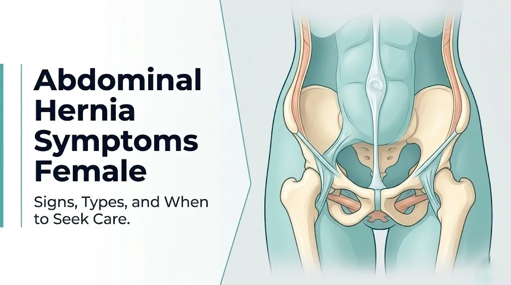
The inguinal canal in women does not transmit the spermatic cord as it does in men. Instead, it carries the round ligament of the uterus, a fibromuscular band that attaches the uterine fundus to the labia majora. This structural difference means that when an inguinal hernia forms in a woman, the peritoneal sac tends to follow the round ligament toward the labia rather than projecting anteriorly as a visible groin bulge.
The femoral ring (the anatomical opening at the top of the femoral canal, through which the femoral vessels pass from the abdomen into the thigh) is measurably wider in the female pelvis due to the greater interischial distance required for the birth canal. A 2019 population study published in the British Journal of Surgery confirmed that approximately 84 percent of all femoral hernias occur in women, precisely because this wider femoral ring creates more anatomical space for peritoneal content to herniate.
Female connective tissue also responds to hormonal fluctuations. The hormone relaxin, produced by the corpus luteum and by the placenta during pregnancy, increases the extensibility of pelvic ligaments and abdominal wall fascia. This reduces the mechanical resistance of fascial layers to herniation, explaining why hernia symptoms often begin or worsen during or after pregnancy. Postmenopausal estrogen loss has a parallel effect on collagen maintenance, discussed in a later section.
Types of Hernia in Women and Their Symptom Patterns
The hernia types that affect women most often are femoral hernia, inguinal hernia (including canal of Nuck hernia), umbilical hernia, hiatal hernia, epigastric hernia, incisional hernia, and sports hernia (athletic pubalgia), each producing a distinct symptom location and character.
| Hernia Type | Primary Location of Symptoms | Visible Bulge in Women | Strangulation Risk |
|---|---|---|---|
| Femoral | Upper inner thigh, groin, knee (referred) | Rare; below inguinal ligament | High (up to 22% within 3 months) |
| Inguinal (indirect) | Groin, labia majora, lower abdomen | Sometimes; in labium or groin | Moderate |
| Canal of Nuck | Labia majora, groin | Often visible in labium | Moderate |
| Umbilical | Around navel | Yes; at umbilicus | Low to moderate |
| Hiatal | Chest, upper abdomen, throat (reflux) | None (internal) | Low (for sliding type) |
| Epigastric | Midline above navel | Small midline lump | Low |
| Incisional | Site of prior surgical scar | Yes; at scar | Moderate |
| Sports hernia | Deep groin, pubic area, inner thigh | None | Low (no peritoneal sac) |
The Society of American Gastrointestinal and Endoscopic Surgeons (SAGES) notes in its patient education resources that femoral and inguinal hernias are the most clinically consequential hernia types in women because their proximity to the femoral vessels and femoral nerve creates significant complication risk if strangulation occurs.
Women with connective tissue disorders, particularly Ehlers-Danlos syndrome (a heritable disorder of collagen structure), are at elevated risk of multiple simultaneous hernia types and tend to develop hernias at younger ages than the general female population. A primary care physician evaluating a young woman with recurrent or multiple hernias should consider connective tissue disorder as an underlying predisposition.
Key Takeaway: Femoral hernias are found in women approximately 84 percent of the time and have a strangulation risk that is nearly 10 times higher than inguinal hernias, making any lower groin pain in a woman a clinical situation that warrants evaluation rather than watchful waiting.
Inguinal Hernia Symptoms in Women
Inguinal hernia symptoms in women typically include deep groin or lower abdominal aching, a sensation of heaviness or pressure in the groin that worsens with prolonged standing, and pain that may radiate into the labia majora or inner thigh, often without any visible external bulge.
The indirect inguinal hernia is the type that occurs in women when a portion of the peritoneum pushes through the deep inguinal ring (an opening in the transversalis fascia) and travels along the inguinal canal. In male patients, this produces a visible anterior bulge in the groin or scrotum. In female patients, the sac follows the round ligament of the uterus and may terminate in the labia majora, producing labial swelling that is often mistaken for a Bartholin gland cyst or lymphadenopathy.
A specific subtype, the canal of Nuck hernia, occurs when a peritoneal diverticulum that normally obliterates during fetal development fails to close. This diverticulum follows the round ligament into the labium majus. Canal of Nuck hernias are exclusively female and produce a soft, reducible labial swelling that disappears when the woman lies down and reappears with standing or straining. According to a 2020 review published in the journal Hernia, canal of Nuck hernias are underdiagnosed because clinicians unfamiliar with the anatomy mistake them for gynecological swelling rather than a hernia requiring surgical repair.
Pain from inguinal hernias in women is often worse during menstruation. The increased pelvic blood flow and prostaglandin-mediated uterine contractility during the menstrual phase increases intra-abdominal pressure and can temporarily enlarge the hernia sac, intensifying the aching sensation at the inguinal ring. This cyclical pattern frequently leads to misdiagnosis as endometriosis or ovarian cyst before the correct diagnosis is established.
Femoral Hernia Symptoms in Women
Femoral hernia symptoms in women include a small, firm bulge or fullness below the crease of the groin (below the inguinal ligament), inner thigh or knee aching, and pain that may be disproportionately severe relative to the visible swelling, particularly because this hernia type strangulates with little warning.
The femoral canal is a narrow fibrous channel medial to the femoral vein at the level of the femoral triangle in the upper thigh. When intra-abdominal pressure pushes peritoneal contents through the femoral ring into this canal, the resulting hernia is constrained on three sides by firm anatomical structures: the inguinal ligament anteriorly, the pectineal ligament (Cooper’s ligament) posteriorly, and the femoral vein laterally. This tight anatomical collar is what makes femoral hernias so prone to incarceration and strangulation.
Think of a femoral hernia like a balloon pushed through a rigid metal ring. The ring does not expand. Any intestinal loop that enters the femoral canal has almost no room to move, and even a small amount of swelling from increased blood flow during physical activity can trap it permanently.
The referred pain pattern is specific and clinically recognizable. The femoral nerve descends lateral to the femoral canal, and its branches supply the anterior and inner thigh down to the medial aspect of the knee via the saphenous nerve. Femoral canal congestion from hernia contents can compress or irritate these branches, producing thigh or knee aching that seems anatomically disconnected from the actual hernia site. Women with femoral hernias occasionally present to orthopedic clinics for knee pain before the hernia is identified.
According to a study published in Annals of Surgery, femoral hernias carry a strangulation rate of approximately 22 percent within 3 months of first presentation, compared to approximately 2 to 3 percent for inguinal hernias in the same period. Any woman with a firm, tender lump below the groin crease requires evaluation by a board-certified general surgeon within 24 to 48 hours.
Umbilical Hernia Symptoms in Women
Umbilical hernia symptoms in women include a soft bulge at or around the navel that increases in size with straining or standing, a dull aching at the umbilicus that worsens in the evening after prolonged upright activity, and skin irritation or discomfort at the umbilical defect from the overlying clothing or belt.
The umbilicus is a natural anatomical weak point in the linea alba, the fibrous midline structure formed by the interlacing aponeuroses (tendinous sheets) of the external oblique, internal oblique, and transversus abdominis muscles. The linea alba at the umbilicus has a circular defect where the umbilical vessels passed during fetal development. After birth this closes, but the closure remains structurally weaker than the surrounding linea alba throughout life.
Women develop umbilical hernias at higher rates than men specifically because pregnancy stretches and thins the linea alba. A 2021 study published in JAMA Surgery found that umbilical hernias are present in approximately 0.5 to 1.5 percent of the general population but occur at higher rates in women who have had multiple pregnancies or who developed diastasis recti (separation of the rectus abdominis muscles along the linea alba) during pregnancy.
Umbilical hernias in women who are obese deserve specific attention. Increased intra-abdominal fat elevates resting intra-abdominal pressure continuously, enlarging the umbilical defect over time. A hernia that was previously small and asymptomatic can become large enough to contain omental fat or a loop of intestine, at which point aching becomes more persistent and the risk of incarceration increases. A woman with a known umbilical hernia who notices a change from intermittent aching to constant pain at the navel should contact her primary care physician the same day.
Key Takeaway: Umbilical hernias in women are directly linked to the structural changes of pregnancy and diastasis recti; a persistent ache at the navel that worsens with any increase in intra-abdominal pressure warrants evaluation rather than watchful waiting.
Hiatal Hernia Symptoms in Women
Hiatal hernia symptoms in women include chronic heartburn, regurgitation of acidic stomach contents into the throat, chest pressure or pain that mimics cardiac symptoms, difficulty swallowing, and a persistent sensation of fullness or bloating in the upper abdomen, particularly after meals.
A hiatal hernia occurs when a portion of the stomach pushes upward through the esophageal hiatus of the diaphragm (the muscular opening through which the esophagus passes from the chest into the abdomen) into the chest cavity. The lower esophageal sphincter, the muscular valve that prevents stomach acid from refluxing into the esophagus, is disrupted when the stomach shifts above the diaphragm, producing gastroesophageal reflux disease (GERD) as the primary symptom complex.
The chest pain produced by a hiatal hernia can be difficult to distinguish from cardiac pain because both activate the same visceral afferent pain pathways via the vagus nerve and the thoracic dorsal horn. Women with hiatal hernias who present with chest pressure, left-sided chest aching, or pain radiating to the left arm or jaw require cardiac evaluation to rule out myocardial ischemia before attributing symptoms to the hernia. The American Heart Association notes that women experiencing chest pain should not delay cardiac evaluation based on assumed gastrointestinal causes.
Women over 50 develop hiatal hernias at higher rates than younger women, partly due to age-related weakening of the diaphragmatic hiatal musculature and partly because the progressive laxity of the phrenoesophageal membrane (the connective tissue securing the gastroesophageal junction below the diaphragm) increases with age. Postmenopausal estrogen loss contributes to this through the same collagen-maintenance pathway described in other hernia types.
Hidden Hernia Symptoms in Women With No Visible Bulge
A hidden hernia in women produces symptoms of groin, pelvic, or lower abdominal pain without any externally visible or palpable lump, and represents one of the most commonly missed diagnoses in female patients presenting with chronic pelvic or groin pain.
The medical term for this presentation is an occult hernia, and it occurs when the hernia defect in the abdominal wall is present but the herniated content (typically preperitoneal fat rather than intestine) is small enough that it does not produce a visible bulge. The pain is generated not by mechanical distension but by traction on the peritoneum and compression or irritation of nearby nerves, particularly the ilioinguinal nerve, the genitofemoral nerve, and the lateral femoral cutaneous nerve, all of which pass through or near the inguinal and femoral regions.
According to a 2022 study published in JAMA Surgery, occult hernias account for a substantial proportion of cases in women with chronic groin pain where standard physical examination and ultrasound are negative. Dynamic MRI of the pelvis in the supine and upright positions, or CT scan of the abdomen and pelvis, is required to identify these defects reliably.
To describe a hidden hernia accurately to a physician or emergency care provider:
- Note the precise location of the pain: is it in the deep groin (above the groin crease), below the groin crease, in the lower abdomen, or in the labia?
- Track whether the pain worsens with standing, walking, coughing, or bearing down (all increase intra-abdominal pressure and enlarge the hernia temporarily).
- Note whether the pain improves significantly when you lie down and relax (reducible hernias typically do).
- Record whether the pain is cyclical with your menstrual cycle (cyclical worsening suggests hormonal influence on the hernia sac).
- Bring this log to your appointment with a primary care physician or general surgeon, along with a list of prior pelvic imaging results and gynecological diagnoses previously assigned to your pain.
Hernia Symptoms in Women: Lower Abdomen and Pelvic Pain
Hernia-related lower abdominal and pelvic pain in women is aching or sharp, located in the lower quadrants, pubic region, or deep pelvis, and typically worsens with upright posture, physical exertion, or any activity that increases intra-abdominal pressure.
This pain pattern is produced by two distinct mechanisms. First, the peritoneum lining the hernia sac is richly innervated by visceral afferent fibers from the hypogastric plexus and the lumbar splanchnic nerves. When the peritoneal sac stretches as hernia contents push through the defect, these fibers generate a poorly localized, deep, aching discomfort that is difficult for the woman herself to pinpoint and equally difficult for a clinician to attribute to a specific structure during physical examination.
Second, the proximity of the inguinal and femoral hernia sites to the ovary, fallopian tube, and broad ligament of the uterus creates a referred pain overlap where hernia pain is interpreted by the brain as originating from pelvic organs. This is because the sensory innervation of the pelvic structures and the inguinal region shares common spinal cord segments (T10 through L2), making it neurologically impossible for the brain to distinguish the source with precision.
Women who report lower abdominal or pelvic pain that worsens specifically with standing up from a seated position, lifting, or walking upstairs, and improves within minutes of lying down, have a symptom pattern that is more consistent with a reducible hernia than with gynecological pathology. A gynecological origin for pelvic pain does not typically show this postural-dependent pattern. A primary care physician evaluating this presentation should include pelvic ultrasound and dynamic abdominal wall imaging in the diagnostic workup, not pelvic ultrasound alone.
Key Takeaway: Lower abdominal and pelvic pain in women that consistently worsens with standing, lifting, or straining and reliably improves with lying down is a hernia pattern, not a gynecological pattern, and warrants abdominal wall imaging, not just pelvic ultrasound.
Hernia Symptoms During Pregnancy
Hernia symptoms during pregnancy include an aching or sharp pain at the navel, a soft bulge appearing at or near the umbilicus, groin discomfort from a pre-existing or new inguinal hernia, and worsening upper abdominal heartburn and regurgitation from increased hiatal hernia pressure.
Pregnancy creates the conditions for hernia formation and symptom exacerbation through two parallel mechanisms. The growing uterus progressively elevates intra-abdominal pressure, from a normal resting pressure of approximately 5 to 7 mmHg to values exceeding 25 to 30 mmHg in the third trimester. This pressure is transmitted uniformly across all abdominal wall fascial layers, pushing against any preexisting structural weakness.
The hormone relaxin, secreted by the corpus luteum in early pregnancy and by the placenta from the first trimester onward, reduces collagen cross-linking in ligamentous and fascial tissue to allow pelvic expansion for delivery. This collagen softening affects not just the pelvic ligaments but the entire abdominal wall fascia, including the linea alba and transversalis fascia, reducing their resistance to herniation.
Diastasis recti (separation of the two bellies of the rectus abdominis along the linea alba) affects approximately 30 to 70 percent of pregnant women by the third trimester, according to research published in the Journal of Orthopaedic and Sports Physical Therapy. A wide diastasis recti exposes the linea alba to direct pressure from the bowel and omentum, creating the anatomical substrate for both umbilical and epigastric hernia formation.
Managing a hernia during pregnancy requires direct guidance from an obstetrician-gynecologist in consultation with a general surgeon. Elective surgical repair is generally deferred until after delivery for reducible hernias; emergency repair is performed at any gestational age if strangulation occurs.
Hernia Symptoms in Postmenopausal Women
Hernia symptoms in postmenopausal women often develop gradually over months, with a dull, progressively worsening groin or abdominal ache that becomes more constant as the hernia defect enlarges, driven by the accelerated loss of collagen in abdominal wall fascia that follows the decline in estrogen production after menopause.
Estrogen receptor alpha is expressed in connective tissue fibroblasts throughout the abdominal wall, including those in the transversalis fascia, the inguinal ligament, and the linea alba. Estrogen signaling through this receptor promotes collagen synthesis and inhibits the activity of matrix metalloproteinases (specifically MMP-1, MMP-2, and MMP-9), the protease enzymes responsible for degrading collagen fibers. After menopause, the steep decline in circulating estrogen reduces this protective effect, accelerating net collagen loss in fascial tissue.
Research published in the British Journal of Surgery found that postmenopausal women have measurably lower collagen content in inguinal hernia sac tissue than premenopausal women, supporting the biological basis for the age-related increase in hernia incidence in female patients after the sixth decade.
Postmenopausal women with hernias also carry a specific risk related to obesity. Visceral adiposity is common after menopause due to the redistribution of fat storage from subcutaneous to visceral compartments that occurs with declining estrogen. This increases baseline intra-abdominal pressure chronically and can accelerate hernia development or enlargement in a woman who had a small, asymptomatic defect before menopause.
Women over 65 with unexplained groin, inner thigh, or pelvic aching who have not had pelvic or abdominal imaging should discuss hernia evaluation with their primary care physician, specifically requesting dynamic abdominal wall ultrasound or CT scan of the abdomen and pelvis in addition to standard pelvic ultrasound.
Hernia After C-Section or Hysterectomy: What Symptoms to Expect
Hernia after a cesarean section (C-section) or hysterectomy produces symptoms at or near the surgical scar: a soft bulge appearing along the incision line, local pain or pulling at the scar that worsens with lifting or straining, and occasionally a burning or sharp sensation along the edge of the defect when the hernia contents press against the scar tissue.
This type of hernia is called an incisional hernia, and it occurs when the abdominal wall closure at the surgical incision fails to maintain its structural integrity. The Pfannenstiel incision used in most C-sections and abdominal hysterectomies runs horizontally just above the pubic hairline. The fascial layers beneath the skin are closed in layers, but if closure is incomplete, tension is too high, wound infection occurs, or the patient returns to heavy activity too early, the fascial closure can fail months to years after surgery.
According to the American College of Surgeons, incisional hernia occurs in approximately 10 to 15 percent of abdominal surgeries, with higher rates in patients who experienced wound complications, are obese, or have had multiple prior abdominal procedures. For women who have had both a C-section and a subsequent hysterectomy through the same incision site, the cumulative risk is higher because repeated surgery disrupts fascial tissue planes.
The symptom pattern of an incisional hernia after these procedures:
- A visible or palpable bulge along the Pfannenstiel incision line, most noticeable when sitting up from lying flat, coughing, or lifting
- A pulling, dragging sensation at the incision site during physical activity
- Pain that develops hours after physical exertion rather than immediately (reflecting delayed inflammatory edema)
- Skin changes at the scar site in large hernias: thinning, dimpling, or redness
Women who notice any new bulging or increasing discomfort at a prior abdominal surgical scar should be evaluated by a board-certified general surgeon, who will assess whether the defect is symptomatic and whether laparoscopic or open mesh repair is appropriate.
Key Takeaway: Incisional hernias after C-section or hysterectomy occur in up to 15 percent of abdominal surgeries and produce a bulge or pulling pain at the scar that is consistently worsened by any increase in abdominal pressure, including sitting up from lying flat.
Incarcerated Hernia Symptoms in Women
An incarcerated hernia in women produces sudden worsening of the pain at the hernia site, a hernia bulge that is firm, tender to touch, and cannot be pushed back in with gentle pressure, often accompanied by nausea and cramping.
Incarceration occurs when the herniated content, whether intestinal loop, omentum, or in rare female cases an ovary, becomes trapped in the hernia defect and cannot return to the abdominal cavity. The trapping itself does not immediately compromise blood supply, distinguishing incarceration from strangulation. However, incarceration is the necessary precursor to strangulation, and in femoral hernias the interval between incarceration and strangulation can be extremely short due to the tight anatomical boundaries of the femoral canal.
The clinical distinction between a reducible hernia and an incarcerated one is both simple and important:
- A reducible hernia disappears or decreases when the woman lies down and relaxes, and the content can be gently pushed back through the defect with mild manual pressure. It is usually associated with intermittent discomfort rather than constant pain.
- An incarcerated hernia remains present regardless of position, is tender to direct pressure, and is associated with escalating rather than intermittent pain. Attempting to force it back can cause injury.
Women with a known hernia who notice that their usual soft, reducible bulge has become hard, persistently present, and more painful should not attempt to reduce it manually. They should contact their primary care physician or general surgeon the same day. If abdominal distension, vomiting, or inability to pass gas or stool accompanies the change, they should go to the emergency room immediately.
Older women with femoral hernias are at particular risk of incarceration presenting without a clearly visible external bulge. In this population, sudden onset of inner thigh or groin pain combined with nausea and inability to pass stool may be the primary presentation of an incarcerated femoral hernia without any palpable lump.
Strangulated Hernia Symptoms in Women
A strangulated hernia in women produces severe, constant, rapidly worsening pain at the hernia site with nausea, vomiting, fever, and a hard, extremely tender bulge that may show skin color changes (redness, darkening, or dusky discoloration), and this is a surgical emergency with a narrow window for saving the affected bowel.
Strangulation occurs when the blood supply to the incarcerated hernia content is cut off by the tight hernia ring compressing the mesenteric blood vessels. The sequence is: venous outflow obstructs first, causing congestion and edema in the bowel wall; arterial inflow is subsequently compromised as edema increases; bowel wall ischemia progresses to necrosis within hours without surgical decompression and resection.
The timeline is not forgiving. Research published in Annals of Surgery has documented that strangulated bowel can become non-viable within 4 to 6 hours of complete arterial occlusion. This is why femoral hernias, with their constrictive anatomical boundaries and high strangulation rate, require emergency surgical evaluation the moment symptoms suggest irreducibility.
Specific symptoms indicating possible strangulation in a woman with a known or suspected hernia:
- Severe, constant pain that does not ease with any position change
- Nausea and vomiting developing alongside or after the onset of pain
- Fever (oral temperature above 38°C / 100.4°F)
- A previously soft hernia bulge becoming hard, hot to the touch, and exquisitely tender
- The skin over the bulge appearing red, purple, or dark
- Abdominal bloating and inability to pass gas or have a bowel movement
Women taking immunosuppressant medications (such as corticosteroids or biologics for autoimmune conditions) may not develop fever even with established bowel ischemia, because their inflammatory response is pharmacologically suppressed. Pain pattern and physical findings are more reliable early indicators of strangulation in this population.
Emergency Symptoms: When to Call 911 or Go to the ER
Certain symptoms associated with abdominal hernias in women require immediate emergency evaluation. Do not wait to see if these resolve on their own.
Call 911 or go to the nearest emergency room immediately if you experience:
- A hernia bulge that was previously soft and reducible has become hard, cannot be pushed back, and is now extremely tender (incarceration requiring emergency evaluation before strangulation occurs)
- Severe, constant, worsening pain at the hernia site not relieved by lying down or rest (possible strangulation)
- Nausea and vomiting combined with groin, lower abdominal, or inner thigh pain in a woman with a known or suspected hernia (incarceration or strangulation)
- Fever above 38°C (100.4°F) alongside hernia site pain and tenderness (systemic infection from ischemic or perforated bowel)
- Skin over the hernia site turning red, purple, or dark (vascular compromise of strangulated tissue)
- Abdominal rigidity (a board-like hard abdomen) combined with any hernia symptom (peritonitis from bowel perforation)
- Inability to pass gas or have a bowel movement for more than 12 hours combined with lower abdominal or groin pain (bowel obstruction from incarcerated hernia)
These presentations can indicate strangulated bowel, peritonitis, or complete intestinal obstruction, all of which require emergency surgical assessment, not an urgent care appointment or a phone call.
Hernia vs. Ovarian Cyst and Other Misdiagnoses in Women
The most common misdiagnoses assigned to women who actually have a hernia include ovarian cyst, round ligament pain, pelvic inflammatory disease, endometriosis, and musculoskeletal groin strain, and the key distinguishing feature of a hernia is the postural relationship between symptom severity and body position.
This diagnostic confusion is not a failure of clinical reasoning in isolation. It is a structural problem: the symptom overlap between these conditions is genuine, and without abdominal wall imaging, the physical examination alone cannot always distinguish them. A 2023 study published in JAMA Surgery found that women with hernias had a median delay to correct diagnosis of approximately 18 months, during which most had received at least one gynecological diagnosis.
| Condition | Pain Pattern | Postural Effect | Visible Lump | Key Differentiator |
|---|---|---|---|---|
| Groin hernia | Worsens standing, lifting; eases lying down | Strong postural effect | Possible | Dynamic imaging confirms defect |
| Ovarian cyst | Not consistently postural | Little postural effect | None externally | Pelvic ultrasound shows cyst |
| Round ligament pain | Sharp, lancinating, brief | Triggered by sudden movement | None | Pregnancy history, bilateral possible |
| Pelvic inflammatory disease | Constant, bilateral, fever | No postural pattern | None | Cervical motion tenderness on exam |
| Endometriosis | Cyclical with menstruation | No postural pattern | None | Diagnostic laparoscopy required |
| Musculoskeletal groin strain | Worsens with specific movements | Movement-dependent | None | Improved with rest, no pressure effect |
When describing symptoms to a primary care physician, specifically state whether the pain is reliably worse when standing and reliably better when lying flat. This postural pattern is the single most clinically distinguishing feature of a hernia from the gynecological alternatives. Request dynamic ultrasound of the inguinal and femoral regions in addition to standard pelvic ultrasound, because a standard pelvic ultrasound does not image the inguinal and femoral canal regions adequately.
Women with obesity or a large pannus (overhanging abdominal fat) may require CT scan of the abdomen and pelvis rather than ultrasound for adequate imaging of the inguinal and femoral regions.
Hernia Diagnosis in Women and What to Expect
Hernia diagnosis in women begins with a physical examination and symptom history, followed by imaging (most commonly dynamic ultrasound or CT scan of the abdomen and pelvis), and is confirmed either radiographically or at the time of laparoscopic surgical exploration when imaging is inconclusive.
The diagnostic process for a suspected hernia in a woman follows this sequence:
- A primary care physician takes a detailed history focusing on the exact location of pain, its postural relationship, any history of prior abdominal surgery, pregnancy history, reproductive status, and any gynecological diagnoses previously assigned to the pain.
- Physical examination is performed with the woman standing upright (not just lying down), because small hernias may only be detectable with the increased intra-abdominal pressure of the upright position and the Valsalva maneuver. The inguinal, femoral, and umbilical regions are all examined.
- Dynamic inguinal and femoral ultrasound is the first-line imaging tool. Dynamic means the ultrasound is performed during Valsalva maneuver in the standing or semi-erect position, which is when the hernia content moves and becomes visible. Standard supine ultrasound misses a significant proportion of occult hernias in women.
- CT scan of the abdomen and pelvis with contrast is ordered when ultrasound is inconclusive, when femoral or obturator hernia is suspected, or when the clinical picture suggests incarceration.
- MRI of the pelvis (using a dynamic protocol with images taken in upright and lying positions) is the most sensitive tool for occult hernias and athletic pubalgia and may be ordered by a general surgeon or sports medicine physician when CT is negative but clinical suspicion remains high.
- Diagnostic laparoscopy is the definitive diagnostic and simultaneously therapeutic option when imaging is negative but symptoms strongly suggest hernia. A board-certified general surgeon performs laparoscopy under general anesthesia; if a hernia defect is found, it is repaired during the same procedure.
Women who have been told their pain is gynecological but whose symptoms show a consistent postural pattern should request a referral to a board-certified general surgeon for a second evaluation, specifically asking whether dynamic inguinal and femoral ultrasound has been performed in the standing position during Valsalva.
Key Takeaway: Dynamic ultrasound in the standing position during Valsalva maneuver is the correct first-line imaging tool for diagnosing hernia in women; a standard supine pelvic ultrasound is insufficient and misses a large proportion of occult inguinal and femoral hernias.
Frequently Asked Questions About Abdominal Hernia Symptoms in Women
What does a hernia feel like in a woman?
A hernia in a woman typically feels like a deep, aching groin, lower abdominal, or pelvic discomfort that worsens when standing, lifting, or coughing and improves when lying down.
Some women feel a soft lump or fullness in the groin, labia, or at the navel that disappears when they lie flat and returns with upright activity.
Women with femoral or occult inguinal hernias frequently feel inner thigh or labial heaviness without any visible external lump, which is why a gynecological cause is often assumed before a hernia is correctly identified.
Can a woman have a hernia with no visible bulge?
A woman can absolutely have a hernia with no visible bulge, and this is more common in women than in men due to the anatomical configuration of the female inguinal and femoral canals.
The femoral hernia, the most common hernia type specific to women, emerges below the inguinal ligament into the femoral triangle where the overlying tissue masks the bulge from external view.
Occult hernias in women are confirmed by dynamic ultrasound or CT imaging rather than physical examination, which is why persistent postural groin or pelvic pain in a woman warrants imaging even when examination appears normal.
What is the most dangerous type of hernia for women?
The femoral hernia is the most dangerous hernia type in women because it carries an incarceration and strangulation rate of approximately 22 percent within 3 months of first presentation, according to data published in Annals of Surgery.
The tight anatomical boundaries of the femoral canal (the inguinal ligament, pectineal ligament, and femoral vein on three sides) leave almost no room for expansion once intestinal content enters the canal.
Any woman with a suspected femoral hernia should be evaluated by a board-certified general surgeon within 24 to 48 hours of diagnosis, without waiting for symptoms to worsen.
How do I know if my groin pain is a hernia or an ovarian cyst?
Groin pain from a hernia is reliably worse when standing or straining and reliably better when lying flat; ovarian cyst pain does not follow this consistent postural pattern.
A hernia may produce a soft lump that disappears with lying down; ovarian cysts do not produce an external lump in the groin.
Only abdominal wall imaging (dynamic ultrasound or CT scan) combined with pelvic ultrasound can definitively distinguish between the two, and a primary care physician or general surgeon should be consulted to order both.
What hernia symptoms in women require emergency care?
Any hernia bulge that becomes hard, persistently tender, and irreducible (cannot be pushed back with gentle pressure) requires emergency room evaluation the same day, because this indicates incarceration that can progress rapidly to strangulation.
Strangulation symptoms, including severe constant pain, fever above 38°C, nausea and vomiting, and skin color changes over the hernia site, require calling 911 or going to the nearest emergency room immediately.
These presentations indicate compromised bowel blood supply, and emergency surgical intervention is time-sensitive.
Can pregnancy cause a hernia?
Pregnancy can directly cause or significantly worsen a hernia by raising intra-abdominal pressure in the third trimester to values exceeding 25 mmHg, combined with relaxin-mediated softening of the abdominal wall fascia.
The most common pregnancy-related hernias are umbilical hernia from stretching of the linea alba and worsening of a pre-existing inguinal hernia due to increased pressure on the deep inguinal ring.
An obstetrician-gynecologist should be informed of any new abdominal bulge, groin discomfort, or labial swelling that develops during pregnancy so that hernia evaluation can be coordinated with obstetric care.
The single most important clinical fact in this article for any woman reading it: the absence of a visible groin bulge does not rule out a hernia, and a hernia in a woman carries a strangulation risk that is far higher than most people, and many clinicians, realize.
If you have been told your groin, lower abdominal, or pelvic pain is gynecological but it reliably worsens when you stand and reliably improves when you lie flat, ask your primary care physician for a referral to a board-certified general surgeon and request dynamic inguinal and femoral ultrasound in the standing position during Valsalva. That single imaging step resolves many years of missed diagnoses.
The woman who gets the right diagnosis gets the right treatment. Start with the right question at your next appointment