Swollen Eyelid Causes: What Is Really Behind the Puffiness
Swollen eyelid causes span a wide clinical range: from simple allergic reactions and blocked oil glands to bacterial infections that can threaten vision. The specific cause matters enormously because management ranges from a warm compress at home to emergency surgery within hours.
The eyelid is a structurally complex tissue with multiple glands, muscle layers, and a critical anatomical boundary called the orbital septum that separates minor, self-limiting problems from potentially dangerous ones. According to the American Academy of Ophthalmology, eyelid and orbital infections account for one of the most common reasons patients present to ophthalmology emergency services, with orbital cellulitis carrying a risk of vision loss and intracranial complication if not treated promptly.
This article covers every major cause of eyelid swelling with its specific physiological mechanism, explains how to read the clinical pattern of your particular swelling, identifies the population-specific risk factors that change the picture, and gives clear guidance on when to monitor at home versus when to seek professional evaluation immediately.
Swollen Eyelid Causes: The Clinical Starting Point
Swollen eyelid causes fall into four broad physiological categories: localized glandular or infectious inflammation, allergic and immune-mediated reactions, structural or traumatic causes, and systemic conditions that produce generalized fluid accumulation around the orbit.
Understanding which category your swelling belongs to requires paying attention to three specific features: whether the swelling is in one eye or both, whether it is painful or painless, and whether it appeared suddenly or gradually over days. These three observations are the starting points clinicians use to narrow the differential diagnosis before any examination begins.
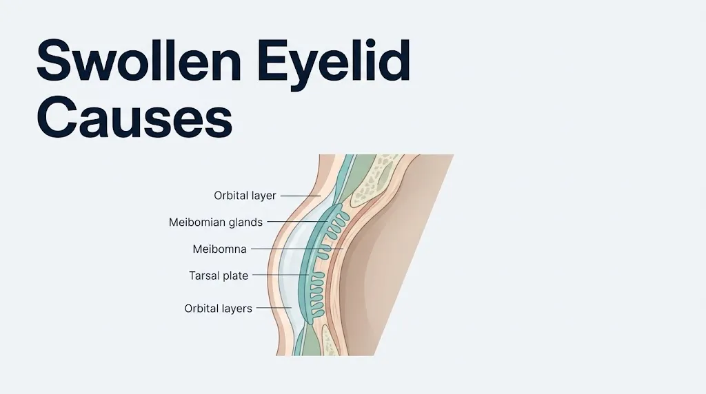
The eyelid’s loose connective tissue makes it unusually susceptible to fluid accumulation compared to skin elsewhere on the body. The subcutaneous layer of the eyelid contains very little supporting fibrous tissue, which means even modest increases in local capillary permeability or systemic fluid retention produce visible swelling quickly. Think of the eyelid’s connective tissue like a kitchen sponge placed under a dripping faucet: it absorbs fluid rapidly and shows it visibly, even when the amount of fluid involved would go unnoticed in a denser tissue.
| Category | Typical Pattern | Painful? | One or Both Eyes? |
|---|---|---|---|
| Localized gland infection (stye) | Sudden, focal, red lump | Yes | One eye, focal |
| Blocked gland (chalazion) | Gradual, firm nodule | Usually no | One eye, focal |
| Allergic reaction | Sudden or gradual, diffuse | Usually no | Often both eyes |
| Orbital cellulitis | Progressive, diffuse, red | Yes, with fever | One eye |
| Systemic cause (cardiac, renal) | Morning puffiness, bilateral | No | Both eyes |
| Angioedema | Sudden, diffuse, non-pitting | No | One or both |
Children under 10 and immunocompromised adults can present atypically, showing swelling without the expected fever or pain even when a serious infection is present.
How Eyelid Swelling Actually Happens in the Body
Eyelid swelling is produced by one of three fundamental physiological events: increased fluid leaving the blood vessels into the eyelid tissue, decreased fluid being removed from that tissue, or physical mass growth (such as a cyst or granuloma) within the eyelid itself.
The most common mechanism is increased vascular permeability. When mast cells in the palpebral tissues release histamine in response to an allergen, histamine binds H1 receptors on the walls of postcapillary venules. This binding causes the endothelial cells lining those vessels to contract, opening gaps between them. Plasma proteins and fluid flow through those gaps into the surrounding tissue. The eyelid swells. The same mechanism is triggered at lower intensity by bacterial toxins, viral proteins, and local trauma.
The second mechanism, decreased fluid removal, explains why people wake up with puffy eyes after sleep. During horizontal recumbency, the lymphatic drainage of the periorbital region slows. The lymphatics of the upper eyelid drain primarily to the preauricular lymph nodes, while the lower eyelid drains to the submandibular nodes. Lying flat reduces the gravitational assist to this drainage. By morning, fluid that accumulated during the night has not cleared, and puffiness is the result. This form of swelling resolves within 30 to 60 minutes of upright activity in most people.
The third mechanism, tissue-based mass growth, is what produces a chalazion. A blocked meibomian gland fills with retained lipid secretion. The body recognizes these lipids as foreign material and mounts a granulomatous inflammatory response, recruiting macrophages and lymphocytes that form a firm, palpable nodule within the tarsal plate. No fluid leak is involved; this is true mass growth within the eyelid tissue.
What Causes a Swollen Eyelid: The Most Common Reasons
The most common causes of a swollen eyelid, in descending order of frequency in clinical presentations, are allergic reactions, styes (hordeola), chalazia, and blepharitis.
Allergic eyelid swelling accounts for the largest proportion of cases seen in primary care and urgent care settings. The National Eye Institute notes that allergic conjunctivitis, which commonly accompanies allergic eyelid swelling, affects approximately 20% of the general population in countries with high allergen exposure. Seasonal allergens (tree pollen, grass pollen, ragweed), pet dander, dust mites, and cosmetic ingredients are the most frequently identified triggers.
Below that, styes and chalazia together account for a large share of presentations involving a visibly swollen eyelid with a palpable lump. These two conditions are distinct despite their surface similarity, and the distinction matters for management. Blepharitis, a chronic inflammatory condition of the eyelid margin, causes lower-grade, recurring swelling that is often bilateral and associated with crusting and flaking at the lash line.
The key causes to recognize clinically:
- Allergic reaction: Diffuse, bilateral or unilateral, itchy, often with associated tearing and redness
- Stye (hordeolum): Focal, painful, red, usually at the eyelid margin
- Chalazion: Firm, round, painless nodule within the eyelid body, not at the margin
- Blepharitis: Bilateral, chronic, scaly eyelid margins with low-grade swelling
- Contact dermatitis: Diffuse, itchy, scaly eyelid skin swelling from a topical irritant or allergen
- Preseptal cellulitis: Diffuse red swelling of the eyelid, often warm, with or without fever
- Orbital cellulitis: All the above plus proptosis, eye movement pain, and possible vision change
People with a personal or family history of atopic conditions (eczema, asthma, hay fever) experience more frequent and more severe allergic eyelid swelling because their mast cells are already primed with higher baseline IgE levels.
Key Takeaway: Most swollen eyelids are caused by allergies, styes, or blocked glands, but the presence of fever, pain on eye movement, or any change in vision changes the clinical picture entirely and requires same-day evaluation.
What Causes Eyelids to Swell from Allergies
Allergic eyelid swelling is caused by IgE-mediated mast cell degranulation in the conjunctival and palpebral tissues, which releases histamine and other inflammatory mediators that increase local capillary permeability and drive fluid into the eyelid interstitium.
The mechanism begins with allergen sensitization. On first exposure to an allergen like cat dander or grass pollen, the immune system produces allergen-specific IgE antibodies, which bind to the surface of mast cells in the eyelid and conjunctival tissues. On re-exposure, the allergen cross-links these IgE molecules, triggering immediate mast cell degranulation. Histamine is released within minutes. It acts on H1 receptors on local blood vessel walls, causing rapid vascular permeability increases. The eyelid swells. This is why allergic eyelid swelling classically appears within minutes of allergen exposure and is almost always accompanied by intense itching and tearing.
According to the American College of Allergy Asthma and Immunology (ACAAI), ocular allergy symptoms affect an estimated 40% of people with seasonal allergic rhinitis, and eyelid swelling is one of the most distressing of these symptoms. Contact allergens (nickel in eye makeup, preservatives in contact lens solutions, and certain botanical extracts in skincare products) can produce a delayed Type IV hypersensitivity reaction, which takes 24 to 72 hours to appear because it is mediated by T lymphocytes rather than IgE. This delayed pattern helps distinguish contact dermatitis of the eyelid from acute allergic conjunctivitis.
Quick Tip:
- Itching is the single most reliable sign that eyelid swelling is allergic in origin. Infectious swellings are painful. Allergic swellings itch.
- Both eyes swelling simultaneously strongly suggests a systemic allergen (airborne or ingested) rather than a localized cause.
- People taking antihistamines regularly may experience blunted swelling even with allergen exposure, which can make the allergic cause harder to identify without an allergy skin prick test.
Patients with atopic dermatitis of the eyelid skin have a compromised skin barrier, which makes them more susceptible to allergen penetration and more likely to develop chronic, lichenified (thickened, leathery) eyelid skin from repeated scratching and inflammation.
How a Stye Causes a Swollen Eyelid
A stye causes a swollen eyelid by producing acute bacterial infection in one of the oil-secreting glands at the eyelid margin, generating a focal abscess that swells, reddens, and becomes tender to touch within 24 to 48 hours.
The specific glands involved determine whether the stye is classified as internal or external. An external hordeolum (the more common form) is an acute bacterial infection of a Zeis gland (a modified sebaceous gland at the base of an eyelash follicle) or a Moll gland (a modified apocrine sweat gland at the eyelid margin). An internal hordeolum is an acute infection of a meibomian gland, which sits deeper within the tarsal plate. Internal hordeola tend to be more painful and produce a more diffuse eyelid swelling because the infection is deeper within the eyelid tissue.
The causative organism in the vast majority of styes is Staphylococcus aureus. Staphylococcal toxins trigger an acute neutrophilic inflammatory response: neutrophils flood the infected gland, release proteolytic enzymes, and begin destroying tissue to contain the infection. The classic result is a pus-filled, tender nodule at or near the eyelid margin that may come to a visible white head.
To describe a stye accurately to a primary care physician or ophthalmologist:
- Note exactly where on the eyelid the lump is located (at the lash line, inside the eyelid when you pull it down, or within the eyelid body away from the margin).
- Record when it first appeared and how quickly it grew.
- Note whether there is pus visible and whether the overlying skin is red and warm.
- State whether you have had a stye before and how the current one differs in location, size, or rate of growth.
- Tell the provider if you are a contact lens wearer, as this affects the likely bacterial profile and the appropriate antibiotic choice.
Most styes resolve spontaneously within 7 to 14 days with warm compress application (10 to 15 minutes, three to four times daily). A stye that has not resolved in two weeks, is growing, or is associated with significant eyelid swelling beyond the focal nodule warrants evaluation by a primary care physician or board-certified ophthalmologist.
Chalazion and Eyelid Swelling: What Is the Difference
A chalazion causes eyelid swelling through granulomatous inflammation of a blocked meibomian gland, producing a firm, painless nodule within the body of the eyelid that can take weeks to months to develop fully.
The meibomian glands are a row of modified sebaceous glands embedded vertically within the tarsal plate of each eyelid. The upper eyelid contains approximately 25 to 30 of these glands; the lower eyelid contains approximately 20 to 25. Their job is to secrete a lipid-rich fluid (meibum) that forms the outermost layer of the tear film and prevents tear evaporation. When a meibomian gland orifice becomes blocked (from thickened secretions, chronic blepharitis, or hyperkeratinization of the ductal opening), retained meibum accumulates inside the gland. The body interprets this retained lipid as a foreign body, mounting a granulomatous response in which macrophages and epithelioid histiocytes form a walled-off nodule within the tarsal plate. This is a chalazion.
The clinical difference between a stye and a chalazion:
| Feature | Stye (Hordeolum) | Chalazion |
|---|---|---|
| Location | Eyelid margin (near lashes) | Within eyelid body, away from margin |
| Pain | Yes, tender on touch | Usually none |
| Texture | Soft, fluctuant if pointing | Firm, rubbery |
| Redness | Yes, skin red and warm | Minimal or none |
| Time to develop | 24 to 48 hours | Weeks to months |
| Cause | Bacterial infection | Glandular obstruction and granuloma |
| Spontaneous resolution | Often, within 2 weeks | Sometimes, but often requires treatment |
Small chalazia (under 5 mm) respond to warm compresses in approximately 25 to 50% of cases. Larger or persistent chalazia are typically treated by a board-certified ophthalmologist via intralesional corticosteroid injection or surgical incision and curettage. People with rosacea or seborrheic dermatitis have significantly higher rates of meibomian gland dysfunction and recurrent chalazia because these conditions alter sebaceous gland secretion viscosity.
Blepharitis Eyelid Swelling Causes
Blepharitis causes eyelid swelling by producing chronic inflammation of the eyelid margins, driven by overgrowth of lid margin bacteria (most commonly Staphylococcus epidermidis and Staphylococcus aureus) and dysfunction of the meibomian glands, creating a persistent low-grade inflammatory cycle that thickens and swells the eyelid margins over time.
Blepharitis is classified as anterior blepharitis (affecting the eyelash follicles and anterior eyelid margin, most commonly staphylococcal or seborrheic in origin) or posterior blepharitis (affecting the meibomian gland openings, also called meibomian gland dysfunction or MGD). Both forms produce eyelid swelling, but the mechanism differs. In anterior blepharitis, bacterial colonization at the base of the eyelashes triggers a low-level immune response: bacterial exotoxins act as superantigens, stimulating T lymphocyte and neutrophil recruitment to the eyelid margin. In posterior blepharitis, the primary problem is meibomian gland obstruction and altered lipid secretion quality, creating the same cycle of retention and granulomatous inflammation that produces chalazia.
According to a 2022 systematic review published in Ophthalmology, blepharitis affects an estimated 37 to 47% of patients seen in ophthalmology practices, making it one of the most common eyelid conditions seen in clinical practice. Despite this frequency, it is consistently underdiagnosed because patients often attribute chronic eyelid puffiness and crusting to tiredness or aging.
The eyelid swelling in blepharitis is characteristically bilateral, worse in the morning, and accompanied by:
- Crusting and flaking at the base of the eyelashes (often described as dandruff of the eyelashes)
- A burning or gritty sensation in the eyes
- Intermittent blurring of vision from tear film instability
- Eyelid margins that appear red, thickened, and irregular
Key Takeaway: Blepharitis is a chronic condition that does not resolve with a single course of treatment. An ophthalmologist can design a long-term lid hygiene program combining warm compresses, eyelid scrubs, and in some cases topical or oral antibiotics, that significantly reduces the recurrence of eyelid swelling.
Conjunctivitis and Swollen Eyelid Causes
Conjunctivitis causes eyelid swelling through inflammation of the palpebral conjunctiva, the transparent mucous membrane lining the inner surface of the eyelids, which produces local vasodilation, chemosis (conjunctival swelling), and secondary eyelid edema in both bacterial and viral forms.
The conjunctiva is highly vascular and densely populated with mast cells and antigen-presenting cells. Bacterial infection (most commonly by Staphylococcus aureus, Streptococcus pneumoniae, or Haemophilus influenzae in children) triggers an acute neutrophilic response with purulent discharge. The profound inflammatory response in the palpebral conjunctiva directly edematizes the overlying eyelid tissue. Viral conjunctivitis, most commonly caused by adenovirus, produces a more watery discharge, follicular changes on the palpebral conjunctiva, and a lymphocytic rather than neutrophilic inflammatory response, which typically produces less eyelid swelling than the bacterial form but more prominent preauricular lymph node enlargement.
Allergic conjunctivitis, the third major type, was covered in the allergy section. The specific clinical features that help distinguish bacterial from viral eyelid swelling in the context of conjunctivitis:
| Feature | Bacterial Conjunctivitis | Viral Conjunctivitis |
|---|---|---|
| Discharge character | Thick, yellow-green, purulent | Watery, clear to light yellow |
| Eyelids stuck together in morning? | Yes, significantly | Mildly |
| Preauricular lymph node tender? | Rarely | Often (adenovirus) |
| Both eyes affected? | Often starts in one, spreads | Usually bilateral |
| Corneal involvement? | Uncommon | Possible (epidemic keratoconjunctivitis) |
| Eyelid swelling severity | Moderate to significant | Mild to moderate |
Newborns and infants represent a specific population requiring urgent attention: conjunctivitis in a newborn (ophthalmia neonatorum), particularly when caused by Neisseria gonorrhoeae or Chlamydia trachomatis, produces rapid, severe eyelid swelling with profuse discharge and can cause corneal perforation and blindness within hours if untreated. Any red, swollen eyelid in a newborn requires same-day evaluation by a pediatrician or pediatric ophthalmologist.
Orbital Cellulitis vs Preseptal Cellulitis: Eyelid Swelling
The single most clinically important distinction in the evaluation of eyelid swelling is whether an infection is preseptal or orbital, because this anatomical boundary determines whether the condition is manageable on oral antibiotics or requires immediate hospitalization and intravenous treatment.
The orbital septum is a thin fibrous membrane that extends from the periosteum of the orbital rim inward to insert on the levator palpebrae superioris muscle in the upper eyelid and the capsulopalpebral fascia in the lower eyelid. It acts as an anatomical barrier between the superficial preseptal tissues (eyelid skin, subcutaneous fat, orbicularis oculi muscle) and the postseptal orbital compartment (orbital fat, extraocular muscles, optic nerve, superior ophthalmic vein).
Preseptal cellulitis (also called periorbital cellulitis) is an infection of the tissues anterior to the orbital septum. The skin is red, warm, and swollen. There is no proptosis (forward protrusion of the globe), no pain on eye movement, and no restriction of ocular motility. Vision is normal. This condition is managed with oral antibiotics in most outpatient settings.
Orbital cellulitis is an infection of the postseptal compartment. It typically arises from direct spread of sinusitis (especially ethmoid sinusitis, because the lamina papyracea, a paper-thin bone separating the ethmoid sinus from the medial orbit, allows bacterial spread with minimal structural resistance). Orbital cellulitis produces:
- Proptosis (the globe is pushed forward by infected orbital fat)
- Pain on moving the eye in any direction (because inflamed tissues surround the extraocular muscles)
- Restricted extraocular motility (the muscles cannot move freely through swollen infected tissue)
- Possible visual change (optic nerve compression or ischemia)
- Fever, often above 102°F (38.9°C)
Untreated orbital cellulitis can produce cavernous sinus thrombosis, subdural empyema, meningitis, and permanent vision loss from optic nerve compression. CT imaging of the orbit and sinuses with intravenous contrast is the standard diagnostic test for establishing this distinction when clinical examination is inconclusive.
Key Takeaway: If a swollen eyelid is accompanied by pain when you move your eye, or you can see that the eye is being pushed forward compared to the other side, this is a possible orbital cellulitis emergency. Go to the emergency room immediately. Do not wait to see if it improves.
Angioedema as a Cause of a Swollen Eyelid
Angioedema causes eyelid swelling through rapid accumulation of fluid in the deep dermis and subcutaneous tissues of the eyelid, driven by either histamine-mediated or bradykinin-mediated mechanisms, producing non-pitting edema that can appear dramatically within minutes to hours.
The two forms of angioedema have distinct mechanisms that determine treatment. Histamine-mediated angioedema is driven by the same IgE-mast cell pathway as allergic conjunctivitis but affects deeper tissue layers and produces more dramatic swelling. It typically responds to antihistamines and, in severe cases, epinephrine. Bradykinin-mediated angioedema does not involve histamine at all, which is why antihistamines provide no relief. Instead, it results from accumulation of bradykinin, a peptide normally degraded by angiotensin-converting enzyme (ACE). In patients taking ACE inhibitors (a commonly prescribed blood pressure medication class including lisinopril, enalapril, and ramipril), ACE is pharmacologically blocked, bradykinin accumulates, and bradykinin binds B2 receptors on vascular endothelium in the deep dermis, producing rapid, severe, non-pitting edema that can affect the eyelids, lips, tongue, and throat.
According to a 2021 review published in the Journal of Allergy and Clinical Immunology, ACE inhibitor-induced angioedema accounts for approximately 30% of all angioedema cases presenting to emergency departments in the United States. It can develop months to years after a patient has been taking the medication without any prior episode, which makes the drug connection non-obvious to patients.
The eyelid swelling in angioedema:
- Appears within minutes to hours
- Is diffuse and puffy rather than firm or nodular
- Does not leave an indentation when pressed (non-pitting)
- Is typically not itchy (distinguishing it from allergic histamine-mediated swelling)
- May be accompanied by swelling of the lips, tongue, or throat
Any angioedema affecting the tongue or throat represents an airway emergency. Call 911 immediately if eyelid angioedema is accompanied by difficulty swallowing, a change in voice quality, or any sensation of throat tightening.
Patients with hereditary angioedema (C1 inhibitor deficiency) are a specific population whose eyelid swelling episodes are triggered by minor trauma, stress, or hormonal changes (particularly estrogen-containing medications) and require specific management with C1 inhibitor concentrate or icatibant rather than epinephrine or antihistamines.
Systemic Causes of Puffy Eyelids
Bilateral periorbital puffiness without redness, pain, or other local signs is most often a systemic cause: the eyelid’s loose connective tissue acts as an early site of fluid accumulation when the body’s fluid balance is disrupted by kidney disease, heart disease, thyroid disease, or liver disease.
The mechanisms by which these systemic conditions produce periorbital edema differ at the physiological level:
| Systemic Condition | Mechanism | Additional Clues |
|---|---|---|
| Nephrotic syndrome | Hypoalbuminemia reduces plasma oncotic pressure; fluid shifts from blood into interstitium | Frothy urine, generalized edema, fatigue |
| Congestive heart failure | Elevated venous hydrostatic pressure drives fluid into periorbital interstitium | Ankle swelling, shortness of breath, worse lying flat |
| Hypothyroidism | Glycosaminoglycans infiltrate periorbital tissues (myxedema); non-pitting edema | Weight gain, cold intolerance, slowed thinking |
| Graves’ ophthalmopathy | Thyroid-stimulating immunoglobulins activate orbital fibroblasts to produce glycosaminoglycans; orbital fat expands | Proptosis, lid retraction, gritty eyes |
| Liver failure (cirrhosis) | Reduced albumin synthesis from damaged hepatocytes decreases oncotic pressure | Jaundice, abdominal swelling, easy bruising |
| Allergic reaction (systemic) | Histamine-mediated permeability increase from systemic allergen | Itching, hives elsewhere on body |
Graves’ ophthalmopathy (also called thyroid-associated orbitopathy) deserves specific attention because it is the most common cause of proptosis in adults and disproportionately affects women between the ages of 30 and 60. The orbital fibroblasts in this condition are stimulated by thyroid-stimulating immunoglobulins to produce large quantities of glycosaminoglycans (particularly hyaluronic acid) within the orbital fat and extraocular muscles, causing these tissues to expand. The eyelids swell, retract, and eventually the globe itself is pushed forward. An endocrinologist and a board-certified ophthalmologist (ideally an oculoplastic specialist with thyroid eye disease expertise) should both be involved in the management of this condition.
Upper Eyelid Swelling Causes
Upper eyelid swelling causes include all the general causes of eyelid swelling, but the upper eyelid has specific anatomical features that make it the more common site for certain conditions and a clinically distinct presentation site for others.
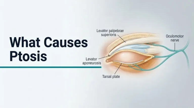
The upper eyelid contains a higher density of meibomian glands (approximately 25 to 30 versus 20 to 25 in the lower eyelid) and a larger tarsal plate, making it the more common site for chalazia and internal hordeola. The levator palpebrae superioris muscle, which elevates the upper eyelid, and its aponeurosis insert into the anterior tarsal plate. Significant swelling within the upper eyelid can mechanically compress the levator aponeurosis, producing secondary ptosis (drooping of the upper eyelid) that is separate from any neurological cause.
The lacrimal gland sits in the superolateral orbit with its palpebral lobe extending beneath the lateral third of the upper eyelid. Inflammation or infection of the lacrimal gland (dacryoadenitis) produces swelling of the lateral portion of the upper eyelid specifically, creating an S-shaped eyelid contour that is a clinical clue to this diagnosis. Dacryoadenitis can be caused by viral infections (mumps, Epstein-Barr virus, cytomegalovirus), bacterial infections, or inflammatory conditions like sarcoidosis and Sjögren’s syndrome.
Upper eyelid swelling that is unilateral, gradual, and painless in a person over 50 warrants evaluation to exclude an eyelid or orbital tumor. Primary eyelid tumors (basal cell carcinoma, squamous cell carcinoma, sebaceous gland carcinoma, Merkel cell carcinoma) are rare but can present as slowly enlarging eyelid masses. Sebaceous gland carcinoma is particularly worth noting because it arises from the meibomian glands or Zeis glands and can masquerade as a recurrent chalazion in older adults, a pattern sometimes called “masquerade syndrome.” Any presumed chalazion that recurs in the same location, bleeds, or does not respond to standard treatment should be evaluated by a board-certified ophthalmologist with biopsy consideration.
Key Takeaway: A swollen upper eyelid that looks like a recurrent chalazion in an older adult, particularly one with loss of eyelashes over the lump, needs a biopsy to exclude sebaceous gland carcinoma, one of the most dangerous eyelid tumors because of its tendency to be misdiagnosed for months.
Swollen Eyelid in One Eye vs Both Eyes: What It Means
A swollen eyelid in one eye almost always points to a localized cause: a stye, chalazion, infection, or trauma specific to that eye. Swelling in both eyes simultaneously shifts the clinical probability strongly toward a systemic or allergic cause.
This distinction holds up consistently in clinical practice because local causes (infected glands, preseptal or orbital cellulitis, insect bites, trauma) affect only the structure or tissues they directly involve. A chalazion forms in one blocked meibomian gland. Preseptal cellulitis typically begins from a local skin break, insect bite, or spread from a nearby sinus on the same side. A stye infects one gland at one eyelash follicle.
Bilateral eyelid swelling that appears simultaneously and symmetrically suggests:
- Airborne or ingested allergen exposure (both eyelids exposed equally)
- A systemic fluid-retention state (nephrotic syndrome, cardiac failure, hypothyroidism)
- Systemic viral infection (bilateral viral conjunctivitis from adenovirus or herpes simplex virus)
- Drug reaction (bilateral eyelid swelling as part of a generalized drug hypersensitivity reaction)
- Angioedema (which can be bilateral even with a localized trigger)
One important exception: bilateral sequential eyelid swelling, where one eye is affected first and the other follows days later, is the classic pattern of adenoviral conjunctivitis. This is not a systemic cause but rather the sequential spread of a highly contagious virus from one eye to the other, usually via hand-to-eye contact.
According to clinical guidelines from the American Academy of Ophthalmology, any patient with bilateral proptosis (both eyes appearing to bulge forward) along with eyelid swelling should be evaluated for thyroid-associated orbitopathy with a thyroid-stimulating hormone (TSH) level and thyroid-stimulating immunoglobulin assay, because Graves’ disease is the most common cause of bilateral proptosis in adults.
What Causes Red Swollen Eyelids
Red swollen eyelids are caused by active inflammation or infection in the eyelid skin or conjunctiva: the redness specifically reflects vasodilation of superficial blood vessels in response to inflammatory cytokines, bacterial toxins, or immune mediators.
Redness distinguishes inflammatory and infectious causes from non-inflammatory causes (like chalazion, myxedema, or simple morning puffiness, which are pale or flesh-colored). When both redness and swelling are present, the differential narrows to:
- Blepharitis with an acute flare: eyelid margins red, scaly, swollen, with crusting
- Stye (hordeolum): focal red, warm, tender lump at eyelid margin
- Preseptal cellulitis: diffuse red swelling of entire eyelid, warm, firm
- Orbital cellulitis: same as preseptal but with eye movement pain and possible proptosis
- Allergic contact dermatitis: red, itchy, scaly eyelid skin from a topical irritant
- Herpes zoster ophthalmicus: vesicular red rash on one side of the forehead and upper eyelid from varicella-zoster virus reactivation along the ophthalmic division of the trigeminal nerve
Herpes zoster ophthalmicus (HZO) warrants specific attention because it is recognizable by its dermatomal distribution (affecting exactly one side of the forehead, nose, and upper eyelid) and carries a risk of serious ocular complications including uveitis, corneal scarring, and acute retinal necrosis. The Hutchinson sign, which is a vesicle on the tip or side of the nose indicating involvement of the nasociliary branch of the trigeminal nerve, predicts a higher risk of intraocular involvement. Anyone with a painful, unilateral red eyelid rash that follows the forehead dermatomal distribution should be evaluated urgently by a primary care physician or ophthalmologist for antiviral therapy, ideally within 72 hours of rash onset.
Adults aged 60 and older, immunocompromised individuals, and people who have not received the recombinant zoster vaccine (Shingrix) are at elevated risk for herpes zoster reactivation and its ocular complications.
Swollen Eyelid in Children: Causes and Concerns
Swollen eyelids in children most commonly result from allergic reactions, styes, or the spread of infection from adjacent sinuses, but children carry a specific elevated risk of complications from orbital cellulitis that makes any eyelid swelling with fever in a child a higher-urgency clinical scenario than the same presentation in a healthy adult.
The most common single cause of periorbital cellulitis in children under 5 is sinusitis, particularly ethmoid sinusitis, because the ethmoid sinus lies immediately adjacent to the medial orbit separated only by the paper-thin lamina papyracea. Bacterial spread from an infected ethmoid sinus can rapidly produce preseptal and then orbital cellulitis. Children under 10 are at higher risk than adults because their sinuses are still developing and the lamina papyracea is thinner and more permeable in early childhood.
According to a 2020 study published in JAMA Ophthalmology, orbital cellulitis in the pediatric population carries a higher rate of intracranial complications, including subperiosteal abscess and epidural abscess, than the same condition in adults, partly because of anatomical differences in sinus structure and partly because children are more likely to have subacute sinusitis that has not yet been diagnosed.
For any child with eyelid swelling, the following features require same-day evaluation by a pediatrician or emergency medicine physician:
- Fever above 100.4°F (38.0°C) in any infant, and above 102°F (38.9°C) in older children
- Swelling that has progressed rapidly (noticeably worse over 6 to 12 hours)
- Pain when the child moves their eye
- The child unwilling to open the eye
- Any change in how clearly the child sees (tested by asking the child to identify familiar objects at a distance)
- Swelling associated with a recent upper respiratory infection or nasal congestion
Neonates (newborns within the first month of life) with eyelid swelling require immediate pediatric evaluation to exclude gonococcal or chlamydial ophthalmia neonatorum regardless of the apparent severity, because these infections can progress to corneal ulceration and perforation within hours.
When Is a Swollen Eyelid an Emergency
A swollen eyelid is an emergency when it is accompanied by any sign suggesting infection posterior to the orbital septum, airway compromise, severe vision change, or in neonates, any eyelid swelling with discharge.
The clinical features that convert a swollen eyelid from a manageable problem to an emergency are specific and learnable. They are worth knowing precisely because the difference between preseptal and orbital cellulitis, or between allergic angioedema and airway-threatening angioedema, is a difference in treatment urgency of hours, not days.
Red flag features requiring emergency evaluation:
- Proptosis: one eye appears more forward than the other when viewed from above or from the front
- Pain on moving the eye in any direction (this reflects inflammation around the extraocular muscles in the postseptal orbit)
- Restricted eye movement (the eye cannot move to one side or upward as it normally can)
- Visual change: blurred vision, double vision, or partial loss of vision in the affected eye
- Fever above 102°F (38.9°C) with eyelid swelling (particularly in a child)
- Swelling that is worsening rapidly over hours, not days
- Difficulty swallowing, change in voice, or throat tightness alongside eyelid swelling (airway angioedema)
- Eyelid swelling in a newborn with any discharge
- Eyelid swelling following trauma with decreased vision or a visible wound
According to the American Academy of Ophthalmology’s clinical guidance, any patient with a swollen eyelid and the combination of proptosis plus restricted eye movement should be treated as having orbital cellulitis until proven otherwise, with CT imaging of the orbit and sinuses with contrast obtained urgently. This is not a presentation to monitor overnight.
For orbital cellulitis, hospitalization with intravenous broad-spectrum antibiotics is typically required. Subperiosteal abscess (a pus collection between the periosteum and the orbital wall) often requires surgical drainage, and cases that progress to cavernous sinus thrombosis require intensive care management. The window from early orbital cellulitis to permanent vision loss can be as short as 24 to 48 hours.
Key Takeaway: The three features that always warrant emergency evaluation of a swollen eyelid are: one eye appearing pushed forward compared to the other, pain when the eye moves, and any loss or blurring of vision in the affected eye. Do not wait for these to worsen before seeking care.
Emergency Symptoms: When to Call 911 or Go to the ER
Certain symptoms associated with eyelid swelling require immediate emergency evaluation. Do not wait to see if these resolve on their own.
Call 911 or go to the nearest emergency room immediately if you or your child experience:
- One eye appearing visibly pushed forward compared to the other (proptosis), which can indicate orbital cellulitis with postseptal infection threatening the optic nerve
- Pain when moving the eye in any direction, suggesting infection or inflammation surrounding the extraocular muscles in the orbital compartment
- Any reduction in vision, blurred vision, or double vision in the affected eye, indicating possible optic nerve compression or ischemia
- Eyelid swelling with fever above 102°F (38.9°C), particularly in a child under 10 with concurrent nasal congestion or recent upper respiratory infection
- Eyelid swelling accompanied by difficulty swallowing, a change in voice quality, or any sensation of throat tightening, indicating possible airway angioedema
- Rapidly progressive eyelid swelling that is measurably worse over a period of 4 to 6 hours
- Eyelid swelling in a newborn within the first month of life with any discharge from the eye
- Eyelid swelling following an injury with an open wound or bleeding on or near the eye
- Any loss of consciousness or confusion alongside eyelid swelling with fever, which can indicate cavernous sinus thrombosis or intracranial extension of orbital infection
These presentations can indicate orbital cellulitis, cavernous sinus thrombosis, airway-threatening angioedema, or serious ocular trauma, all of which require emergency medical assessment, not an urgent care appointment or a phone call to see if it clears up.
Frequently Asked Questions About Swollen Eyelid Causes
What is the most common cause of a swollen eyelid?
The most common cause of a swollen eyelid is an allergic reaction, typically triggered by airborne allergens like pollen, pet dander, or dust mites acting on IgE-sensitized mast cells in the palpebral and conjunctival tissues.
Styes (hordeola) and chalazia are the second most common cause, particularly in people with chronic blepharitis or meibomian gland dysfunction.
A primary care physician can distinguish between these causes based on clinical examination, and an allergist-immunologist can confirm the specific allergen trigger with skin prick testing if the cause is allergic.
Can allergies cause only one eyelid to swell?
Yes, allergies can cause swelling in one eyelid if the allergen contact was localized, such as touching one eye after handling a pet or applying a product to one side of the face.
Bilateral (both eyes) swelling is more typical of airborne allergen exposure, while unilateral swelling suggests a contact allergen or physical transfer of the allergen from the hand to one eye.
If swelling remains unilateral and does not itch or improve with antihistamines, evaluation by a primary care physician or board-certified ophthalmologist is appropriate to exclude other causes like a stye or preseptal cellulitis.
How do I know if my swollen eyelid is a stye or a chalazion?
A stye is painful, red, and located at the eyelid margin near the eyelashes, typically appearing within 24 to 48 hours as an acute bacterial infection of a lid gland.
A chalazion is firm, painless, and located within the body of the eyelid away from the margin, developing gradually over weeks as granulomatous inflammation around a blocked meibomian gland.
A board-certified ophthalmologist can confirm the diagnosis by direct eyelid examination, and this distinction matters because large or persistent chalazia often require intralesional steroid injection or surgical removal rather than the warm compresses that resolve most styes.
What causes puffy eyelids every morning?
Morning eyelid puffiness is most commonly caused by fluid accumulation in the loose periorbital connective tissue during sleep, when lymphatic drainage of the eyelids slows in the horizontal position and fluid that would normally clear overnight does not.
Other causes of persistent morning puffiness include salt-heavy diet increasing fluid retention, poor sleep quality, allergic rhinitis (the sinuses and eyelid lymphatics share drainage pathways), and systemic conditions like hypothyroidism, kidney disease, or heart failure in people over 50.
Morning puffiness that is bilateral, consistent, and accompanied by other symptoms like weight gain, fatigue, shortness of breath, or ankle swelling warrants evaluation by a primary care physician to exclude systemic disease.
Can a swollen eyelid be a sign of something serious?
A swollen eyelid can be a sign of something serious, particularly when it is accompanied by fever, pain on eye movement, proptosis (one eye pushed forward), or any change in vision.
These features together suggest orbital cellulitis, which is a bacterial infection posterior to the orbital septum that can cause cavernous sinus thrombosis, optic nerve damage, and permanent vision loss if not treated with intravenous antibiotics in a hospital setting.
Systemic causes like nephrotic syndrome, Graves’ ophthalmopathy, and heart failure can also initially present as bilateral eyelid puffiness, making evaluation appropriate when puffiness is new, persistent, and unexplained.
Should I go to the emergency room for a swollen eyelid?
Go to the emergency room immediately for a swollen eyelid if you have proptosis, pain on eye movement, restricted eye movement, any vision change, fever above 102°F, difficulty swallowing or throat tightness, or eyelid swelling in a newborn.
Most swollen eyelids from allergies, styes, or chalazia do not require emergency care and can be assessed by a primary care physician or board-certified ophthalmologist within 24 to 48 hours.
The decision point is clear: if the swelling is painful when you move your eye or your vision has changed in any way, treat it as an emergency and go to the nearest emergency room or call 911.
Closing
Most swollen eyelids have a benign cause: an allergic reaction, a blocked gland, or the temporary fluid that settles around the eye during sleep. Knowing the mechanism behind each cause lets you read your own situation accurately rather than guessing.
Watch the swelling for these three specific features. Does the eyelid look pushed forward compared to the other side? Does moving your eye hurt? Has your vision changed at all in that eye? If the answer to any of those three questions is yes, go to the nearest emergency room immediately. Every other cause, from a stye to morning puffiness to a chalazion, can be assessed more calmly, though persistent, unexplained, or recurrent swelling always warrants evaluation by a board-certified ophthalmologist or your primary care physician.
You now have the anatomical and clinical framework to recognize where your swelling falls on that spectrum.