Tooth Infection Spread to Brain: Symptoms and Signs
A tooth infection spreading to the brain is a genuine, life-threatening medical emergency that produces a recognizable set of neurological and systemic symptoms. If you have an active or recent tooth infection and are experiencing severe progressive headache, high fever, neck stiffness, eye swelling, vision changes, confusion, or any weakness on one side of your body, stop reading and call 911 or go to the nearest emergency room immediately.
The intracranial complications of dental infection are rare but carry serious outcomes. According to a 2021 review published in the Journal of Neurosurgery, odontogenic sources account for approximately 10 to 14 percent of all cerebral brain abscesses in adults, and the case fatality rate for untreated intracranial infection from any source remains above 20 percent even with modern antibiotic therapy.
This article covers every clinically recognized symptom of dental infection spreading to the brain, the three distinct anatomical routes by which this spread occurs, how to distinguish a brain abscess from meningitis, the specific symptoms that require a 911 call, and who is at highest risk of a rapid or atypical progression.
Tooth Infection Spread to Brain Symptoms: What You Need to Know
When tooth infection spread to brain symptoms occur, they typically include a combination of worsening headache, fever, confusion, and specific neurological changes that are absent in an ordinary dental abscess. Understanding this distinction is not academic. It is the difference between calling a dentist in the morning and calling 911 tonight.
A standard dental abscess produces localized pain, swelling of the gum or jaw, and sometimes fever. The moment neurological symptoms appear alongside dental infection, the clinical situation has changed category entirely. The central nervous system is involved, which means the infection has moved from the oral cavity into structures that control consciousness, movement, vision, and thought.
The three life-threatening intracranial complications that can result from a dental infection are: cerebral brain abscess (a localized pocket of pus forming within brain tissue), bacterial meningitis (infection of the three membranes surrounding the brain and spinal cord: the pia mater, arachnoid mater, and dura mater), and cavernous sinus thrombosis (septic clotting of the venous sinus at the base of the skull). Each produces overlapping but distinct symptoms. Recognizing the differences matters because each complication follows its own timeline and requires specific emergency treatment.
Key clinical signs that distinguish spreading infection from a contained dental abscess:
- Headache that is progressively worsening, not fluctuating
- Fever above 38.5°C (101.3°F) that does not respond to acetaminophen or ibuprofen
- Any change in consciousness: confusion, unusual drowsiness, or inability to focus
- Stiff neck that limits forward bending of the chin toward the chest
- Any eye symptom: bulging, double vision, or drooping eyelid
- New weakness, numbness, or coordination problem on one side of the body
Older adults may not develop fever even with confirmed intracranial infection, according to research published in Annals of Emergency Medicine. In this population, confusion or sudden behavioral change may be the first and only warning sign.
How Does a Tooth Infection Spread to the Brain?
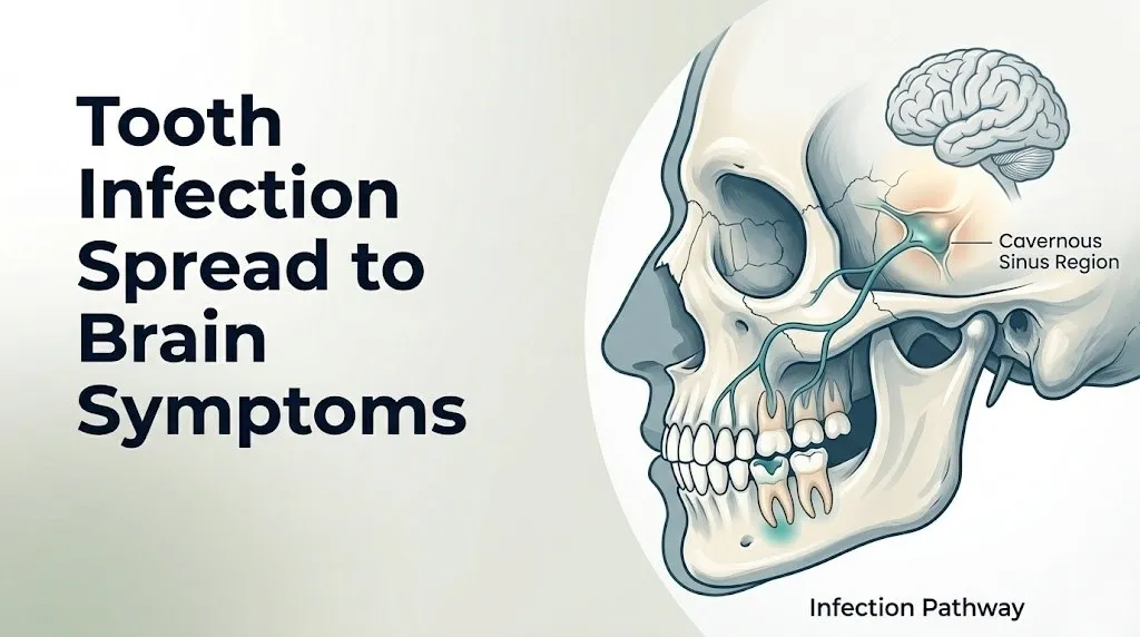
A tooth infection spreads to the brain through three distinct anatomical routes: direct extension through the fascial planes of the head and neck, retrograde spread through the valveless venous system connecting facial veins to the cavernous sinus, and hematogenous dissemination through the bloodstream to the cerebral vasculature.
Understanding the anatomy makes the urgency concrete. Picture the venous drainage of your face as a highway without traffic lights. Blood and anything carried in it moves freely in either direction, because the veins of the face have no valves. The pterygoid venous plexus, a network of veins surrounding the pterygoid muscles just behind the upper molars, connects directly with the angular vein and facial vein on one side, and communicates with the cavernous sinus on the other. The cavernous sinus is a venous chamber sitting at the base of the skull, immediately adjacent to the pituitary gland and the cranial nerves that control eye movement.
When bacteria from an abscessed upper molar or lower molar enter this venous network, they have an unobstructed path to the base of the brain. This is Route 1, the venous route, and it is the pathway responsible for cavernous sinus thrombosis from dental infection.
Route 2 is the fascial plane route. The head and neck contain a series of connected anatomical compartments called fascial spaces. Infection beginning in the submandibular space (below the jaw) can spread to the sublingual space (under the tongue), then to the parapharyngeal space (beside the throat), and from there to the retropharyngeal and danger spaces, which lead directly to the skull base and the mediastinum. This is how Ludwig’s angina, a rapidly spreading floor-of-mouth infection, can become a brain emergency.
Route 3 is hematogenous, meaning bacteria enter the bloodstream during bacteremia (a transient or sustained bacterial blood stream presence), travel to the brain’s microvascular supply, and use inflammatory disruption of the blood-brain barrier to establish infection within brain tissue itself.
The specific bacteria most frequently identified in odontogenic brain infections include the Streptococcus anginosus group (also called the Streptococcus milleri group), anaerobic species including Fusobacterium nucleatum, and Prevotella species, according to published microbiological series in the British Journal of Oral and Maxillofacial Surgery.
Symptoms of Tooth Infection Spreading to Brain: Early Warning Signs
The symptoms of a tooth infection spreading to the brain appear in two phases. The first phase involves systemic signs of spreading infection that have not yet reached the central nervous system. The second phase involves neurological symptoms indicating the brain is already affected. Recognizing the first phase is the window for preventing the second.
Early warning signs of spread, before neurological involvement:
- Fever above 38.5°C (101.3°F) developing or worsening after dental symptoms began
- Swelling that extends beyond the gum or cheek into the neck, under the chin, or toward the ear
- Difficulty swallowing (dysphagia) due to swelling pushing against the throat
- Trismus (inability to fully open the mouth, typically defined as less than 30mm interincisal opening) indicating masticator space involvement
- General feeling of being seriously unwell (malaise, rigors, shaking chills) disproportionate to what a tooth problem should produce
These are not symptoms to monitor at home. They are emergency symptoms on their own. The reason they matter here is that they signal active spreading infection before the brain is involved, and treating the infection at this stage can prevent intracranial extension entirely.
According to a 2019 study published in JAMA Otolaryngology-Head and Neck Surgery, patients with deep neck space infections from odontogenic sources who presented within 24 hours of symptom onset had significantly lower rates of intracranial extension compared to patients who waited more than 48 hours before seeking emergency care.
Patients with uncontrolled type 2 diabetes mellitus are at particularly high risk of rapid spread. Hyperglycemia impairs neutrophil function, reducing the body’s ability to contain the infection, and these patients may develop deep neck space infection or intracranial extension without the degree of pain or fever that would alarm a person with a functioning immune response.
Quick Tip:
- If your tooth pain suddenly decreases while swelling increases, this does not mean the infection is resolving. It can mean the abscess has ruptured into deeper tissue spaces.
- Worsening symptoms 48 to 72 hours after a dental procedure (extraction, root canal) can indicate post-procedural bacteremia seeding a new site.
- Patients who are immunocompromised due to corticosteroid therapy, organ transplant medications, or HIV should treat any worsening dental infection as a potential emergency without waiting for classic systemic symptoms to develop.
Key Takeaway: Swelling extending from the tooth into the neck, jaw, or floor of the mouth combined with high fever and difficulty swallowing is a medical emergency that warrants an immediate trip to the emergency room, not a call to the dental office in the morning.
Brain Infection From Tooth Symptoms: The Full Clinical Picture
Brain infection from tooth sources produces three clinically distinct patterns depending on which intracranial structure is affected: the brain parenchyma (abscess), the meningeal membranes (meningitis), or the cavernous sinus (thrombosis). Each pattern has a different leading symptom and a different emergency timeline.
Cerebral brain abscess from dental infection presents primarily with:
- Progressive headache, typically described as the worst or most persistent headache the person has ever had, building over days
- Focal neurological deficits: weakness or numbness on one side of the face or body, speech difficulty (aphasia or dysarthria), coordination problems
- Fever present in approximately 50 percent of confirmed cases, according to data published in the Journal of Neurosurgery
- Altered consciousness ranging from mild confusion to stupor in advanced cases
- Nausea and vomiting caused by rising intracranial pressure
Bacterial meningitis from dental origin presents primarily with:
- The classic triad: fever, severe headache, and nuchal rigidity (neck stiffness)
- Photophobia (pain on exposure to light)
- Phonophobia (discomfort with loud sounds)
- Positive Kernig sign and Brudzinski sign (clinical signs of meningeal irritation that an examining physician tests at bedside)
- Rapid deterioration over hours
Cavernous sinus thrombosis from dental infection presents primarily with:
- Periorbital edema (swelling around one or both eyes)
- Proptosis (bulging of the eyeball outward from the orbit)
- Chemosis (conjunctival swelling)
- Diplopia (double vision) from cranial nerve involvement
- Fever and severe headache
These three patterns are not mutually exclusive. A patient can present with features of two or all three simultaneously, particularly in rapidly progressing cases.
| CNS Complication | Leading Symptom | Fever Present | Eye Involvement | Neck Stiffness |
|---|---|---|---|---|
| Cerebral Brain Abscess | Progressive focal headache | ~50% of cases | Possible late sign | Uncommon |
| Bacterial Meningitis | Severe diffuse headache plus fever | >90% of cases | Uncommon | Classic sign |
| Cavernous Sinus Thrombosis | Periorbital swelling with headache | >90% of cases | Defining feature | Variable |
Brain Abscess From Dental Infection: What It Feels Like
A brain abscess from dental infection typically develops over 5 to 14 days after the initial dental infection takes hold, which is one reason it can be mistakenly attributed to other causes during early symptoms. The gradual onset distinguishes it from the faster collapse seen in acute bacterial meningitis.
The most reported early symptom is headache. This is not an ordinary headache. It is typically described by patients as deep, constant, and worsening over days, often not responding to over-the-counter pain relievers. Research published in the Journal of Neurosurgery across multiple case series identifies headache as the presenting complaint in 70 to 90 percent of confirmed brain abscess cases.
What follows depends on where in the brain the abscess has formed. An abscess in the frontal lobe causes personality changes, difficulty with planning or judgment, and frontal headache. An abscess in the temporal lobe causes word-finding difficulty, hearing disturbance, or receptive aphasia (difficulty understanding speech). An abscess in the cerebellum causes coordination loss, gait instability, and dizziness. These focal symptoms are produced because the abscess acts like a space-occupying lesion, compressing the surrounding brain tissue and disrupting the function of the specific region it occupies.
Think of a cerebral abscess like a slowly inflating balloon inside a rigid box. The skull cannot expand. As the abscess grows, pressure inside the skull rises. That rising pressure pushes on surrounding brain structures, eventually displacing them. This is called cerebral herniation, and it is the mechanism that makes an untreated brain abscess fatal.
Fever is present in approximately half of confirmed cases and is sometimes the least prominent symptom. The National Institutes of Health National Institute of Neurological Disorders and Stroke (NINDS) notes that the absence of fever does not exclude brain abscess and that reliance on fever as the primary alert symptom can cause dangerous diagnostic delay.
Pediatric patients present differently. In children, seizure is frequently the first symptom of cerebral abscess, appearing before headache or fever becomes prominent. A child with a known dental abscess who experiences a first-ever seizure requires immediate emergency evaluation.
Symptoms of Brain Infection From Tooth: Neurological Red Flags
The neurological red flags of brain infection from tooth sources are symptoms that indicate the central nervous system is under active attack. Each red flag listed below corresponds to a specific mechanism occurring within or around the brain.
Focal neurological deficits occur because the abscess, inflamed tissue, or septic thrombosis is disrupting function in a specific brain region or compressing a specific cranial nerve. Weakness on one side of the face or body, sudden clumsiness, or one-sided numbness all fall into this category.
Papilledema (swelling of the optic disc visible on fundoscopic examination) indicates elevated intracranial pressure. Patients may notice this as visual blurring or dimming before a physician identifies it on examination.
Aphasia, meaning difficulty producing or understanding spoken language, indicates involvement of the dominant hemisphere, typically the left temporal or frontal lobe.
Dysarthria, meaning slurred or difficult speech with preserved language comprehension, indicates cerebellar or brainstem involvement.
Ataxia, meaning loss of coordination, unsteady gait, or inability to perform smooth voluntary movements, indicates cerebellar involvement, which is the structure that sits directly above the posterior jaw and skull base.
The neurological red flags from tooth infection brain spread, organized by urgency:
| Symptom | Anatomical Structure Involved | Action Required |
|---|---|---|
| Progressive worsening headache | Rising intracranial pressure, meninges | Emergency room now |
| Neck stiffness preventing chin-to-chest | Meningeal irritation, CSF inflammation | Emergency room now |
| Confusion or disorientation | Cortical function disruption | Call 911 now |
| One-sided weakness or facial droop | Focal cortical or subcortical compression | Call 911 now |
| Seizure (new onset) | Cortical irritation from abscess or edema | Call 911 now |
| Eye bulging or severe double vision | Cavernous sinus or cranial nerve III/VI | Call 911 now |
| Loss of consciousness | Brainstem compression or herniation | Call 911 now |
Pregnant women face a specific challenge. The physiological immunomodulation of pregnancy means that fever and white cell count may be less reliably elevated. A pregnant patient with active dental infection and any new neurological symptom requires emergency evaluation without delay and without the assumption that the symptom is pregnancy-related.
Key Takeaway: Any focal neurological symptom (one-sided weakness, slurred speech, coordination loss, or new seizure) occurring in a person with an active or recent tooth infection is a neurological emergency until proven otherwise by imaging and clinical examination.
Meningitis From Tooth Infection Symptoms
Meningitis from tooth infection symptoms follow a pattern that is faster and more dramatically systemic than the symptom progression of cerebral abscess. Bacterial meningitis is among the fastest-deteriorating infectious emergencies in medicine.
The Centers for Disease Control and Prevention states that bacterial meningitis can cause death within 24 to 48 hours of symptom onset without appropriate antibiotic treatment, and that survivors have a 10 to 15 percent rate of long-term neurological disability including hearing loss, cognitive impairment, or limb loss from associated septicemia.
The classic symptom triad of bacterial meningitis is:
- Severe headache (often described as the worst headache of a person’s life, sudden or rapidly building)
- High fever (typically above 38.5°C / 101.3°F, with rigors or shaking chills)
- Nuchal rigidity (neck stiffness so severe that the chin cannot be brought forward toward the chest without intense pain)
Beyond the triad, these symptoms indicate meningeal infection from dental origin:
- Photophobia: pain or extreme discomfort when looking at light, caused by meningeal nociceptors being irritated by the inflammatory process in the CSF
- Phonophobia: distress in response to ordinary sounds
- Kernig sign: inability to straighten the knee when the hip is bent at 90 degrees, due to stretch of inflamed lumbosacral nerve roots through the meninges
- Brudzinski sign: involuntary flexion of the knees when the neck is bent forward, for the same reason
- Petechial or purpuric rash: present in meningococcal meningitis specifically, but can appear in severe bacteremic states from oral organism spread and indicates disseminated intravascular coagulation, an immediately life-threatening complication
Older adults over age 65 frequently lack Kernig and Brudzinski signs despite confirmed bacterial meningitis, according to research published in Clinical Infectious Diseases. In this age group, confusion or acute delirium may be the first and only prominent symptom. A frail older adult with a recent tooth infection who suddenly becomes confused or agitated requires emergency evaluation without waiting for classic meningeal signs to appear.
Cavernous Sinus Thrombosis From Tooth Infection
Cavernous sinus thrombosis from tooth infection is a specific and particularly dangerous complication in which septic blood clotting occurs within the cavernous sinus, the paired venous sinuses sitting at the base of the skull on either side of the pituitary gland.
The cavernous sinus is the anatomical crossroads between the face and the brain. Six major cranial nerves pass through or adjacent to it: the oculomotor nerve (cranial nerve III), the trochlear nerve (cranial nerve IV), the abducens nerve (cranial nerve VI), and branches of the trigeminal nerve (cranial nerves V1 and V2). The internal carotid artery passes directly through it. When infection reaches the cavernous sinus from facial veins (which drain the region around the upper teeth and midface through the angular and ophthalmic veins), septic thrombus forms within the sinus. This thrombus directly compresses or inflames the cranial nerves running through it.
The characteristic symptom cluster of cavernous sinus thrombosis from a dental source includes:
- Periorbital edema: swelling of the tissues around one or both eyes
- Proptosis: forward displacement of the eyeball from the orbit as venous outflow from the orbit is obstructed
- Chemosis: bright red swelling of the conjunctiva (the white membrane around the eye)
- Diplopia: double vision from palsy of cranial nerve VI (abducens), which controls lateral eye movement
- Ptosis: drooping of the upper eyelid from cranial nerve III involvement in advanced cases
- Ophthalmoplegia: restricted or absent eye movement from combined cranial nerve palsy
- Severe headache and high fever: present in more than 90 percent of confirmed cases, according to a review in Annals of Emergency Medicine
The most alarming feature of cavernous sinus thrombosis is that it spreads rapidly. Because the two cavernous sinuses communicate through the intercavernous sinuses, infection beginning on one side frequently spreads to the contralateral sinus within 24 to 48 hours. Bilateral eye involvement after unilateral onset is a specific warning sign of this progression.
Patients with a history of facial skin infections, dental procedures, or nasal furuncles are at particular risk of cavernous sinus thrombosis because of the shared venous drainage. Any person with a dental infection who develops eye swelling, a bulging eye, or double vision should call 911 immediately.
Key Takeaway: Cavernous sinus thrombosis from dental infection is identified by the combination of eye swelling, bulging or restricted eye movement, and high fever occurring in a person with an active facial or dental infection. This requires 911, not an optometrist appointment.
Ludwig’s Angina Symptoms and Spread to the Brain
Ludwig’s angina is a rapidly spreading bacterial infection involving the submandibular, sublingual, and submental fascial spaces simultaneously, most commonly arising from an abscessed lower second or third molar. Despite its name, it has nothing to do with the heart. It is a life-threatening infection of the floor of the mouth and deep neck tissues.
The Mayo Clinic describes Ludwig’s angina as a clinical diagnosis characterized by a rapidly expanding, woody-firm (board-like in texture) swelling of the floor of the mouth and upper neck that does not fluctuate the way a typical abscess does, because the infection is spreading through tissue planes rather than collecting in one pocket.
Symptoms of Ludwig’s angina:
- Severe pain and swelling under the chin and along the floor of the mouth
- Elevation of the tongue (the floor of the mouth swells upward, pushing the tongue toward the palate)
- Drooling from inability to swallow due to tongue displacement and pharyngeal swelling
- Stridor (a high-pitched breathing sound) indicating airway narrowing
- Change in voice quality to a “hot potato” muffled sound indicating supraglottic edema
- High fever and systemic toxicity (rigors, diaphoresis)
- Neck swelling that feels indurated (hardened) and extends toward the clavicle in severe cases
The connection to brain infection occurs through two routes. First, the deep cervical fascial spaces communicate with the retropharyngeal and danger spaces, which extend to the skull base. Second, bacteremia from Ludwig’s angina can seed the cerebral microvasculature, producing hematogenous brain abscess.
According to case series published in the British Journal of Oral and Maxillofacial Surgery, approximately 3 to 5 percent of Ludwig’s angina cases develop intracranial complications including cerebral abscess, subdural empyema, or meningitis. This sounds low, but given that Ludwig’s angina itself carries a mortality risk of up to 10 percent without prompt intervention, these rates represent a substantial absolute risk in a rapidly deteriorating clinical scenario.
Patients with dental anxiety who delay dental care longer than the general population are disproportionately represented in Ludwig’s angina case series. A molar infection that has been present for weeks rather than days carries a significantly higher risk of involving fascial spaces.
Tooth Infection Sepsis Symptoms
Tooth infection sepsis symptoms represent the systemic stage of spread, occurring when the body’s immune response to the dental infection becomes dysregulated and begins causing damage to the person’s own organs rather than just fighting the bacteria.
Sepsis is defined clinically as life-threatening organ dysfunction caused by a dysregulated host response to infection. The Sepsis-3 definition, established in JAMA in 2016 by the Society of Critical Care Medicine Task Force, uses an increase of 2 or more points on the Sequential Organ Failure Assessment (SOFA) score as the clinical threshold, though bedside recognition relies on specific physical signs.
Recognizable symptoms of sepsis from a dental source:
- Fever above 38.5°C (101.3°F) or, paradoxically, temperature below 36°C (96.8°F) (hypothermia in sepsis signals overwhelming bacterial load overwhelming thermoregulatory capacity)
- Heart rate above 90 beats per minute (tachycardia driven by the sympathetic nervous system response to cytokine release and falling vascular resistance)
- Breathing rate above 20 breaths per minute (compensatory respiratory alkalosis as the body attempts to buffer metabolic acidosis from tissue hypoperfusion)
- Altered mental status: confusion, agitation, or unexpected drowsiness from cerebral hypoperfusion
- Skin changes: mottled skin (a lace-like blue-purple discoloration of the skin), cold and clammy extremities, or flushed warm skin in early sepsis
- Reduced or absent urine output indicating kidney hypoperfusion
The critical point about tooth infection sepsis is that it can coexist with brain infection. A patient presenting to the emergency room with both brain abscess and sepsis is dealing with two organ systems in simultaneous failure. This combined presentation is associated with a substantially higher mortality rate than either condition in isolation.
The Infectious Diseases Society of America guidelines on sepsis management emphasize that source control (meaning surgical drainage of the dental infection, root canal, or tooth extraction) must occur within 6 to 12 hours of sepsis identification, not after stabilization alone.
Patients over age 65 account for a disproportionate number of sepsis deaths from dental sources because delayed immune response, multiple chronic medications, and reduced physiological reserve create a narrower window between early sepsis and septic shock.
How Quickly Can a Tooth Infection Spread to the Brain?
A tooth infection can spread to the brain in as little as 2 to 3 days from the onset of deep tissue involvement, though the more typical progression from untreated periapical abscess to cerebral complication occurs over 1 to 3 weeks.
The timeline depends on three factors: the anatomical route of spread, the virulence of the specific bacteria involved, and the immune status of the person. The venous route to the cavernous sinus can produce cavernous sinus thrombosis within 48 to 72 hours of the infection entering the facial venous system. The fascial plane route through Ludwig’s angina can produce airway compromise within 12 to 24 hours and skull base extension within days. The hematogenous route to cerebral abscess typically produces a slower-building lesion over 5 to 14 days.
A published case series in JAMA Otolaryngology-Head and Neck Surgery identified several cases where patients went from an initial dental abscess to confirmed brain abscess within 5 days. In each case, the patient had dismissed early systemic symptoms (fever, neck tenderness) as a worsening dental problem and delayed seeking care.
The spread timeline by complication type:
| Complication | Typical Spread Timeline | Fastest Documented Cases |
|---|---|---|
| Cavernous Sinus Thrombosis | 48 hours to 7 days | 48 hours from facial infection |
| Ludwig’s Angina | 24 to 72 hours from lower molar abscess | Less than 24 hours in virulent cases |
| Cerebral Brain Abscess | 5 to 21 days | As short as 3 to 5 days |
| Bacterial Meningitis | 24 to 96 hours from bacteremia | 24 hours in severe bacteremia |
| Subdural Empyema | 7 to 21 days | 4 to 5 days |
Children under age 12 tend to have faster spread timelines for odontogenic infections due to thinner cortical bone in the jaw, which allows bacteria to penetrate into surrounding soft tissue more readily, according to pediatric oral surgery data published in the journal Oral Surgery, Oral Medicine, Oral Pathology, Oral Radiology.
Key Takeaway: There is no safe holding period when a tooth infection is producing systemic or neurological symptoms. The question is not how long you have. The question is whether the spread has already begun. Emergency evaluation answers that question. Waiting at home does not.
Signs of Spreading Tooth Infection in the Jaw and Neck
The signs of a tooth infection spreading beyond the tooth and gum into the jaw, floor of mouth, and neck are the physical evidence that the infection is tracking through fascial planes and approaching the structures that lead to the brain and chest.
Specific observable signs that indicate spread beyond the tooth:
- Swelling below the jawline or under the chin that was not present when the tooth pain first started
- Swelling that crosses the midline of the neck or floor of mouth (single-space infections tend to stay on one side; multi-space infections spread across)
- Firmness or hardness of the swollen tissue rather than fluctuance (a soft, fluid-filled feel), because fascial space infections spread through tissue planes rather than forming a discrete pocket
- Progressive trismus (worsening inability to open the mouth fully)
- Neck swelling that extends visibly below the level of the chin toward the collarbone
- A change in the quality of the voice to a “hot potato” or muffled character, indicating pharyngeal wall swelling
- Difficulty breathing or a sensation of throat tightening, indicating the airway is being compressed
These signs represent Ludwig’s angina or deep cervical space infection until proven otherwise. They are airway emergencies and brain-spread emergencies simultaneously.
The fascial spaces communicate in a predictable anatomical sequence. The submandibular space leads to the parapharyngeal space, which leads to the retropharyngeal space and the danger space, which leads to the superior mediastinum. Simultaneously, the parapharyngeal space communicates with the skull base. This means a floor-of-mouth infection that looks like “severe dental swelling” has already placed the patient within one anatomical compartment of both brain involvement and a descending mediastinitis, which has a mortality rate of 30 to 40 percent even with aggressive surgical treatment, according to data published in Surgical Infections.
Patients taking NSAIDs (non-steroidal anti-inflammatory drugs) such as ibuprofen for tooth pain may have partial masking of fever and swelling, which can create a falsely reassuring clinical picture. Pain relief from ibuprofen does not indicate the infection is contained. The inflammatory response being suppressed by an NSAID is the body’s visible warning system, and pharmacologically muting it delays recognition.
Tooth Infection Brain Abscess vs Meningitis Symptoms
The difference between tooth infection brain abscess and meningitis symptoms is the difference between a slow-growing focal problem and a rapidly spreading diffuse inflammation, and the distinction has practical emergency implications.
A cerebral brain abscess from dental infection is a contained, walled-off collection of pus forming inside brain tissue. It compresses locally. A bacterial meningitis from dental infection is a diffuse infection of the CSF and meningeal membranes. It attacks globally. Both are emergencies. But their symptom timelines, leading symptoms, and emergency presentations differ enough that a clinician uses them to guide immediate treatment decisions.
| Feature | Brain Abscess | Bacterial Meningitis |
|---|---|---|
| Onset speed | Days to weeks | Hours to days |
| Primary headache character | Focal, progressive, may be localized | Diffuse, severe, often sudden |
| Fever presence | ~50% of cases | >90% of cases |
| Neck stiffness | Uncommon (unless abscess ruptures into CSF) | Classic defining sign |
| Photophobia | Uncommon | Common |
| Focal neurological deficit | Common (weakness, aphasia, ataxia by location) | Uncommon unless complicated |
| Seizures | Present in ~25% of cases | Possible in severe cases |
| Altered consciousness | Late sign, correlates with ICP rise | Can appear within hours |
| Risk of fatal herniation | High if untreated and growing | High if untreated |
| Required emergency imaging | CT with contrast or MRI with gadolinium | CT before lumbar puncture, then CSF analysis |
The critical overlap point: when a brain abscess ruptures into the ventricular system or subarachnoid space, it converts to a meningitis-like picture instantly. A patient with a known brain abscess who suddenly develops neck stiffness and photophobia may have just experienced abscess rupture. That is a neurological catastrophe requiring emergency neurosurgical intervention within hours.
Immunocompromised patients (those on high-dose corticosteroids, biological therapies, or chemotherapy) may present with neither classic abscess nor classic meningitis features. Their immune systems cannot mount the typical inflammatory response. Brain imaging rather than symptom pattern is the most reliable diagnostic tool in this population.
Vision and Eye Symptoms From Tooth Infection Spread
Eye symptoms from tooth infection spread are among the most specific indicators that infection has reached the cavernous sinus or the orbit, and they should be treated as neurological emergencies regardless of how mild they seem on first appearance.
The eye is connected to the venous drainage of the face and the cavernous sinus through the superior ophthalmic vein, which flows posteriorly from the orbit directly into the cavernous sinus without valves. This means any infection in the facial venous system can reach the orbital structures through this vein.
Specific eye and vision symptoms from tooth infection spread:
- Periorbital edema: swelling around one eye that is warm, red, and tender, distinct from allergic puffiness which tends to be bilateral and not warm
- Proptosis: one eye appearing to protrude forward from the orbit more than the other, caused by rising venous pressure within the orbital veins obstructing orbital venous drainage
- Chemosis: dramatic redness and swelling of the conjunctival membrane covering the white of the eye
- Diplopia: double vision, particularly on lateral gaze (looking to the affected side), caused by abducens nerve (cranial nerve VI) palsy from cavernous sinus inflammation
- Ptosis: drooping of one upper eyelid from oculomotor nerve (cranial nerve III) pressure
- Fixed or dilated pupil on one side: a neurosurgical emergency indicating oculomotor nerve compression severe enough to compromise pupillary constriction
- Visual blurring or dimming: can indicate papilledema from raised intracranial pressure or direct optic nerve involvement
A 2022 review in Annals of Emergency Medicine documented that unilateral periorbital swelling with high fever and a recent history of dental infection should be evaluated with contrast CT of the head and orbits in the emergency setting, with ophthalmology and neurosurgery consultations requested simultaneously.
Contact lens wearers who develop periorbital swelling around the time of a dental infection face a specific diagnostic challenge, because corneal abrasions and contact-lens-related periorbital inflammation can mimic early orbital cellulitis. Any concurrent fever, headache, or proptosis immediately shifts this away from contact-lens-related etiology and toward cavernous sinus involvement.
Key Takeaway: Any new eye swelling, bulging, double vision, or drooping eyelid occurring alongside dental infection symptoms is cavernous sinus thrombosis or orbital cellulitis until a CT scan and emergency physician examination say otherwise.
Altered Mental Status and Seizures From Dental Abscess
Altered mental status and seizures from dental abscess are among the most serious CNS symptoms that can occur and represent clear evidence that the brain is being directly affected by infection, elevated pressure, or inflammatory disruption of cortical neurons.
Altered mental status in this context means any change from a person’s normal baseline cognitive function: confusion about where they are or what day it is, difficulty following a simple conversation, unusual agitation or aggression, extreme drowsiness or difficulty being kept awake, or a Glasgow Coma Scale score below their normal baseline. The Glasgow Coma Scale (GCS) measures eye opening, verbal response, and motor response. A score of 14 to 15 is normal. A score of 13 or below in the setting of infection warrants immediate emergency evaluation. A score of 8 or below indicates severe impairment and constitutes an immediate life threat.
The mechanism of altered mental status in dental-source intracranial infection is threefold:
- Cerebral edema from the inflammatory response around a brain abscess increases intracranial pressure, compressing cortical tissue and the reticular activating system (which controls wakefulness)
- Bacteremia-associated sepsis causes cerebral hypoperfusion and metabolic disruption affecting neurotransmitter function throughout the brain globally
- In meningitis, direct inflammation of the meningeal membranes and CSF disrupts cerebrospinal fluid dynamics and oxygen delivery to cortical neurons
Seizures from dental-source brain infection are typically focal at onset, beginning with rhythmic jerking of one hand, face, or arm on the side opposite the abscess, reflecting the cortical irritation caused by the focal inflammatory lesion. They may secondarily generalize to involve the entire body. Approximately 25 to 35 percent of patients with confirmed cerebral brain abscess experience a seizure, according to clinical series published in Neurology.
A single first-ever seizure in a person who has had a dental infection in the preceding weeks is a medical emergency. It is not a diagnosis of epilepsy. It is a signal that something is actively irritating the brain, and intracranial infection is one of the first diagnoses an emergency physician will rule out.
Patients already taking anti-seizure medications for epilepsy may not experience a visible convulsive seizure even if cortical irritation from a brain abscess is present. They may present instead with a prolonged post-ictal confusion or an unexplained behavioral change. Any person with a seizure disorder who has a recent dental infection and presents with unexplained confusion should be evaluated for intracranial infection as a precipitating cause.
Emergency Symptoms: When to Call 911 or Go to the ER
Certain symptoms associated with tooth infection spreading to the brain require immediate emergency evaluation. Do not wait to see if these resolve on their own. Do not drive yourself to an urgent care clinic. Do not call your dentist in the morning.
Call 911 or go to the nearest emergency room immediately if you have an active or recent tooth infection and experience any of the following:
- Severe headache that is the worst of your life or that has been progressively worsening over days and is not relieved by any over-the-counter medication
- Neck stiffness so severe that you cannot bring your chin toward your chest
- Fever above 38.5°C (101.3°F) combined with neck stiffness or any neurological symptom
- Confusion, disorientation, or any unexplained change in mental status
- Weakness or numbness on one side of the face or body
- Difficulty speaking, slurred speech, or inability to find words
- A new seizure
- Swelling around one or both eyes, a bulging eye, or double vision
- Swelling of the neck or floor of the mouth that is rapidly growing or causing difficulty breathing or swallowing
- Skin that is mottled, blotchy, or turning a grayish or purple color
- Loss of consciousness for any period of time
These presentations can indicate cerebral brain abscess, bacterial meningitis, cavernous sinus thrombosis, Ludwig’s angina, or septic shock from odontogenic origin. All of these require emergency medical and surgical assessment in a hospital setting, not an urgent care appointment or a phone call.
The emergency team will typically involve an emergency medicine physician, a neurosurgeon, an infectious disease specialist, and in cases of Ludwig’s angina, an otolaryngologist (ENT surgeon). Computed tomography (CT scan) with contrast will typically be obtained within minutes of arrival, followed by MRI with gadolinium if CT is not conclusive. A lumbar puncture (spinal tap) will be performed if meningitis is suspected and no contraindication (such as elevated intracranial pressure) exists on imaging.
Frequently Asked Questions About Tooth Infection Spreading to the Brain
Can a tooth infection really spread to your brain?
Yes, a tooth infection can spread to the brain through three anatomical routes: the venous system connecting facial veins to the cavernous sinus, the fascial planes of the head and neck leading to the skull base, and the bloodstream carrying bacteria to the cerebral vasculature.
This complication is not common but it is well-documented, with odontogenic sources accounting for approximately 10 to 14 percent of all brain abscesses in adults according to data published in the Journal of Neurosurgery.
The risk is highest in untreated or inadequately treated dental infections, particularly in people who are immunocompromised, have uncontrolled diabetes mellitus, or have delayed dental care for weeks or months.
How do you know if a tooth infection has spread to your brain?
A tooth infection has likely spread to intracranial structures if you are experiencing a combination of severe or progressively worsening headache, high fever, neck stiffness, confusion, eye swelling, double vision, or any weakness or numbness on one side of the body.
These symptoms are distinct from the localized pain, swelling, and low-grade fever of an ordinary dental abscess, and their appearance alongside dental infection symptoms should prompt an immediate 911 call or emergency room visit.
Only emergency imaging (CT scan or MRI with contrast) and clinical evaluation by an emergency physician can confirm intracranial involvement.
What does a brain abscess from a tooth infection feel like?
A brain abscess from a tooth infection most commonly begins as a deep, persistent, progressively worsening headache that does not respond to over-the-counter pain medication.
Over days, focal neurological symptoms develop depending on the abscess location: one-sided weakness, difficulty speaking, coordination problems, or seizures indicate different areas of brain involvement.
Fever is present in approximately half of confirmed brain abscess cases, meaning its absence does not rule out the diagnosis.
How quickly can a tooth infection spread to the brain?
A tooth infection can progress to cavernous sinus thrombosis in as little as 48 to 72 hours from when bacteria enter the facial venous system, and to cerebral brain abscess in as few as 3 to 5 days in aggressive cases.
The typical progression from untreated periapical abscess to confirmed intracranial complication takes 1 to 3 weeks, but rapid spread is documented in children, immunocompromised patients, and those infected with highly virulent bacterial strains.
There is no safe observation period for dental infection accompanied by systemic or neurological symptoms.
What is the difference between meningitis and a brain abscess from a tooth infection?
Bacterial meningitis from a dental source is a diffuse infection of the fluid and membranes surrounding the brain, presenting with rapid onset of severe headache, high fever, and neck stiffness over hours.
A brain abscess from a dental source is a localized pocket of pus forming within brain tissue, developing more slowly over days to weeks and producing focal neurological symptoms such as one-sided weakness, speech difficulty, or seizures.
Both are life-threatening emergencies requiring immediate hospital care, but they are diagnosed and treated differently: meningitis requires lumbar puncture and intravenous antibiotics, while brain abscess typically requires neurosurgical drainage.
What should I do if I think my tooth infection has spread to my brain?
Call 911 or go to the nearest emergency room immediately if you have a current or recent tooth infection and are experiencing severe headache, fever, neck stiffness, confusion, eye swelling, vision changes, or any neurological symptoms.
Do not attempt to drive yourself if you are confused, weak, or having vision disturbances.
Tell the emergency team immediately that you have an active or recent dental infection, because this information directly guides the imaging and diagnostic tests they will order first.
Closing
A tooth infection that reaches the brain does not announce itself with certainty. It announces itself with symptoms that overlap with a bad headache day or a worsening cold, which is exactly why people delay care. That delay is what converts a treatable infection into a fatal one.
The single most actionable thing you can take from this article: if you have a current or recent dental infection and any of these symptoms develop, severe or worsening headache, fever, neck stiffness, eye swelling, confusion, weakness on one side of the body, or a new seizure, the correct response is 911 or the nearest emergency room. Not the dentist. Not urgent care. The emergency room, where CT imaging, lumbar puncture, neurosurgical consultation, and intravenous antibiotics are available within the time window that determines whether you survive this with your neurological function intact.
An emergency physician who hears “I have a tooth infection and now I have the worst headache of my life” knows exactly what to do next. Give them the information and let them act.