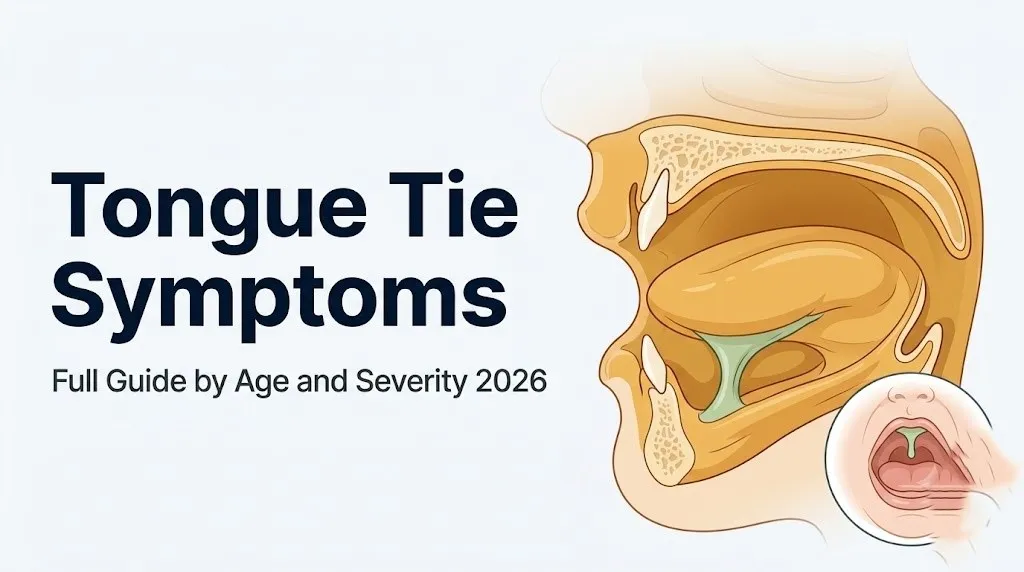
Tongue Tie Symptoms: Full Guide by Age and Severity 2026
Tongue tie symptoms range from a newborn’s inability to latch effectively at the breast to a toddler’s difficulty producing certain speech sounds to an adult’s unexplained jaw tension and neck pain, and the specific signs vary so dramatically by age and severity that many cases go unrecognized for years. Understanding what to look for at each life stage, and why the symptoms occur at an anatomical level, gives you the clinical context to decide whether what you’re observing warrants professional evaluation.
According to the American Academy of Pediatrics (AAP), ankyloglossia (the clinical term for tongue tie) affects an estimated 4% to 11% of newborns, with male infants affected at approximately three times the rate of female infants. A systematic review published in Pediatrics found that tongue tie is among the most consistently identified anatomical factors in breastfeeding failure during the newborn period, yet it remains underdiagnosed across all age groups because the clinical presentation varies so widely between individuals.
This guide covers every recognized tongue tie symptom from birth through adulthood, the specific physiological mechanism producing each symptom, how to distinguish functional tongue tie from normal frenulum variation, the critical difference between anterior and posterior tongue tie, how clinical assessment works, and the specific threshold that turns a scheduled pediatrician visit into a same-day urgency.
What Is Tongue Tie and What Causes It
Tongue tie, clinically termed ankyloglossia (ICD-10 Q38.1), is a congenital condition in which the lingual frenulum (the midline band of mucosal tissue connecting the underside of the tongue to the floor of the mouth) is abnormally short, thick, tight, or anteriorly positioned, restricting the tongue’s range of motion in ways that can affect feeding, speech, swallowing, and oral posture.
The lingual frenulum develops embryologically from midline mesenchymal tissue in the floor of the developing oral cavity during the first trimester of pregnancy. Normally, the frenulum thins and retracts from the tongue tip as development progresses. When this thinning is incomplete, an abnormally persistent frenulum band remains that tethers the tongue to the floor of the mouth.
Research has found an association between ankyloglossia and variants in the FOX gene family, particularly FOXE1 and FOXD2, which are involved in orofacial developmental patterning. A positive family history of tongue tie is present in a proportion of affected individuals, supporting a genetic component to the condition’s occurrence.
The condition is not caused by anything the parent did during pregnancy. It is a developmental anatomical variation reflecting incomplete resolution of a midline tissue band that is part of normal fetal oral development.
According to the Academy of Breastfeeding Medicine (ABM), ankyloglossia should be assessed as a potential contributing factor in any breastfeeding dyad experiencing feeding difficulty in the newborn period, rather than waiting for obvious visual confirmation of a tight frenulum, because functional impact does not always correlate with visible appearance.
Male infants are affected approximately three times more frequently than females, a sex disparity that has been replicated across multiple epidemiological studies and likely reflects the sex-linked nature of the FOX gene variants associated with the condition.
Tongue Tie Symptoms: The Complete Clinical Picture
Tongue tie symptoms span four functional domains: feeding, speech, swallowing, and orofacial posture, and they differ substantially by the patient’s age and the degree of frenulum restriction.
Here is the complete clinical symptom inventory across all age groups and functional domains:
| Functional Domain | Age Group | Observable Symptom | Mechanism |
|---|---|---|---|
| Feeding | Neonate | Ineffective latch, frequent feeding, weight loss | Impaired peristaltic tongue wave, reduced negative pressure |
| Feeding | Infant | Clicking sound while nursing, fatigue at breast | Air ingestion from poor seal, reduced suction efficiency |
| Feeding (maternal) | Nursing mother | Nipple pain, blanching, bleeding, low supply | Compensatory jaw clamping, poor nipple positioning |
| Swallowing | All ages | Difficulty with thick textures, gagging on lumpy foods | Reduced tongue base elevation during pharyngeal phase |
| Speech | Toddler/child | Distorted /t/, /d/, /n/, /l/, /r/ sounds | Inability to elevate tongue apex to alveolar ridge |
| Oral posture | Child/adult | Mouth breathing, low tongue rest posture | Tongue unable to rest on palate, airway narrowing |
| Dental/skeletal | Child/adult | High-arched palate, dental crowding, open bite | Chronic low tongue pressure on palatal vault |
| Myofascial | Adult | Jaw tension, neck pain, headache | Increased suprahyoid and infrahyoid muscle tension |
| Airway | Adult | Sleep-disordered breathing, snoring | Reduced nasopharyngeal airway from low tongue posture |
Premature infants with concurrent low suckling tone may have symptoms that overlap with tongue tie, making clinical differentiation from neurological feeding difficulties important. Evaluation by both a lactation consultant and a pediatric neurologist may be appropriate in this population.
Newborn Tongue Tie Symptoms
Newborn tongue tie symptoms appear within the first hours to days of life and center almost entirely on the ability to feed effectively, whether breastfeeding or bottle feeding.
A newborn with tongue tie typically cannot create adequate negative intraoral pressure during feeding because the restricted lingual frenulum prevents the tongue from elevating and cupping the nipple correctly. The result is a feeding session that is long (often exceeding 45 minutes), effortful, and ineffective in terms of actual milk transfer. The baby may appear to be feeding actively, with visible jaw movement, but very little milk is actually being consumed.
Observable signs that a newborn may have tongue tie include:
- Inability to maintain latch without sliding off the nipple or breast repeatedly
- A clicking or smacking sound during feeding (indicating air entering because the lip and tongue seal is incomplete)
- Visible flattening, notching, or a heart-shape at the tongue tip when the baby cries or the tongue is elevated
- Visible inability to protrude the tongue beyond the lower lip
- Excessive rooting and apparent hunger immediately after long feeding sessions
- Weight loss exceeding 7% to 10% of birth weight by day 3 to 4 of life (or failure to regain birth weight by day 10 to 14)
- Fewer than the expected number of wet diapers (fewer than 6 per 24 hours after day 5)
The Academy of Breastfeeding Medicine’s Clinical Protocol on Ankyloglossia and Frenotomy states that clinical assessment of the lingual frenulum should be part of the standard newborn oral examination, because early identification and, where indicated, frenotomy in the first two weeks of life is associated with significantly improved breastfeeding outcomes compared to later intervention.
Bottle-fed newborns with tongue tie may show less dramatic symptoms than breastfed infants because bottle nipples are more rigid and require less tongue peristalsis. However, these infants may still take longer to feed, fatigue during feeding, or swallow excessive air.
Tongue Tie Symptoms During Breastfeeding
Tongue tie symptoms during breastfeeding are the most frequently recognized presentation of the condition in the newborn period, and they reflect the specific demands that breastfeeding places on tongue range of motion and coordination.
Normal breastfeeding requires the infant’s tongue to perform a complex peristaltic movement: the tongue apex rises to cup the nipple against the hard palate, then a wave of muscular contraction moves from the anterior tongue to the posterior tongue, compressing the lactiferous sinuses (the milk-containing ducts within the breast) and transferring milk toward the pharynx. This action requires the tongue to elevate freely, which is precisely the movement restricted by an abnormal lingual frenulum.
Think of the tongue’s role in breastfeeding like a peristaltic pump. In a healthy infant, the tongue squeezes the breast from front to back in a coordinated wave, drawing milk efficiently into the throat. In an infant with tongue tie, the anterior anchoring of the tongue interrupts this wave. The infant compensates by biting or clamping with the jaw, which extracts some milk but transfers compressive force directly to the nipple rather than the breast tissue, producing the pain and nipple trauma so frequently reported by nursing mothers.
Specific breastfeeding-related symptoms of tongue tie in the infant include:
- Difficulty sustaining latch beyond the first 5 to 10 minutes
- Progressive feeding fatigue and falling asleep before adequate intake
- Excessive hunger and demanding cluster feeding that seems inconsistent with the time spent at the breast
- Audible clicking or gulping sounds indicating air swallowing (aerophagia)
- Visible compression stripe across the nipple (a horizontal white line indicating where the infant is biting rather than cupping)
A study published in the Journal of Human Lactation found that infants with ankyloglossia who underwent frenotomy showed measurable improvement in latch quality and maternal reported nipple pain compared to a control group, with the most substantial improvements seen in infants treated within the first four weeks of life.
Tongue Tie Symptoms in the Nursing Mother
The nursing mother experiences her own set of tongue tie symptoms driven by the compensatory jaw clamping her infant uses to extract milk when tongue-mediated suction is impaired.
Nipple pain is the primary maternal symptom. It is not the normal initial tenderness that many new mothers experience in the first few days of breastfeeding. It is persistent, often described as sharp or burning during feeding, and typically does not improve with correct latch techniques or nipple positioning adjustments because the root cause is the infant’s tongue mechanics, not the mother’s positioning.
Specific maternal symptoms associated with an infant’s tongue tie include:
- Nipple blanching (turning white) during or after feeding, caused by compressive ischemia from jaw clamping
- Nipple cracking, blistering, or bleeding from repeated trauma at the same compression point
- Nipple appearing pinched, wedge-shaped, or lipstick-shaped after feeding (indicating the nipple was compressed rather than cupped)
- Breast engorgement developing despite frequent feeding sessions, because ineffective milk removal signals reduced production over time
- Low milk supply developing within the first 2 to 3 weeks postpartum, driven by insufficient breast stimulation from a baby who cannot effectively drain the breast
- Mastitis (breast tissue inflammation and infection) from milk stasis in incompletely drained ducts
- Emotional distress, feeding aversion, and decisions to discontinue breastfeeding earlier than intended
According to the International Breastfeeding Journal, maternal nipple pain that persists beyond the first two weeks of breastfeeding and does not respond to latch correction by a trained lactation consultant should prompt evaluation of the infant for tongue tie, among other oral anatomical factors.
Mothers who have previously breastfed successfully and find a subsequent infant substantially more difficult to feed should specifically mention this pattern to their pediatrician or lactation consultant, because comparison to a prior successful feeding experience is clinically informative.
Key Takeaway: The nursing mother’s symptoms (persistent nipple pain, blanching, breast engorgement, and declining milk supply) are often the first clinically identifiable sign that the infant has tongue tie, making maternal symptoms as diagnostically relevant as the infant’s own feeding behavior.
Tongue Tie Symptoms in Babies and Infants Beyond the Newborn Period
As a tongue-tied infant moves past the newborn stage into the second and third months of life, the feeding-related symptoms may subtly shift while new functional challenges emerge.
Some infants compensate well enough for a mild-to-moderate tongue tie that breastfeeding becomes more manageable as the mother’s milk supply stabilizes and the baby grows stronger. But other tongue-tied infants continue to show feeding inefficiency, and additional symptoms emerge that reflect the broader impact of restricted tongue movement on oral function.
Aerophagia (excessive air swallowing during feeding) is more apparent in this period. It produces visible abdominal distension after feeds, frequent and forceful burping requirements, and gassiness that can mimic infant colic. Some infants are diagnosed with gastroesophageal reflux (GERD) and treated with acid-reducing medications when the actual driver of their symptoms is swallowed air from poor latch mechanics rather than true acid hypersecretion.
Gagging on thickened or pureed foods introduced at 4 to 6 months of age is another emerging symptom. Normal swallowing requires the tongue base to elevate during the pharyngeal phase of the swallow, pushing the food bolus past the epiglottis toward the esophagus. Restricted tongue elevation reduces this posterior propulsion, making thick textures more likely to pool near the pharynx and trigger the gag reflex.
The American Academy of Pediatric Dentistry (AAPD) recommends that infants with persistent feeding difficulty, recurrent reflux-like symptoms, or documented poor weight gain in the absence of other identified medical cause should receive oral anatomical assessment including frenulum evaluation as part of the diagnostic process.
Infants with concurrent hypotonia (reduced muscle tone) from neurological conditions including Down syndrome or cerebral palsy may show similar feeding symptoms to tongue tie, and clinical differentiation between tongue tie and neurologically mediated feeding dysfunction requires evaluation by both a feeding specialist and a developmental pediatrician.
Tongue Tie Symptoms in Toddlers and Speech Development
Tongue tie symptoms in toddlers center primarily on speech articulation, because the period between 18 months and 3 years is when children are rapidly acquiring consonant sounds that require precise tongue placement.
The tongue must elevate the apex to the alveolar ridge (the bony ridge immediately behind the upper front teeth) to produce the consonants /t/, /d/, /n/, and /l/ accurately. These are alveolar consonants, so named because they require alveolar ridge contact. The /r/ sound (a rhotic consonant) requires complex tongue body elevation and retraction that also depends on frenulum freedom. A restricted frenulum that prevents the tongue tip from reaching the alveolar ridge produces predictable articulatory errors in these specific sounds.
Observable speech symptoms in a toddler with tongue tie include:
- Substituting /d/ for /l/ (“dion” for “lion”)
- Producing /t/ and /d/ with a more posterior tongue contact, giving them a slightly indistinct quality
- Difficulty with /n/ at the end of words
- Persistent lisping or imprecise /s/ and /z/ sounds from altered tongue placement
- Difficulty producing words that require the tongue tip to leave the lower dental arch
It is important not to assume that all speech delays in toddlers reflect tongue tie. The relationship between mild-to-moderate tongue tie and speech delay is contested in the speech pathology literature. Research published in Pediatrics suggests that severe tongue tie (Kotlow Class III or IV, with free tongue length under 11mm) is more consistently associated with articulation errors than mild restriction. A speech-language pathologist (SLP) should evaluate any toddler with speech delay before tongue tie is assumed to be the primary cause.
Toddler girls with speech concerns and suspected tongue tie may be more readily evaluated at younger ages because speech milestones in girls are often tracked more closely by pediatricians due to earlier typical speech onset in biological females, despite the lower prevalence of tongue tie in females.
Tongue Tie Symptoms in Older Children
Tongue tie symptoms in older children expand beyond feeding and early speech to include dental development, oral hygiene challenges, and social-emotional impacts from speech differences.
The tongue at rest normally presses gently against the hard palate, exerting outward pressure that guides palatal arch development. A child with tongue tie typically has a habitual low tongue rest posture because elevating the tongue to the palate is uncomfortable or limited. Without this upward palatal pressure, the palatal vault develops with a higher, narrower arch than normal, a condition called high-arched palate. This change in palatal geometry reduces nasal airway volume and may promote mouth breathing.
Dental effects in older children include:
- High-arched, narrow palate with reduced transverse width
- Dental crowding from insufficient arch space
- An anterior open bite (upper and lower front teeth not meeting) in some cases
- Difficulty cleaning the lingual surfaces of the lower front teeth with the tongue, contributing to plaque accumulation and increased cavity risk at these sites
According to the American Academy of Pediatric Dentistry, the dental and skeletal impacts of prolonged altered tongue rest posture and oral function make it appropriate to assess older children with these findings for tongue tie as a contributing factor, even if the immediate concern is orthodontic rather than functional.
Mouth breathing from habitual open-mouth posture in tongue-tied children can also affect sleep quality. Nasal breathing during sleep is important for normal airway resistance and is protective against upper airway collapse. Children who breathe through their mouths habitually are more likely to snore and may have disrupted sleep architecture, though distinguishing tongue tie-related mouth breathing from adenotonsillar hypertrophy as the primary driver requires evaluation by a pediatric otolaryngologist.
Tongue Tie Symptoms in Adults
Tongue tie symptoms in adults represent either the continuation of untreated childhood tongue tie or the first recognition of a lifelong anatomical variation that has been compensated for until cumulative functional demands produce symptoms.
The most consistently reported adult tongue tie symptoms include:
- Jaw tension and temporomandibular dysfunction (TMD): Chronic restriction of tongue elevation increases resting tension in the suprahyoid muscle group, particularly the digastric (anterior belly), mylohyoid, and geniohyoid muscles. These muscles attach to both the tongue apparatus and the mandible; chronically elevated tension in this group increases compressive loading on the temporomandibular joint.
- Neck and upper shoulder tension: The suprahyoid muscles connect to the hyoid bone, which is mechanically linked through the infrahyoid muscles and cervical fascia to the cervical spine. Sustained suprahyoid hypertonicity can contribute to cervical myofascial tension and posterior cervical discomfort.
- Difficulty with certain oral functions: Licking food from lips, licking an ice cream cone, playing wind instruments, and kissing may all be noticeably limited.
- Speech sound distortions: Adults may have compensated for articulation limitations so effectively that only an SLP evaluation reveals residual distortions that the person has normalized.
- Dysphagia (swallowing difficulty): Particularly with thick or dry foods requiring substantial tongue base propulsion.
- Sleep-disordered breathing or snoring: The relationship between adult tongue tie and obstructive sleep apnea is based primarily on observational data and mechanistic reasoning (low tongue rest posture reducing posterior airway dimensions) rather than controlled trial evidence. This association is emerging rather than established.
Evidence quality varies significantly across adult tongue tie associations. The articulation and oral function limitations are well-established clinically. The TMD and myofascial associations are supported by clinical observation and small case series. The sleep-disordered breathing association is preliminary and requires controlled study confirmation before it can be presented as established.
Key Takeaway: Adult tongue tie symptoms reflect decades of compensatory muscle adaptation around a restricted frenulum, producing jaw tension, neck pain, and swallowing difficulty as the suprahyoid and infrahyoid muscle groups work harder than they should to compensate for limited tongue mobility.
Posterior Tongue Tie Symptoms and Why It Is Frequently Missed
Posterior tongue tie is a form of ankyloglossia in which the restrictive frenulum tissue is located at the base of the tongue rather than at or near the tongue tip, producing a submucosal (beneath the visible mucosa) tethering band that is not readily visible during routine oral examination.
Think of posterior tongue tie as a restriction buried under the surface. Unlike anterior tongue tie, where the frenulum band is visible as a tight cord connecting the tongue tip to the floor of the mouth, a posterior tie appears as normal mucosa. The tongue tip looks free. A clinician who only looks for a visible anterior frenulum band will miss a posterior tie entirely. The restriction is only detectable by elevating the tongue and palpating the floor of the mouth for a tight, cord-like submucusal band, or by watching the tongue move and noting the limited elevation despite an apparently normal appearance.
This anatomical difference explains the frequent misdiagnosis. Parents of a posterior tongue-tied infant are often told “the tongue looks normal” because the visual cue that most clinicians expect (a visible short frenulum) is absent.
Functional symptoms of posterior tongue tie are often indistinguishable from anterior tongue tie symptoms:
- Ineffective breastfeeding with poor milk transfer
- Maternal nipple pain and trauma from compensatory jaw clamping
- Clicking during feeding from incomplete lip and tongue seal
- Limited tongue elevation on clinical assessment despite normal visual appearance
- The characteristic bunching or humping of the tongue body when the infant attempts elevation (because the posterior tethering prevents the posterior body from retracting while the tip is free, producing a hump shape)
Assessment of posterior tongue tie requires a trained examiner who performs a complete functional evaluation including both visual inspection and digital palpation of the floor of the mouth. The Hazelbaker Assessment Tool for Lingual Frenulum Function (HATLFF) assesses both appearance and function, making it more sensitive to posterior ties than a visual-only examination.
Mild vs Severe Tongue Tie Symptoms: How Restriction Level Changes the Picture
The severity of tongue tie symptoms corresponds broadly, though not perfectly, to the degree of frenulum restriction, and clinical classification systems exist to standardize this assessment.
The Kotlow classification grades tongue tie by the length of free tongue (the portion of tongue anterior to the frenulum attachment point):
| Kotlow Class | Free Tongue Length | Clinical Significance |
|---|---|---|
| Class I (mild) | 12 to 16mm | Minimal functional restriction; symptoms may be absent or mild |
| Class II (moderate) | 8 to 11mm | Moderate restriction; breastfeeding difficulty likely; speech monitoring needed |
| Class III (severe) | 3 to 7mm | Marked restriction; breastfeeding impairment expected; speech intervention typically required |
| Class IV (complete) | Less than 3mm | Complete tethering to floor of mouth; severe feeding, speech, and oral function impact |
The Coryllos classification adds morphological description to this framework, distinguishing between anterior membrane-like ties (Type 1), submucosal anterior ties (Type 2), posterior muscular ties (Type 3), and posterior submucosal ties (Type 4).
Mild tongue tie may produce no symptoms at all, particularly in bottle-fed infants where the bottle nipple compensates for reduced tongue peristalsis. It may produce mild breastfeeding difficulty that resolves with skilled lactation support without frenotomy. In many mild cases, the infant’s growing tongue strength and improved coordination over weeks reduces the functional gap between tongue range of motion and feeding demand.
Severe tongue tie (Kotlow Class III or IV) rarely resolves with support alone and typically requires frenotomy for feeding to improve adequately. According to a randomized controlled trial published in Pediatrics (Hogan, 2005), frenotomy in infants with severe tongue tie resulted in immediate improvement in maternal nipple pain and was safe and well-tolerated in the neonatal period.
Children with only mild restriction who develop speech sound errors should be evaluated by a speech-language pathologist before assuming frenotomy is indicated, because many mild articulatory errors respond to targeted speech therapy without surgical intervention.
How Tongue Tie Causes These Symptoms: The Anatomy and Physiology
Every tongue tie symptom traces back to the mechanical restriction imposed by the abnormal lingual frenulum on the tongue’s range and pattern of motion, and understanding the specific anatomy involved makes the symptom pattern logical rather than puzzling.
The tongue is a muscular organ composed of four intrinsic muscles (superior longitudinal, inferior longitudinal, transverse, and vertical fibers) that change the tongue’s shape, and four extrinsic muscles (genioglossus, hyoglossus, styloglossus, and palatoglossus) that move the tongue within the oral cavity. The genioglossus, the largest extrinsic tongue muscle, runs from the inner surface of the mandibular symphysis to the tongue body and is the primary tongue protruder and elevator. It is innervated by the hypoglossal nerve (cranial nerve XII).
The lingual frenulum normally attaches near the midpoint of the tongue’s inferior surface. When it attaches too anteriorly or is too short, it creates a tethering force on the tongue apex that resists the genioglossus-driven elevation. The tongue can move, but it cannot move freely through the full range required for feeding, articulation, and palatal pressure.
In infant breastfeeding, the tongue must complete a peristaltic wave (front-to-back muscular contraction) to generate negative intraoral pressure and milk transfer. A restricted frenulum interrupts this wave at the anterior tongue, causing the infant to recruit the jaw muscles (masseter, pterygoids) to compensate. The masseter and medial pterygoid apply compressive force to the nipple that the tongue should instead be cupping, producing the pain, blanching, and nipple trauma characteristic of tongue-tied breastfeeding.
In speech, the tongue tip must contact the alveolar ridge (the bony prominence immediately posterior to the upper incisors) to produce /t/, /d/, /n/, and /l/. A frenulum that prevents full tongue elevation physically prevents this contact point, producing the substitution and distortion patterns described in the toddler and child sections. This is not a learning problem; it is a mechanical one.
Tongue Tie Symptoms vs Normal Frenulum Variation
Not every visible lingual frenulum represents a pathological tongue tie, and distinguishing clinically significant ankyloglossia from normal frenulum anatomy is one of the most contested areas in the field.
Every person has a lingual frenulum. It is a normal anatomical structure. Its size, thickness, and attachment point vary considerably among individuals with completely normal tongue function. A short-appearing frenulum that allows full tongue elevation, full tongue protrusion, and effective oral function is not clinically significant regardless of its appearance.
The critical distinction is functional, not visual. A frenulum is clinically significant when it restricts tongue function to a degree that produces symptoms. The same frenulum morphology may be entirely asymptomatic in one infant and functionally impairing in another, depending on individual variation in tongue muscle strength, jaw anatomy, and the specific demands of feeding or speech in that child.
To assess whether a frenulum is causing functional restriction rather than merely being visible:
- Observe the tongue’s ability to elevate the tip to the hard palate with the mouth open (a healthy tongue should touch the palate easily with the mouth open wide).
- Observe tongue protrusion: the tongue tip should extend beyond the lower lip without visible notching, heart-shaping, or lateral curvature.
- Observe the tongue’s ability to touch the inner surface of each upper molar (lateral movement toward each cheek).
- Observe the tongue’s shape during elevation attempts: a tongue that “cups” or shows a visible midline groove pulling the tip down is demonstrating tethering rather than free movement.
- In infants, observe the feeding pattern, milk transfer effectiveness (pre- and post-feed weight on a clinical scale), and maternal nipple condition as functional indicators rather than relying on visual appearance alone.
The Bristol Tongue Assessment Tool (BTAT), a validated four-point clinical assessment tool developed in the United Kingdom and published in peer-reviewed pediatric literature, scores four observable tongue characteristics (ability to lift tongue, reach outside the mouth, fit in the gum line, and the tongue’s appearance on elevation) to produce an objective severity score rather than relying on individual clinical impression.
How Tongue Tie Is Diagnosed and Assessed Clinically
Diagnosis of clinically significant tongue tie involves a structured clinical examination that assesses both the anatomical characteristics of the lingual frenulum and the functional impact on feeding, speech, or oral function.
The diagnostic process at a specialist assessment:
- Medical history review: Feeding history (breastfeeding success, weight gain, feeding duration, maternal nipple pain), speech history in toddlers and children (specific sound difficulties, intelligibility), oral function concerns in adults (jaw pain, neck tension, swallowing difficulty), and family history of tongue tie.
- Oral visual inspection: Observation of the tongue at rest, during protrusion, and during voluntary elevation. Assessment of frenulum visibility, attachment point, and thickness. Assessment of palate shape (high-arched palate, palatal width).
- Digital palpation: For posterior tongue tie specifically, the examiner runs a finger along the floor of the mouth to feel for a submucous cord of tissue that is not visible on surface inspection.
- Validated assessment tool application: The Hazelbaker Assessment Tool for Lingual Frenulum Function (HATLFF) scores both appearance items (length of frenulum when tongue is lifted, attachment to tongue, attachment to ridge) and function items (lateralization, lift, extension, spread of tongue, cupping, peristalsis). The Bristol Tongue Assessment Tool (BTAT) provides a simpler four-point scoring system suitable for rapid neonatal assessment.
- Feeding observation: In infants, the assessment is incomplete without watching the infant attempt to feed, either at the breast or with a bottle, and ideally including a pre- and post-feed weight measurement to quantify milk transfer.
- Speech assessment: In toddlers and children, referral to a speech-language pathologist for formal articulation assessment provides data on which specific phonemes are affected and the degree of functional impact on intelligibility.
The relevant specialist providers for tongue tie diagnosis include a board-certified international lactation consultant (IBCLC) for feeding assessment, a pediatric dentist or pediatric otolaryngologist for anatomical classification and frenotomy decision-making, and a speech-language pathologist for speech functional assessment in older children and adults.
Tongue Tie Treatment: Frenotomy, Frenuloplasty, and What Comes After
Treatment for clinically significant tongue tie is a procedure that divides the restrictive lingual frenulum, either by frenotomy (a simple division with scissors or laser) or frenuloplasty (a more involved surgical revision with closure for thicker or posterior ties).
Frenotomy (also called frenulotomy) involves dividing the frenulum with sterile scissors or a laser with no sutures. It is typically performed in the office setting for neonates and young infants. In experienced hands, it is fast, well-tolerated, and associated with low complication rates. The Academy of Breastfeeding Medicine supports frenotomy for breastfeeding infants with confirmed tongue tie and significant feeding impairment, based on randomized controlled trial evidence showing improved maternal pain and feeding outcomes.
Frenuloplasty involves more extensive tissue revision, often using a Z-plasty technique to release a thicker or more muscular posterior tie and close the wound with absorbable sutures. It is more commonly used for older children and adults with thicker, more muscular tethering that cannot be adequately addressed by simple division.
Laser frenectomy using a diode or CO2 laser is offered by some pediatric dentists and ENTs. The evidence comparing laser to scissors frenotomy in infants does not consistently favor one technique over the other in terms of outcomes; both are used by practitioners experienced in the procedure.
Treatment outcomes depend on the specific indication:
- For breastfeeding improvement: most improvement is seen within the first 24 to 48 hours of frenotomy in neonates.
- For speech: frenotomy alone does not teach correct articulation. Post-procedure speech therapy with a speech-language pathologist is typically required for children with established compensatory speech patterns.
- For adults: orofacial myofunctional therapy before and after frenuloplasty is recommended to retrain the tongue muscles that have compensated for decades, because releasing the frenulum without retraining does not automatically restore normal tongue posture and function.
When Tongue Tie Symptoms Require Prompt Professional Evaluation
Certain tongue tie symptom patterns warrant evaluation within 24 to 48 hours rather than waiting for a routine scheduled appointment.
For newborns, the urgency threshold is clear:
Seek same-day or next-day medical evaluation with a pediatrician if your newborn shows:
- Weight loss exceeding 10% of birth weight at any point in the first week of life
- Failure to regain birth weight by day 10 to 14
- Fewer than 6 wet diapers in 24 hours after day 5 of life
- Signs of dehydration: sunken anterior fontanelle (the soft spot on the top of the head), absent or minimal tears when crying, very dry mouth and lips, dark yellow or orange-tinged urine
- Feeding sessions consistently longer than 45 to 60 minutes with the infant still appearing hungry
- Complete inability to latch to the breast despite sustained attempts across multiple feeds
These findings in a newborn represent potential failure to thrive from feeding failure, which is a medical urgency. Dehydration in a neonate progresses rapidly and can cause electrolyte imbalances, jaundice, and neurological injury.
For older children, prompt evaluation (within 1 to 2 weeks) is appropriate when:
- A toddler’s speech development has stalled or regressed following a period of normal progress
- A school-age child’s speech intelligibility is causing significant communication difficulty or social distress at school
For adults, prompt evaluation is appropriate when dysphagia (difficulty swallowing) is causing weight loss or aspiration concerns, which warrants referral to an otolaryngologist and a speech-language pathologist for formal swallowing assessment rather than being attributed to tongue tie without ruling out other dysphagia causes.
When a Newborn’s Tongue Tie Symptoms Require Urgent Evaluation
Certain presentations in newborns with tongue tie symptoms represent a medical urgency that goes beyond scheduling a lactation consultation.
Call your pediatrician immediately or go to the emergency room if your newborn shows any of the following:
- Weight loss of more than 10% of birth weight before day 5, or any weight loss after day 5 (birth weight should be regaining by this point)
- No wet diapers for 8 or more hours in a newborn under 2 weeks of age
- A sunken soft spot (anterior fontanelle) on the top of the head, indicating dehydration
- Absence of visible tears when crying, with dry or sticky mucous membranes in the mouth
- Orange or brick-red crystals in the diaper (uric acid crystals from concentrated, inadequate urine output)
- Extreme lethargy, difficulty waking for feeds, or floppy muscle tone in the first 2 weeks of life
- Jaundice (yellowing of skin and whites of the eyes) developing or worsening rather than improving after day 5 of life, particularly in a baby who is not feeding well
These presentations indicate that inadequate feeding from any cause, including tongue tie, has progressed to a level of dehydration or nutritional deficit that constitutes a neonatal medical emergency.
Do not wait for a scheduled pediatrician visit if your newborn is showing any of these signs. Neonatal dehydration from feeding failure can progress to hypernatremia (elevated sodium from free water deficit), hyperbilirubinemia requiring treatment, and in severe cases, neurological injury. This requires immediate medical evaluation, not home monitoring or an urgent care visit.
Frequently Asked Questions About Tongue Tie Symptoms
What are the first signs of tongue tie in a newborn?
The first signs of tongue tie in a newborn are typically poor latch at the breast, ineffective milk transfer despite prolonged feeding sessions, and a clicking or smacking sound during feeding indicating air entry from an incomplete oral seal.
A newborn with tongue tie often appears hungry immediately after a long feed because the tongue mechanics were not effective enough to transfer adequate milk volume.
Additional early signs include inability to protrude the tongue tip beyond the lower lip and a heart-shaped or notched appearance of the tongue tip when the baby cries or attempts to lift the tongue.
Can tongue tie cause speech problems in toddlers?
Tongue tie can cause specific articulation errors in toddlers, particularly with consonants that require the tongue tip to elevate to the alveolar ridge, including /t/, /d/, /n/, /l/, and /r/.
Research published in Pediatrics indicates that the association between tongue tie and speech difficulty is most consistently documented in severe ankyloglossia (Kotlow Class III or IV), while the evidence for speech impact in mild-to-moderate cases is less definitive.
Any toddler with persistent speech sound errors should be evaluated by a speech-language pathologist for a functional articulation assessment before assuming tongue tie is the primary cause, because speech therapy alone resolves many toddler articulation errors regardless of frenulum anatomy.
What does a posterior tongue tie look like or feel like?
Posterior tongue tie does not look like anything distinctive from the outside of the mouth, which is precisely why it is frequently missed on routine examination.
Unlike anterior tongue tie, where a visible cord connects the tongue tip to the floor of the mouth, a posterior tie is submucosal and feels like a firm cord or band running along the floor of the mouth when an examiner palpates beneath an elevated tongue.
A parent may notice that their baby’s tongue bunches or humps upward in the middle when the baby tries to elevate it, rather than lifting smoothly at the tip, which is a functional clue to posterior tethering even when nothing looks visibly abnormal.
Can adults have tongue tie symptoms without knowing it?
Yes, adults can have clinically relevant tongue tie that was never diagnosed in childhood, often because they compensated sufficiently to function but at a cost of chronically elevated suprahyoid muscle tension, limited tongue range of motion, and habitual low tongue rest posture.
Adult symptoms of undiagnosed tongue tie include jaw pain and temporomandibular dysfunction from increased suprahyoid muscle loading, neck and upper shoulder myofascial tension, difficulty with oral functions like licking the lips or playing wind instruments, and in some individuals, sleep-disordered breathing associated with low tongue rest posture.
The association between adult tongue tie and temporomandibular dysfunction is supported primarily by clinical observation and case series rather than controlled trials, so adults considering evaluation should work with an orofacial myofunctional therapist and an oral surgeon or ENT experienced in adult ankyloglossia.
How do I know if my baby’s tongue tie is affecting breastfeeding?
The most reliable indicators that tongue tie is affecting breastfeeding are persistent maternal nipple pain that does not resolve with repositioning, nipple blanching or a lipstick-shaped nipple after feeds, feeding sessions lasting more than 45 minutes that leave the baby seeming unsatisfied, and inadequate infant weight gain.
A pre- and post-feed weight measurement on a clinical scale (performed in a lactation consultant or pediatrician’s office) provides objective evidence of how much milk the infant is transferring per feeding session.
If your baby is gaining less than the expected 0.5 to 1 ounce per day in the first month and you are experiencing significant nipple pain, request evaluation by an international board-certified lactation consultant (IBCLC) and ask for the infant’s oral anatomy including the lingual frenulum to be specifically assessed.
Does tongue tie go away without treatment?
True ankyloglossia does not reliably resolve without intervention, though some mild, thin anterior frenula do thin and retract somewhat with normal oral movement during early infancy.
The Academy of Breastfeeding Medicine does not recommend watchful waiting as a primary strategy when tongue tie is producing significant breastfeeding impairment, because the feeding window for establishing maternal milk supply is narrow and delays can result in permanent supply reduction.
Posterior and muscular tongue ties are particularly unlikely to resolve spontaneously because the tethering involves denser connective tissue and muscle fiber rather than a thin mucosal membrane.
Closing
Tongue tie symptoms are not one thing. A newborn who cannot latch, a three-year-old who cannot produce /l/ clearly, a ten-year-old with a narrow palate, and an adult with unexplained jaw tension are all potentially expressing the same underlying anatomical restriction at completely different life stages.
The most practical next step depends entirely on the age of the person you are concerned about. For a newborn with feeding difficulty, contact an international board-certified lactation consultant and your pediatrician within 24 to 48 hours. For a toddler with speech concerns, request a speech-language pathologist evaluation and ask specifically whether tongue tie assessment is included. For an adult, seek out an orofacial myofunctional therapist or a dentist, oral surgeon, or ENT with documented experience in adult ankyloglossia evaluation.
Ask your provider to use a validated assessment tool, specifically the Hazelbaker Assessment Tool for Lingual Frenulum Function or the Bristol Tongue Assessment Tool, rather than relying on visual impression alone. These tools exist because clinical impression without structure misses posterior tongue tie repeatedly, and that missed diagnosis is the single most preventable gap between this condition and effective treatment.