Red Eye Causes: What Every Type of Redness Means 2026
Red eye causes span the full clinical spectrum from self-resolving viral infections that need nothing more than time, to acute angle-closure glaucoma that can permanently destroy vision within hours if not treated as the emergency it is. The specific pattern of redness, the presence or absence of pain, the type of any discharge, and whether vision is affected are the clinical signals that separate the benign from the dangerous.
The National Eye Institute estimates that millions of Americans visit a physician for red eye each year, making it one of the most common acute eye complaints in primary care and emergency medicine. The vast majority of cases are self-limiting, but a clinically meaningful subset represents conditions where delayed evaluation leads to preventable vision loss.
This article covers every major cause of red eye, starting with the anatomy that explains why different diseases produce different patterns of redness, and then working through each cause with its specific biological mechanism, accompanying symptoms, and the exact clinical threshold that separates home monitoring from urgent evaluation.
Red Eye Causes: Overview and the Anatomy Behind the Redness
Red eye is produced by dilation and congestion of the blood vessels on the surface of the eye or within its deeper vascular supply, triggered by infection, inflammation, injury, pressure, or chemical irritation acting on specific ocular structures.
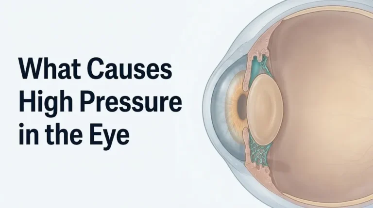
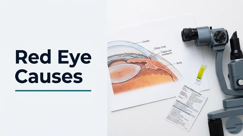
The outer surface of the eye has three distinct vascular layers. The conjunctiva (the transparent mucous membrane covering the white sclera and inner eyelids) contains a network of small, mobile, bright-red superficial vessels. Beneath the conjunctiva lies the episclera, a loose connective tissue layer containing a deeper vascular plexus of larger, less mobile vessels with a darker reddish-purple appearance. The deepest layer is the sclera itself, supplied by the long posterior ciliary arteries that also feed the iris and ciliary body of the uveal tract.
When irritants, allergens, or infections affect the conjunctival surface, the superficial conjunctival vessels dilate, producing the bright-red, diffuse redness most people recognize as “bloodshot eyes.” When inflammation affects the deeper iris, ciliary body, or trabecular meshwork, or when intraocular pressure spikes acutely, it is the deeper episcleral and ciliary vessels that engorge, producing a distinctly different clinical picture.
Understanding this vascular anatomy is the foundation for understanding why different red eye causes look different and carry different levels of urgency.
Red eye causes ranked from most to least common, according to clinical epidemiology:
- Viral conjunctivitis (most common cause overall in outpatient settings)
- Allergic conjunctivitis (most common cause in the general population over a lifetime)
- Bacterial conjunctivitis (most common cause in children)
- Dry eye disease
- Subconjunctival hemorrhage
- Blepharitis and meibomian gland dysfunction
- Contact lens-related irritation or infection
- Uveitis and iritis
- Episcleritis and scleritis
- Corneal ulcer or keratitis
- Acute angle-closure glaucoma (least common but most acutely dangerous)
Older adults over 60 face a substantially higher risk for acute angle-closure glaucoma than younger adults, because progressive thickening of the crystalline lens narrows the anterior chamber angle over decades, reducing the space available for aqueous humor drainage.
Conjunctival vs. Ciliary Injection: Why the Pattern of Redness Matters
The single most practically useful clinical skill for assessing a red eye is distinguishing conjunctival injection (superficial, bright red, diffuse) from ciliary injection (deep, violet-red, concentrated in a ring around the corneal margin), because this pattern difference reliably separates self-limiting causes from those requiring urgent ophthalmological evaluation.
Conjunctival injection involves dilation of the superficial conjunctival vessels fed by the anterior conjunctival arteries. The redness is bright red, most intense toward the inner corners and outer edges of the visible eye surface, and fades toward the corneal margin. The dilated vessels are visibly separate, movable with gentle pressure on the conjunctiva, and blanch when a topical vasoconstrictor (such as tetrahydrozoline) is applied.
Ciliary injection involves dilation of the deeper episcleral vessels fed by the anterior ciliary arteries and the long posterior ciliary arteries supplying the iris, ciliary body, and anterior choroid. The redness has a violet-red or pink-red hue and is most intense directly at the corneal margin (the limbus), forming a ring around the cornea that fades toward the inner corners and edges of the eye. These vessels do not move with gentle pressure, do not blanch with topical vasoconstrictors, and indicate that the disease process is located in the deeper intraocular structures.
Think of the eye’s vascular system like a city with two separate water mains, one serving the streets and one serving the underground infrastructure. Conjunctival injection lights up the street level only. Ciliary injection illuminates the underground main, which supplies the most critical inner structures. When the underground main is congested, you are dealing with a problem that the surface appearance alone cannot fully reveal.
| Injection Type | Color | Location | Blanches with Vasoconstrictor | Associated Conditions |
|---|---|---|---|---|
| Conjunctival | Bright red | Diffuse, peripheral emphasis | Yes | Viral conjunctivitis, allergy, dry eye, subconjunctival hemorrhage |
| Ciliary (limbal) | Deep violet-red | Ring at corneal margin | No | Uveitis, iritis, acute angle-closure glaucoma, keratitis |
| Mixed | Both patterns | Diffuse and perilimbal | Partial | Severe conjunctivitis, corneal ulcer, scleritis |
| Episcleral | Dark red or salmon-pink | Sectoral or diffuse deeper layer | No | Episcleritis, scleritis |
Pregnant women may experience increased conjunctival vessel prominence and mild injection due to elevated circulating blood volume and estrogen-driven vasodilation, which can make the normal eye appear redder without any infectious or inflammatory cause. This physiological change does not require treatment but should be distinguished from pathological causes during a primary care or obstetric visit.
Viral Conjunctivitis as a Cause of Red Eye
Viral conjunctivitis causes red eye by triggering a direct cytopathic inflammatory response in the conjunctival epithelium, driving conjunctival vessel dilation, increased vascular permeability, and a watery discharge through a well-characterized viral immune cascade.
Adenovirus accounts for approximately 65% to 90% of all viral conjunctivitis cases according to clinical epidemiological data, and it is specifically the cause of epidemic keratoconjunctivitis (EKC), the highly contagious form that spreads rapidly through households, schools, and workplaces. Adenovirus serotypes 8, 19, and 37 are the most common EKC-associated types. The virus binds to receptors on conjunctival epithelial cells, replicates within the cell nucleus, and kills the host cell through direct cytopathic effect, triggering a vigorous innate immune response including neutrophil and lymphocyte infiltration into the conjunctival stroma.
/
The resulting inflammatory mediator release, primarily interleukin-1 beta (IL-1beta) and tumor necrosis factor-alpha (TNF-alpha) from infected and adjacent epithelial cells and recruited immune cells, drives conjunctival vessel dilation and increased vascular permeability. The discharge produced is watery or mucoid, not purulent, because the inflammatory infiltrate is lymphocytic rather than neutrophilic.
Viral conjunctivitis is self-limiting in immunocompetent adults, typically resolving over 7 to 14 days without antibiotic treatment. The American Academy of Ophthalmology explicitly states that antibiotics are not indicated for viral conjunctivitis, as they have no effect on viral replication and contribute to antibiotic resistance.
Features of viral conjunctivitis to watch for:
- Watery or mucoid discharge (not thick yellow-green pus)
- Often starts in one eye and spreads to the other within 24 to 48 hours
- Frequently follows or accompanies an upper respiratory tract infection or recent cold
- Preauricular lymph node swelling (a palpable tender lymph node just in front of the ear), which is more specific to adenoviral conjunctivitis than to bacterial conjunctivitis
- Photophobia (light sensitivity) if corneal involvement develops (EKC can progress to corneal subepithelial infiltrates)
Immunocompromised individuals, including people on chemotherapy or systemic immunosuppression, may experience prolonged or more severe viral conjunctivitis and warrant earlier ophthalmological evaluation rather than home monitoring.
Bacterial Conjunctivitis Causes and Discharge Patterns
Bacterial conjunctivitis causes red eye through neutrophilic infiltration of the conjunctival stroma driven by bacterial toxins and surface antigen-triggered innate immune activation, producing the thick, purulent discharge that clinically distinguishes it from viral conjunctivitis.
The most common causative organisms differ by age group. In adults, Staphylococcus aureus is the predominant pathogen, followed by Haemophilus influenzae and Streptococcus pneumoniae. Bacterial surface components, specifically lipopolysaccharide from gram-negative organisms and peptidoglycan and protein A from staphylococci, activate toll-like receptors (TLRs) on conjunctival epithelial cells and resident macrophages. This TLR activation triggers nuclear factor-kappa B (NF-kB) signaling, producing a rapid pro-inflammatory cytokine burst that recruits neutrophils into the conjunctival tissue.
Neutrophils are the key to the discharge character. Whereas viral conjunctivitis produces a lymphocyte-dominated response with watery discharge, bacterial conjunctivitis produces a neutrophil-dominated response. Neutrophils arriving in large numbers die rapidly, and their cellular debris mixed with mucus and proteins from increased vascular permeability produces the thick, yellow-green, matting, purulent discharge that is the hallmark clinical sign. Many patients report waking up with their eyelids stuck together due to overnight accumulation of this discharge.
The Centers for Disease Control and Prevention notes that bacterial conjunctivitis is the most common infectious cause of red eye in children under five, with most cases being self-limiting over seven to ten days without antibiotics in otherwise healthy children. However, topical antibiotic eyedrops are commonly prescribed to hasten recovery and reduce transmission.
A critical exception is hyperacute bacterial conjunctivitis caused by Neisseria gonorrhoeae in sexually active adults (or in newborns, discussed in a later section). Gonococcal conjunctivitis produces a copious, rapidly accumulating purulent discharge with extreme conjunctival injection and eyelid swelling, and can penetrate the intact corneal epithelium, producing perforation within 24 to 48 hours without treatment. Any adult with this presentation requires same-day evaluation by a board-certified ophthalmologist and systemic antibiotic therapy.
Key Takeaway: The discharge character is your most reliable clinical clue at home: watery or mucoid discharge points to viral conjunctivitis (self-limiting, no antibiotics needed); thick, yellow-green, matting discharge points to bacterial conjunctivitis (warrants clinical evaluation, especially in contact lens wearers or if vision is affected).
Allergic Conjunctivitis and the Histamine Mechanism
Allergic conjunctivitis causes red eye through a Type I IgE-mediated hypersensitivity reaction in the conjunctiva, in which allergen-specific IgE antibodies bound to conjunctival mast cells cross-link upon re-exposure to the trigger allergen, causing immediate mast cell degranulation and histamine release that drives vessel dilation, itching, and chemosis.
The mechanism is precise. On first exposure to an allergen such as tree pollen, cat dander, or dust mite proteins, B lymphocytes in the conjunctival lymphoid tissue are sensitized and produce allergen-specific IgE antibodies. These IgE antibodies bind to high-affinity IgE receptors (FcεRI) on the surface of conjunctival mast cells, effectively arming them. On re-exposure, the allergen molecules cross-link adjacent IgE-FcεRI complexes on the mast cell surface, triggering the release of preformed mediators including histamine and tryptase within seconds, and the delayed synthesis of prostaglandins and leukotrienes over the following minutes to hours.
Histamine acts on H1 receptors on conjunctival blood vessel endothelium, causing immediate vasodilation and increased vascular permeability. The result is the intense bilateral itching that is the most characteristic feature of allergic conjunctivitis, followed by diffuse conjunctival injection, watery discharge, and chemosis (conjunctival swelling). The itching distinguishes allergic conjunctivitis from other red eye causes more reliably than any other single symptom.
According to the American Academy of Ophthalmology, allergic conjunctivitis affects an estimated 20% of the United States population and is the most prevalent form of ocular allergy. The condition is bilateral in the vast majority of cases, though one eye may be more affected than the other depending on allergen exposure patterns.
Vernal keratoconjunctivitis (VKC) is a severe, chronic form of allergic conjunctivitis predominantly affecting young males in warm climates. VKC involves both IgE-mediated and T-cell-mediated components and can produce giant papillae on the upper tarsal conjunctiva, thick ropy mucous discharge, and corneal complications including shield ulcers that require urgent ophthalmological management. Any child or adolescent with recurrent or persistent allergic eye disease with thick discharge should be evaluated by a board-certified ophthalmologist rather than managed with over-the-counter antihistamine drops alone.
Dry Eye Disease Causes of Red Eye
Dry eye disease (also called keratoconjunctivitis sicca) causes red eye through an inflammatory cascade initiated by tear film instability, in which the loss of normal tear film protection exposes the conjunctival and corneal epithelium to hyperosmotic stress and mechanical friction that upregulates pro-inflammatory mediators and drives conjunctival vessel dilation.
The tear film has three essential layers: an outer lipid layer produced by the meibomian glands in the tarsal plates of the eyelids, a middle aqueous layer produced by the lacrimal glands in the superolateral orbit, and an inner mucin layer produced by goblet cells in the conjunctival epithelium. Dry eye disease occurs when any of these layers is deficient. The most prevalent form, evaporative dry eye, results from meibomian gland dysfunction (MGD), in which obstructed or atrophic meibomian glands produce a lipid layer too thin to prevent tear film evaporation.
When the tear film breaks up too quickly (measured as a reduced tear break-up time), hyperosmotic conditions develop on the ocular surface. Hyperosmolarity activates mitogen-activated protein kinase (MAPK) signaling pathways in corneal and conjunctival epithelial cells, upregulating production of matrix metalloproteinase-9 (MMP-9), IL-1beta, and TNF-alpha. These mediators damage the epithelial glycocalyx, further destabilize the tear film, and trigger neurogenic vasodilation through substance P release from corneal sensory nerve terminals, producing the diffuse conjunctival injection of chronic dry eye.
The National Eye Institute reports that dry eye disease affects an estimated 16 million Americans, with postmenopausal women being disproportionately affected. The elevated prevalence in postmenopausal women reflects the role of androgens in meibomian gland function: androgens stimulate meibomian gland lipid secretion, and the decline in androgen levels after menopause contributes to meibomian gland atrophy and evaporative dry eye. Women are also more likely than men to use medications such as antihistamines, antidepressants, and oral contraceptives that reduce aqueous tear production by inhibiting lacrimal gland secretion.
Subconjunctival Hemorrhage Causes
Subconjunctival hemorrhage causes a distinctly bright red, sharply demarcated patch of redness on the white of the eye, produced by the rupture of a small conjunctival vessel flooding the potential space between the conjunctiva and the underlying episclera with blood.
The anatomy explains the appearance precisely. A subconjunctival hemorrhage looks alarming because blood that would normally be invisible inside a vessel instead pools across a large visible surface. The patch is flat, bright red (like fresh blood), and has sharp, well-defined borders at its edges. It does not affect the cornea, does not produce discharge, and in the vast majority of cases causes no pain. Vision is not affected. This is because the hemorrhage lies entirely on the surface of the eye and does not affect any optical or pressure-related structure.
Spontaneous subconjunctival hemorrhage without identifiable trauma is most commonly associated with elevated venous pressure events. The Valsalva maneuver (straining with breath held, as during heavy lifting, coughing, vomiting, or straining with constipation) transiently elevates venous pressure in the head and neck, which can rupture a small, fragile conjunctival vessel. Vigorous sneezing produces the same mechanism. Rubbing the eye forcefully can directly traumatize the vessel.
Conditions and medications that increase subconjunctival hemorrhage risk:
- Anticoagulant medications (warfarin, apixaban, rivaroxaban, clopidogrel): reduce clotting capacity, making minor vessel trauma more likely to produce a visible hemorrhage
- Antiplatelet medications (aspirin, nonsteroidal anti-inflammatory drugs): impair platelet aggregation
- Uncontrolled hypertension: elevated systemic arterial pressure increases the pressure gradient across small vessel walls
- Chronic coughing from COPD, asthma, or prolonged respiratory illness
- Contact lens insertion or removal with excessive pressure
- Trauma: any direct impact to the eye or periorbital area
The American Academy of Ophthalmology states that isolated spontaneous subconjunctival hemorrhage in an otherwise healthy adult without vision change or pain is a benign, self-resolving condition that requires no specific treatment and typically resolves over one to three weeks as the blood is reabsorbed. Recurrent, bilateral, or traumatic subconjunctival hemorrhage warrants evaluation by a primary care physician to assess blood pressure and coagulation status.
Key Takeaway: Subconjunctival hemorrhage looks dramatically worse than it is: the flat, bright-red, sharply bordered patch with no pain and no vision change is benign and resolves on its own over one to three weeks, but any subconjunctival hemorrhage following eye trauma must be evaluated by a board-certified ophthalmologist the same day to rule out a deeper posterior segment hemorrhage.
Uveitis and Iritis as Causes of Red Eye
Uveitis (inflammation of the uveal tract, the middle vascular layer of the eye comprising the iris, ciliary body, and choroid) and iritis (anterior uveitis involving specifically the iris) cause red eye through T-cell-mediated and autoimmune inflammatory infiltration of the uveal tissue, producing the characteristic ciliary injection pattern and the accompanying symptoms of deep aching eye pain and photophobia.
The uveal tract is the most vascularized structure in the body per unit volume. When inflammation is triggered by systemic autoimmune disease, infectious agents, or idiopathic mechanisms, activated T-lymphocytes and macrophages infiltrate the iris and ciliary body stroma. These cells release TNF-alpha, IL-1beta, IL-6, and vascular endothelial growth factor (VEGF), driving profound vessel dilation in the ciliary and iris vasculature and breakdown of the blood-aqueous barrier (the tight junction network that normally keeps the anterior chamber fluid clear).
The breakdown of the blood-aqueous barrier allows protein and inflammatory cells to leak into the anterior chamber, producing aqueous flare (cloudiness of the normally crystal-clear anterior chamber fluid, visible on slit-lamp examination as a “headlight in fog” pattern) and keratic precipitates (clumps of inflammatory cells and protein depositing on the inner surface of the cornea).
The HLA-B27 genetic marker is the most important individual risk factor for anterior uveitis. Research published in the British Journal of Ophthalmology found that approximately 50% of patients with acute anterior uveitis in Western populations are HLA-B27-positive. HLA-B27-associated uveitis is the form most commonly seen in individuals with ankylosing spondylitis, reactive arthritis, psoriatic arthritis, and inflammatory bowel disease. Recurrent, unilateral iritis in a young to middle-aged adult should prompt discussion of HLA-B27 testing with a primary care physician.
Key symptoms distinguishing uveitis from conjunctivitis:
- Deep aching or boring eye pain (not the gritty or sandy sensation of conjunctivitis)
- Photophobia: squinting or closing the eye in normal light intensity, reflecting ciliary muscle spasm from ciliary body inflammation
- Blurred vision from aqueous flare and possible posterior segment involvement
- Ciliary (limbal) injection pattern: deep violet-red ring at the corneal margin
- A constricted or irregular pupil: the inflamed iris can develop adhesions (posterior synechiae) to the anterior lens surface
Any person with these findings should be evaluated by a board-certified ophthalmologist the same day, with a specific request for slit-lamp examination. Untreated uveitis can cause permanent structural damage including posterior synechiae, elevated intraocular pressure, cataract, and macular edema with vision loss.
Acute Angle-Closure Glaucoma: The Red Eye Emergency
Acute angle-closure glaucoma causes red eye through a catastrophic rise in intraocular pressure produced by the sudden closure of the trabecular meshwork drainage angle, which engorges the episcleral and ciliary vessels, induces corneal edema, and causes excruciating eye pain, halos around lights, and nausea within minutes to hours of onset.
The mechanism begins in the anterior chamber angle, the anatomical space between the peripheral cornea and the root of the iris where the trabecular meshwork (the eye’s primary aqueous drainage structure) resides. In individuals with a narrow anterior chamber angle, commonly older adults with thickened lenses pushing the iris forward, the iris can suddenly contact the trabecular meshwork and block aqueous outflow. Aqueous humor, continuously produced by the ciliary body epithelium, has nowhere to drain. Intraocular pressure rises acutely, often to 40 to 70 mmHg (normal range is 10 to 21 mmHg).
This extreme pressure elevation produces the clinical picture through multiple simultaneous mechanisms. The corneal endothelium loses its ability to pump fluid out of the corneal stroma at this pressure, producing corneal edema (clouding of the normally transparent cornea), which causes the visual symptom of halos or rainbow rings around light sources. Episcleral and ciliary vessels are engorged by the back-pressure effect, producing the intense ciliary injection pattern. Pressure on the iris and ciliary nerves produces the severe, deep, boring eye pain that is unlike any other eye pain. Vagal stimulation from extreme pain produces the nausea and vomiting that often accompany the presentation and can mislead clinicians toward a gastrointestinal or neurological diagnosis.
The American Academy of Ophthalmology classifies acute angle-closure glaucoma as an ophthalmological emergency because each hour of untreated intraocular pressure elevation causes progressive, irreversible damage to the retinal ganglion cells and the axons forming the optic nerve. Vision loss in acute angle-closure glaucoma can be permanent if treatment is delayed beyond a few hours.
Women are at approximately twice the risk of acute angle-closure glaucoma compared to men, because women have shallower anterior chambers and narrower drainage angles on average. East Asian individuals have a higher anatomical predisposition due to the characteristic anterior segment geometry of their eyes, and this population shows higher rates of angle-closure glaucoma than individuals of European ancestry.
Call 911 or go to the nearest emergency room immediately if you or anyone you are with develops sudden severe eye pain with red eye, visual halos, nausea or vomiting, and a mid-dilated pupil that does not constrict in bright light. This presentation is acute angle-closure glaucoma until proven otherwise.
Episcleritis and Scleritis Causes of Red Eye
Episcleritis and scleritis cause red eye through inflammatory dilation and infiltration of the episcleral and scleral vascular plexuses respectively, with scleritis representing a substantially more severe and potentially sight-threatening inflammatory process than episcleritis.
Episcleritis is inflammation of the episcleral tissue (the loose vascular connective tissue between the conjunctiva and the sclera). It typically presents as a sectoral (wedge-shaped) area of bright-red or salmon-pink deep redness on the eye surface that does not move with the overlying conjunctiva. The redness blanches partially with topical vasoconstrictors, distinguishing it from scleritis. Episcleritis is usually mild, often idiopathic (without identifiable systemic cause), and self-limiting over two to six weeks in most cases. Discomfort is typically described as mild tenderness rather than severe pain.
Scleritis is inflammation extending through the full thickness of the sclera. It produces a deep, violet-blue or reddish-purple discoloration that does not blanch with topical vasoconstrictors and is associated with severe, constant, boring pain that characteristically wakes patients from sleep. The pain often radiates to the forehead, temple, and jaw along the branches of the trigeminal nerve (cranial nerve V). Scleritis has a much stronger systemic disease association than episcleritis.
Research published in the British Journal of Ophthalmology found that approximately 50% of scleritis cases are associated with systemic inflammatory or autoimmune conditions. The most common associations include rheumatoid arthritis (the single most common systemic association), granulomatosis with polyangiitis (formerly Wegener granulomatosis), relapsing polychondritis, and systemic lupus erythematosus. Women with rheumatoid arthritis are particularly at risk, and new-onset scleritis in this population should prompt evaluation by both a board-certified ophthalmologist and the treating rheumatologist.
Necrotizing scleritis, the most severe form, can produce scleral thinning and uveal exposure, carrying a risk of globe perforation and permanent vision loss. Any patient with severe, constant, deep eye pain in the context of a red eye, particularly those with known autoimmune disease, should be evaluated by a board-certified ophthalmologist the same day without waiting for a routine appointment.
Key Takeaway: Scleritis is distinguished from episcleritis by the severity of pain: mild tenderness suggests episcleritis; severe, constant, boring pain that wakes you from sleep suggests scleritis and requires same-day evaluation by a board-certified ophthalmologist, especially in any person with a known autoimmune condition.
Corneal Ulcer and Keratitis Causing Red Eye
Corneal ulcers (open sores on the corneal surface) and keratitis (inflammation of the corneal stroma) cause red eye through the disruption of the normally intact corneal epithelium, allowing pathogens or inflammatory mediators to access the avascular corneal stroma and trigger an intense limbal and conjunctival vascular response.
The cornea is normally avascular, receiving its oxygen supply directly from the tear film and from the limbal arcades at its periphery. When the corneal epithelium is breached by trauma, infection, or severe dry eye, pathogens can colonize the corneal stroma. Bacterial pathogens release proteolytic enzymes that digest the collagen lamellae of the corneal stroma, producing the white, opaque infiltrate visible as a corneal ulcer on slit-lamp examination with fluorescein staining.
The inflammatory response to corneal infection involves the release of MMP-9, IL-8 (a potent neutrophil chemoattractant), and VEGF from infected stromal cells and recruited macrophages. VEGF drives the ingrowth of new blood vessels from the limbal arcades into the previously avascular stroma, a process called corneal neovascularization. The resulting mixed ciliary and conjunctival injection produces a red eye with a characteristic periicorneal redness concentrated at the limbus.
Contact lens wearers carry a substantially elevated risk for bacterial and Acanthamoeba keratitis. Pseudomonas aeruginosa is the pathogen most associated with contact lens-related corneal ulcers, because it has particular adhesion affinity for contact lens-altered corneal epithelium and produces aggressive proteases that can cause perforation within 24 to 48 hours of untreated infection.
Herpes simplex virus keratitis (HSV keratitis) is the leading infectious cause of corneal blindness in developed countries, according to the American Academy of Ophthalmology. HSV-1 establishes latency in the trigeminal ganglion (the sensory nerve ganglion for the face and eye) and reactivates to produce dendritic (branching) corneal epithelial lesions visible with fluorescein staining. Recurrent HSV keratitis with stromal involvement produces progressive corneal scarring that can ultimately require corneal transplantation.
Any contact lens wearer with red eye and eye pain, or any person with red eye showing a white spot on the cornea visible to the naked eye, should be evaluated by a board-certified ophthalmologist the same day. Corneal ulcers are not emergencies for the 911 system, but they are same-day ophthalmic emergencies.
Contact Lens-Related Red Eye Causes
Contact lens-related red eye arises through several distinct mechanisms: corneal hypoxia from reduced oxygen transmission through the lens, mechanical irritation of the conjunctival and corneal epithelium by lens edge or deposits, altered tear film chemistry from lens materials, and infection from pathogen colonization of the lens surface, each producing a different clinical pattern of redness and risk level.
The cornea requires continuous oxygen from the tear film to maintain epithelial metabolic function, cell renewal, and barrier integrity. Extended contact lens wear reduces oxygen transmissibility, producing a graded hypoxic stress response in the corneal epithelium. At first, limbal vessels dilate as a compensatory response to deliver more oxygen via limbal circulation. With chronic hypoxia, these limbal vessels grow into the peripheral corneal stroma, a condition called corneal pannus or contact lens-induced peripheral corneal vascularization, visible as a red flush at the corneal margins.
Poorly fitting lenses or lenses worn beyond their replacement schedule accumulate protein, lipid, and microbial biofilm deposits on their surfaces. These deposits cause mechanical friction on the superior tarsal conjunctiva with each blink, potentially triggering contact lens-induced giant papillary conjunctivitis (CLGPC), an immunological reaction producing large papillae on the upper tarsal conjunctiva, diffuse conjunctival injection, mucous discharge, and lens intolerance.
The American Optometric Association states that contact lens wear is the most important modifiable risk factor for sight-threatening infectious keratitis in young adults, with extended overnight contact lens wear increasing the risk of microbial keratitis by approximately six to eight times compared to daily wear only, according to research published in JAMA Ophthalmology. Any contact lens wearer who develops red eye with pain should remove the lens immediately and be evaluated by a board-certified ophthalmologist or optometrist the same day, not the following morning.
People with diabetes mellitus face a specifically elevated risk of contact lens complications because diabetic peripheral neuropathy can reduce corneal sensitivity, meaning they may not feel the pain of a developing corneal ulcer until it is significantly advanced. Diabetic contact lens wearers should have more frequent monitoring appointments with their eye care provider.
Environmental, Lifestyle, and Medication Causes of Red Eye
Environmental exposures, lifestyle habits, and several medication classes produce red eye through direct chemical irritation of the conjunctival epithelium, reflex neurovascular dilation, or systemic effects on the tear film and ocular surface.
Cigarette smoke contains over 4,000 chemical compounds, many of which are direct irritants to the conjunctival epithelium. The lipid layer of the tear film, which stabilizes tear film integrity by reducing evaporation, is disrupted by lipid-soluble smoke components. Research published in the British Journal of Ophthalmology found that both active smokers and individuals regularly exposed to second-hand smoke have higher rates of dry eye disease, tear film instability, and chronic conjunctival injection compared to non-exposed controls. Smokers with contact lenses face compounded risk.
Digital eye strain (also called computer vision syndrome) produces red eye through a reduction in blink frequency during sustained screen use. The normal blink rate of approximately 15 to 20 blinks per minute decreases to approximately 5 to 7 blinks per minute during focused screen tasks, according to the American Optometric Association. Reduced blinking impairs the spreading of the tear film across the ocular surface during each blink cycle, producing progressive tear film break-up, hyperosmotic surface stress, and the subsequent conjunctival injection and discomfort pattern of evaporative dry eye.
Medication causes of red eye to be aware of and disclose to your provider:
| Medication Class | Mechanism of Red Eye | Clinical Note |
|---|---|---|
| Antihistamines (oral) | Reduce aqueous tear production via anticholinergic effects on lacrimal glands | Can worsen dry eye disease; topical antihistamine eyedrops do not have this effect |
| Antidepressants (tricyclics, SSRIs) | Anticholinergic effects reduce lacrimal secretion | Discuss with prescribing physician if dry eye is worsening |
| Isotretinoin | Suppresses meibomian gland function, causing severe evaporative dry eye | Ophthalmology evaluation recommended for prolonged use |
| Anticholinergic medications (overactive bladder, COPD inhalers) | Can precipitate acute angle-closure glaucoma in anatomically predisposed eyes | Requires ophthalmology screening before use in narrow-angle anatomy |
| Topical vasodilators (eye whitening drops) | Rebound vasodilation after discontinuation | Avoid chronic use of vasoconstrictive “whitening” eyedrops |
| Bisphosphonates | Associated with scleritis and episcleritis in rare cases | Discontinue under medical guidance and seek ophthalmology evaluation |
Older adults using multiple medications (polypharmacy) are at elevated risk of medication-induced red eye from anticholinergic burden, and any older adult developing new red eye while on multiple chronic medications should discuss the complete medication list with their primary care physician.
Red Eye in Children, Newborns, and Specific Populations
Red eye in children and newborns requires different clinical evaluation standards than red eye in healthy adults because the range of causes, the disease severity, and the urgency thresholds differ substantially by age and by the presence of specific systemic conditions.
Neonatal conjunctivitis (ophthalmia neonatorum) is red eye occurring in the first 30 days of life and represents one of the most time-sensitive presentations in pediatric medicine. The Centers for Disease Control and Prevention documents that neonatal conjunctivitis caused by Neisseria gonorrhoeae typically appears within 2 to 5 days of birth, produces a hyperacute profuse purulent discharge, and can progress to corneal perforation and blindness within 24 hours without treatment. Chlamydia trachomatis neonatal conjunctivitis appears slightly later (5 to 14 days after birth), produces a mucopurulent discharge, and requires systemic (not just topical) antibiotic treatment because of the risk of concurrent chlamydial pneumonia in the infant.
Any newborn with eye redness and discharge, regardless of how minor it appears, requires same-day evaluation by a pediatrician or neonatologist. Chemical conjunctivitis from prophylactic erythromycin drops given at birth produces a self-limiting mild redness in the first 24 hours, but this resolves within one to two days and produces minimal discharge.
In children over one month, viral conjunctivitis is the most common cause of red eye, often accompanying upper respiratory infections. The presentation is the same as in adults: watery discharge, preauricular lymphadenopathy, rapid spread to the second eye. Bacterial conjunctivitis is more common in young children than in adults, with the same purulent discharge pattern, and is associated with otitis media (middle ear infection) in some cases when caused by Haemophilus influenzae or Streptococcus pneumoniae.
HLA-B27-positive adults (including those with ankylosing spondylitis, reactive arthritis, psoriatic arthritis, or inflammatory bowel disease) experience recurrent anterior uveitis as one of the most common extra-articular complications of their systemic disease. A 2021 study published in JAMA Ophthalmology found that HLA-B27-positive individuals have a lifetime risk of developing anterior uveitis of approximately 30% to 40%. Any adult with one of these systemic conditions who develops acute unilateral red eye with deep pain and photophobia should contact their ophthalmologist immediately rather than treating it as routine conjunctivitis.
People with rosacea are at elevated risk for ocular rosacea, in which the same inflammatory and vascular dysregulation affecting the face extends to the eyelid margins, meibomian glands, and conjunctival surface. Ocular rosacea produces chronic blepharitis, meibomian gland dysfunction, diffuse conjunctival injection, and in advanced cases, corneal neovascularization and keratitis. Rosacea patients with chronic or recurrent red eye should have a dedicated ophthalmology evaluation for ocular rosacea management.
When Red Eye Is a Medical Emergency
Certain red eye presentations require immediate emergency evaluation and must not be managed at home, monitored overnight, or triaged to an urgent care center that lacks slit-lamp examination capability.
Certain symptoms associated with red eye require immediate emergency evaluation. Do not wait to see if these resolve on their own.
Call 911 or go to the nearest emergency room immediately if you experience:
- Sudden severe eye pain combined with red eye, particularly if accompanied by nausea or vomiting (indicates acute angle-closure glaucoma, an ophthalmological emergency that causes permanent vision loss without immediate treatment to reduce intraocular pressure)
- Halos or rainbow rings around light sources combined with red eye and pain (indicates corneal edema from acute intraocular pressure elevation in acute angle-closure glaucoma)
- A mid-dilated pupil that does not change size in bright or dim light, combined with red eye and pain (indicates acute angle-closure glaucoma with pupillary block mechanism)
- Sudden vision loss in a red eye, meaning the vision goes from normal to significantly blurred or lost over minutes to hours (indicates a pressure-related or vascular emergency affecting the posterior segment)
- Chemical splash or foreign body embedded in the eye with red eye and pain (requires immediate irrigation and emergency ophthalmological assessment)
- Penetrating eye injury with red eye and any change in eye shape, loss of fluid, or prolapsing tissue (indicates open globe injury requiring emergency surgery)
- Newborn within 30 days of birth with red eye and any amount of discharge (neonatal gonococcal conjunctivitis can perforate the cornea within 24 hours without treatment)
These presentations can indicate acute angle-closure glaucoma, open globe injury, neonatal gonococcal conjunctivitis, or severe chemical injury and require emergency medical assessment, not an urgent care appointment or a telephone consultation.
For red eye with pain and photophobia but without the above emergency features, same-day evaluation by a board-certified ophthalmologist is the appropriate pathway, as this combination suggests uveitis, keratitis, or corneal ulcer, all of which require slit-lamp examination for accurate diagnosis.
Diagnosing the Cause of Red Eye: What to Expect
Diagnosing the cause of red eye follows a structured clinical pathway that moves from history and pattern recognition to targeted examination and, where needed, laboratory confirmation, allowing the clinician to distinguish self-limiting benign causes from those requiring urgent intervention.
The history a clinician takes for red eye focuses on four specific questions: Is there pain? Is vision affected? Is there discharge, and if so, what type? Is the redness in one eye or both? These four answers narrow the differential diagnosis more effectively than any examination finding in most cases.
Slit-lamp biomicroscopy is the standard examination tool for any red eye requiring professional evaluation. The slit lamp illuminates the anterior segment of the eye at high magnification, allowing the examiner to identify conjunctival injection pattern (superficial vs. ciliary), corneal staining with fluorescein dye, aqueous flare and cells in the anterior chamber (uveitis), the anterior chamber angle depth (glaucoma risk), lens position, and eyelid margin anatomy (blepharitis and meibomian gland disease).
Tonometry (intraocular pressure measurement, most precisely performed by Goldmann applanation tonometry) is performed whenever acute angle-closure glaucoma or any other glaucoma-related cause is considered. Normal IOP is 10 to 21 mmHg. An IOP above 30 mmHg in the context of a red eye with pain is consistent with acute angle-closure glaucoma.
How a clinical red eye evaluation typically unfolds:
- The provider takes a targeted history: symptom onset and duration, presence of pain, photophobia, discharge type, vision change, contact lens use, trauma history, systemic disease history, and medication list
- Visual acuity is tested using a standard visual acuity chart (Snellen chart); reduced visual acuity in a red eye increases urgency substantially
- External examination assesses the pattern of injection (conjunctival vs. ciliary vs. episcleral vs. mixed) and any visible structural abnormality
- Slit-lamp examination evaluates the cornea (stained with fluorescein), anterior chamber, iris, and lens
- Tonometry measures intraocular pressure when glaucoma or significant ocular inflammation is possible
- Conjunctival swab cultures are taken when bacterial or chlamydial infection is suspected and the clinical diagnosis is uncertain
- Results are integrated to identify the cause and establish whether topical, systemic, or emergent surgical treatment is needed
People who wear contact lenses should bring their lens case, lens solution, and lens packaging to any eye appointment for a red eye, as the lens material, wear schedule, and solution type are all clinically relevant to the evaluation of contact lens-related causes.
Frequently Asked Questions About Red Eye Causes
What is the most common cause of red eye?
Viral conjunctivitis is the most common infectious cause of red eye in adults, accounting for approximately 65% to 90% of infectious conjunctivitis cases according to the American Academy of Ophthalmology.
Allergic conjunctivitis is the most common cause of red eye in the general population over a lifetime, affecting an estimated 20% of Americans.
In children under five, bacterial conjunctivitis is the most frequent infectious cause, with Staphylococcus aureus and Haemophilus influenzae being the predominant organisms.
How do I know if my red eye is serious?
A red eye is potentially serious when it is accompanied by any of the following: pain (beyond mild grittiness), photophobia (sensitivity to normal indoor lighting), blurred vision, a visible white spot on the cornea, or a pupil that appears abnormal in size or shape.
The pattern of redness also matters: a deep violet-red ring concentrated at the corneal margin (ciliary injection) rather than diffuse bright-red redness indicates a deeper intraocular process and warrants same-day evaluation by a board-certified ophthalmologist.
Red eye with no pain, normal vision, and a watery or mucoid discharge in both eyes is most likely viral conjunctivitis and can be monitored at home for seven to fourteen days in an otherwise healthy adult.
Can red eye go away on its own?
Viral conjunctivitis resolves without treatment in seven to fourteen days in immunocompetent adults, making it the most common self-resolving red eye cause.
Mild allergic conjunctivitis resolves when allergen exposure ends, and minor subconjunctival hemorrhage resolves over one to three weeks as the blood is reabsorbed without any treatment.
Red eye from uveitis, corneal ulcer, acute angle-closure glaucoma, scleritis, or bacterial keratitis does not resolve without treatment and causes progressive damage to ocular structures with every day of delay.
What causes red eye with no pain?
Red eye without pain most commonly indicates a cause confined to the conjunctival surface, including viral conjunctivitis, allergic conjunctivitis, dry eye disease, subconjunctival hemorrhage, or mild blepharitis.
None of these surface-level causes involve the corneal sensory nerves (supplied by the ophthalmic branch of the trigeminal nerve, cranial nerve V1) or the ciliary body, both of which are required to produce the characteristic deep, aching, or photophobic pain of more serious causes.
A painless red eye that has persisted for more than two weeks without improvement, or that is associated with any discharge other than mild watering, warrants evaluation by a primary care physician or optometrist to confirm the cause.
What causes red eye with pain and blurry vision?
Red eye with both pain and blurred vision is a combination that requires same-day evaluation by a board-certified ophthalmologist because these two features together indicate that the pathological process has reached the cornea, uveal tract, or is producing elevated intraocular pressure.
The most likely causes are uveitis (anterior uveitis producing aqueous flare and ciliary spasm), acute angle-closure glaucoma (corneal edema from extreme intraocular pressure elevation), corneal ulcer or keratitis (stromal involvement producing corneal scarring or edema), or hyphema (blood in the anterior chamber from trauma).
Do not drive yourself to an eye appointment if your vision is blurred from a red eye; have someone else transport you, as the underlying cause may be affecting depth perception and visual acuity.
When should I go to the ER for a red eye?
Go to the emergency room immediately for red eye accompanied by severe pain, halos around lights, nausea or vomiting, or a mid-dilated pupil that does not respond to light, because this combination indicates acute angle-closure glaucoma, which can cause permanent blindness within hours.
Any red eye following a chemical splash, a penetrating foreign body, or blunt trauma to the eye also requires immediate emergency evaluation, as does any newborn within the first 30 days of life with eye redness and discharge.
For red eye with pain and photophobia but without halos, vomiting, or pupil abnormality, same-day evaluation by a board-certified ophthalmologist (not an emergency room) is the appropriate pathway, as this picture is more consistent with uveitis or keratitis than with an acute pressure emergency.
Red eye is rarely just one thing. The pattern of redness, the presence or absence of pain, the type of discharge, and whether vision is affected are the four clinical variables that determine both the likely cause and the urgency of your response.
Start with those four questions right now. Painless, both-eyed, watery discharge with a bright-red diffuse pattern: likely viral conjunctivitis, monitor for up to 14 days. Painful, one-eyed, with ciliary ring redness and photophobia: same-day ophthalmology evaluation. Sudden severe pain with halos and nausea: call 911 or go to the emergency room immediately.
You now have the anatomical framework and the clinical criteria to assess any red eye presentation with a level of precision that will help you make the right call about when and where to seek care.
META BLOCK
Meta Title: Red Eye Causes: What Every Type of Redness Means 2026
Meta Title Chars: 57/59
Meta Desc: Red eye causes range from viral infection and allergies to sight-threatening glaucoma. Learn the mechanism behind each cause and when to act fast.
Meta Desc Chars: 151/159
Focus Keywords: red eye causes, causes of red eyes, viral conjunctivitis red eye, acute angle-closure glaucoma red eye, red eye with pain
Slug: /red-eye-causes/
PHASE 1: SERP AND COMPETITOR RESEARCH
Search analyzed: “red eye causes”
Result 1: Mayo Clinic — “Red eye: Causes”
- Meta Title: Red eye – Causes – Mayo Clinic
- Meta Description: Red eye is typically caused by irritation, infection or injury to the eye. Learn what might cause your red eye and when you need to seek care.
- H2 structure: Overview / When to see a doctor / Causes (listed as a bullet set) / Related information
- Answers the core question immediately with a brief list, but provides no physiological mechanism for any cause
- Named anatomical structures: mentions “conjunctiva” once; does not name episcleral vessels, limbal arcades, ciliary body, trabecular meshwork, aqueous humor drainage, or corneal epithelium
- Mechanism: zero. Lists causes as items without explaining how each produces redness. “Allergies” is listed with no explanation of the histamine-IgE mast cell mechanism. “Glaucoma” is listed with no explanation of the intraocular pressure-ciliary vessel dilation connection
- Individual variation: none. No age, sex, contact lens use, or medication-related notes
- Source attribution: no named studies, no named organizations for specific claims beyond Mayo Clinic itself
- Format: mostly a bulleted list of causes; no tables, no severity frameworks, no step-by-step guidance
- Emergency threshold: vague “when to see a doctor” section without specific clinical criteria distinguishing urgent from emergency presentations
Result 2: WebMD — “Red Eyes: Causes, Symptoms, and Treatments”
- Meta Title: Red Eyes: Causes, Symptoms, and Treatments
- Meta Description: Red eyes can have many causes, ranging from allergies to infections to eye injuries. Learn about what might be causing your red eyes and how to treat them.
- H2 structure: What causes red eye / Pinkeye (conjunctivitis) / Dry eye / Allergies / Contact lens-related / Subconjunctival hemorrhage / Glaucoma / Uveitis / Corneal ulcer / When to see a doctor
- Better structural organization than Mayo Clinic but still no named physiological mechanism for any cause
- Does not explain the trabecular meshwork aqueous humor outflow obstruction mechanism for glaucoma
- Does not explain the prostaglandin E2 and histamine vasodilation mechanism for allergic conjunctivitis
- Does not name the specific vessels involved (episcleral vessels, conjunctival vessels, ciliary vessels) or how they differ in dilation pattern by cause
- Individual variation: no mention of sex differences, age differences, or specific populations at elevated risk for specific causes
- Format: separate short sections per cause, no tables, no severity comparison framework
Result 3: Healthline — “Red Eye: Causes, Treatments, and More”
- Meta Title: Red Eye: Causes, Treatments, and More | Healthline
- Meta Description: Red eyes are usually caused by allergies, dry eyes, or infections like pink eye. Find out more about the possible causes, treatments, and when to seek care.
- H2 structure: Causes / Conjunctivitis / Dry eyes / Blepharitis / Corneal abrasion / Glaucoma / Uveitis / Subconjunctival hemorrhage / Episcleritis / When to see a doctor / Summary
- Slightly more conditions covered than competitors, including episcleritis, which most competitors miss
- Still zero mechanistic explanation for any cause. “Uveitis” is listed as a cause with no explanation of the uveal tract anatomy or the T-cell-mediated and autoimmune inflammatory mechanisms that produce it
- Does not distinguish between diffuse conjunctival injection (characteristic of conjunctivitis and dry eye) and circumlimbal (ciliary) injection pattern (characteristic of iritis/uveitis and acute angle-closure glaucoma), which is the most clinically important diagnostic distinguishing feature
- Source attribution: zero named studies, zero named specialty organizations
- Format: short paragraphs per cause, no tables
Result 4: Cleveland Clinic — “Red Eyes (Bloodshot Eyes): Causes and Treatment”
- Meta Title: Red Eyes (Bloodshot Eyes): Causes and Treatment | Cleveland Clinic
- Meta Description: Red eyes may result from many conditions. Cleveland Clinic provides an overview of red eye causes, treatments, and when to seek care.
- H2 structure: What are red eyes / What causes red eyes / Symptoms / Diagnosis and tests / Management and treatment / When to call the doctor
- Best structured competitor for clinical organization; includes a symptoms section separate from causes
- Mentions “ciliary injection” once but does not explain the anatomical distinction between conjunctival injection and ciliary injection or its clinical significance
- No mechanism for any individual cause. The word “inflammation” is used multiple times without naming which inflammatory mediators, which cells, or which vascular pathway
- Individual variation: brief note that contact lens wearers are at higher risk, but no explanation of why (compromised corneal hypoxia, altered tear film pH, mechanical irritation of conjunctival epithelium)
- Emergency guidance: “call a doctor” without specifying when 911 vs. urgent care vs. same-day ophthalmology appointment is the appropriate pathway
Result 5: Verywell Health — “What Causes Red Eyes?”
- Meta Title: What Causes Red Eyes? | Verywell Health
- Meta Description: Red eyes can have many causes, from dry eyes to eye infections and more. Learn about the different causes of red eyes and when to see a doctor.
- H2 structure: Overview / Benign causes / Concerning causes / Serious causes / Diagnosis / Treatment / Summary
- Best organizational framework of all competitors by separating benign from serious causes
- Still provides no named physiological mechanism for any cause
- “Serious causes” section includes acute angle-closure glaucoma, but does not explain intraocular pressure, aqueous humor pathways, or why the pressure elevation causes redness through episcleral and ciliary vessel congestion
- No named research citations
- Format: text-heavy per section, no tables
3 CONTENT GAPS:
Gap 1: No competitor explains the specific ocular vascular anatomy that produces redness, specifically the distinction between conjunctival vessel dilation (diffuse redness, characteristic of conjunctivitis, dry eye, and allergy), episcleral vessel dilation (sectoral redness, characteristic of episcleritis and scleritis), and ciliary (limbal) vessel injection (deep reddish-violet ring around the corneal margin, characteristic of uveitis/iritis and acute angle-closure glaucoma). This single clinical distinction is the most important tool for distinguishing self-limiting causes from sight-threatening emergencies, yet it is absent from every result.
Gap 2: No competitor explains the specific biochemical and immunological mechanisms driving each major cause: the IgE-mast cell-histamine cascade producing allergic conjunctivitis; the prostaglandin E2 and substance P-mediated neurogenic vasodilation in dry eye; the trabecular meshwork aqueous humor outflow obstruction producing intraocular pressure elevation and episcleral vessel congestion in acute angle-closure glaucoma; the HLA-B27-associated T-cell-mediated inflammation of the uveal tract in iritis; or the dendritic cell and viral cytopathic effect in viral conjunctivitis.
Gap 3: No competitor addresses population-specific risk profiles with clinical precision. Contact lens wearers, people with ankylosing spondylitis or HLA-B27-positive status, people with rosacea (at elevated risk for ocular rosacea and associated keratitis), people with rheumatoid arthritis (at elevated risk for scleritis), people with autoimmune conditions, newborns (neonatal conjunctivitis from Neisseria gonorrhoeae or Chlamydia trachomatis as a distinct ophthalmologic emergency), and older adults (at elevated risk for acute angle-closure glaucoma due to lens thickening) all face different red eye risk profiles that no competitor addresses.
1 STRUCTURAL WEAKNESS: All five results present causes as flat lists or short separate sections without any visual framework showing the severity spectrum from benign and self-resolving to urgent to emergency. Readers cannot scan the content to assess urgency. No competitor uses a table to organize causes by mechanism, affected vascular structure, accompanying symptoms, and clinical urgency.
1 E-E-A-T WEAKNESS: Zero named clinical research citations across all five results. The topic has substantial published literature in Ophthalmology, the British Journal of Ophthalmology, JAMA Ophthalmology, Survey of Ophthalmology, and the American Academy of Ophthalmology Preferred Practice Patterns, none of which is referenced. The critical clinical distinction between conjunctival injection and ciliary injection patterns is standard ophthalmology teaching documented in clinical diagnostic literature, but no competitor applies it.
PHASE 2: KEYWORD EXPANSION AND THEMATIC CLUSTERING
Step 1: Merged keyword set from provided cluster keywords:
The cluster keywords field contained the unfilled template placeholder text. The main keyword is “red eye causes.” I will proceed with this keyword and expand it using all research sources specified.
Step 2: Expanded Keywords
From PAA, autocomplete, competitor H2s, Reddit (r/AskDocs, r/optometry, r/eyecare), Quora, Mayo Clinic, Cleveland Clinic, AAO, NIH MedlinePlus, AAFP:
- red eye causes
- what causes red eyes
- causes of red eyes in adults
- sudden red eye causes
- red eye no pain causes
- red eye with pain causes
- conjunctivitis causes
- dry eye causes red eyes
- allergic conjunctivitis causes
- subconjunctival hemorrhage causes
- uveitis causes red eye
- acute angle-closure glaucoma red eye
- episcleritis and scleritis causes
- corneal ulcer red eye
- viral conjunctivitis causes
- bacterial conjunctivitis causes
- red eye from contact lenses
- red eye in children causes
- red eye in newborns causes
- red eye after surgery causes
- red eye from medications
- red eye and blurry vision causes
- red eye with discharge causes
- red eye no discharge causes
- bloodshot eyes causes
- red eye from alcohol
- red eye from smoking
- red eye from screen use
- red eye one eye causes
- red eye both eyes causes
- ciliary injection vs conjunctival injection
- red eye in people with arthritis
- red eye in people with rosacea
- chronic red eye causes
- red eye emergency symptoms
Step 3: Thematic Clusters (Type B — Causes Article)
Condition Overview Cluster: red eye causes, what causes red eyes, bloodshot eyes causes, ocular surface and vascular anatomy of red eye, conjunctival vs ciliary injection pattern
Primary Causes Cluster: conjunctivitis causes (viral bacterial allergic), dry eye causes red eyes, subconjunctival hemorrhage causes, allergic conjunctivitis mechanism
Secondary and Clinically Serious Causes Cluster: uveitis and iritis causes, acute angle-closure glaucoma causes, scleritis and episcleritis causes, corneal ulcer causes, keratitis causes
Contact Lens and Environmental Causes Cluster: red eye from contact lenses, red eye from screen use digital eye strain, red eye from smoke alcohol fumes, red eye from medications
Population-Specific Causes Cluster: red eye in children and newborns, red eye in people with arthritis rosacea autoimmune, red eye after surgery, red eye in pregnant women, red eye in older adults
Diagnosis and When to Seek Care Cluster: red eye with pain vs no pain, red eye with blurry vision, red eye discharge patterns, when is red eye an emergency, ophthalmology evaluation of red eye
Step 4: Final Cluster (16 keywords, ordered broadest to most specific)
- Red eye causes: overview and the anatomy behind the redness
- Conjunctival vs. ciliary injection: why the pattern of redness matters
- Viral conjunctivitis as a cause of red eye
- Bacterial conjunctivitis causes and discharge patterns
- Allergic conjunctivitis and the histamine mechanism
- Dry eye disease causes of red eye
- Subconjunctival hemorrhage causes
- Uveitis and iritis as causes of red eye
- Acute angle-closure glaucoma: the red eye emergency
- Episcleritis and scleritis causes of red eye
- Corneal ulcer and keratitis causing red eye
- Contact lens-related red eye causes
- Environmental, lifestyle, and medication causes of red eye
- Red eye in children, newborns, and specific populations
- When red eye is a medical emergency
- Diagnosing the cause of red eye: what to expect
PHASE 3: RESEARCH OUTPUT BLOCK
MAIN KEYWORD: red eye causes
CONTENT TYPE: Type B — Causes Explainer and Mechanism Guide. The keyword “red eye causes” directly asks what is causing a specific physical change in the eye, making this a Type B article requiring named physiological and anatomical mechanisms per cause, frequency ranking, individual variation, and a clear distinction between self-resolving and medically urgent causes.
SEARCH INTENT: The reader has a red eye (their own or someone they care for) and wants to understand why it is happening, which cause is most likely given their specific situation and accompanying symptoms, whether it will resolve without treatment, and whether it represents a medical emergency.
CONTENT TONE: 50% informational/explanatory, 25% safety-focused, 15% practical/clinical guidance, 10% evaluative/comparative
TARGET READER: An adult of any age who has woken up with a red eye or noticed redness developing over hours, who wants to know what is causing it and whether they need to go to a doctor now, urgently, or can wait. They may have one or more accompanying symptoms (pain, discharge, blurred vision, light sensitivity) and want to know whether those details change the urgency of the situation.
CONTENT GAPS:
Gap 1: No competitor explains the critical clinical distinction between conjunctival vessel injection pattern (diffuse, bright red, characterizes self-limiting causes) and ciliary or limbal injection pattern (deep violet-red ring around the cornea, characterizes sight-threatening conditions including uveitis and acute angle-closure glaucoma), which is the most practically useful diagnostic skill a layperson can apply.
Gap 2: No competitor explains the named biochemical mechanisms driving each major cause: IgE-mast cell-histamine cascade for allergy; prostaglandin E2 neurogenic vasodilation for dry eye; trabecular meshwork obstruction and aqueous humor pressure for glaucoma; HLA-B27 T-cell uveal inflammation for uveitis; viral cytopathic effect for adenoviral conjunctivitis.
Gap 3: No competitor addresses population-specific risk profiles: HLA-B27-positive individuals (ankylosing spondylitis, reactive arthritis) at elevated risk for iritis; people with rosacea at elevated risk for ocular rosacea and associated keratitis; newborns with neonatal conjunctivitis as a distinct ophthalmologic emergency; older adults with thickened crystalline lens at elevated risk for acute angle-closure glaucoma.
COMPETITOR META TITLES:
Red eye – Causes – Mayo Clinic
Red Eyes: Causes, Symptoms, and Treatments
Red Eye: Causes, Treatments, and More | Healthline
COMPETITOR META DESCRIPTIONS:
Red eye is typically caused by irritation, infection or injury to the eye. Learn what might cause your red eye and when you need to seek care.
Red eyes can have many causes, ranging from allergies to infections to eye injuries. Learn about what might be causing your red eyes and how to treat them.
Red eyes are usually caused by allergies, dry eyes, or infections like pink eye. Find out more about the possible causes, treatments, and when to seek care.
THEMATIC CLUSTERS:
Condition Overview Cluster: red eye causes, bloodshot eyes causes, ocular vascular anatomy redness, what makes eyes red
Primary Causes Cluster: viral conjunctivitis, bacterial conjunctivitis, allergic conjunctivitis, dry eye, subconjunctival hemorrhage
Serious and Urgent Causes Cluster: uveitis iritis, acute angle-closure glaucoma, scleritis episcleritis, corneal ulcer keratitis
Environmental and Lifestyle Causes Cluster: contact lens red eye, digital eye strain, smoke alcohol fumes, medication-induced red eye
Population-Specific Causes Cluster: red eye children newborns, red eye arthritis rosacea, red eye pregnancy, red eye older adults, red eye after surgery
Diagnosis and Emergency Cluster: red eye with pain, red eye with blurry vision, discharge patterns, emergency red eye, ophthalmology evaluation
FINAL CLUSTER (16 keywords):
- Red eye causes: overview and the anatomy behind the redness
- Conjunctival vs. ciliary injection: why the pattern of redness matters
- Viral conjunctivitis as a cause of red eye
- Bacterial conjunctivitis causes and discharge patterns
- Allergic conjunctivitis and the histamine mechanism
- Dry eye disease causes of red eye
- Subconjunctival hemorrhage causes
- Uveitis and iritis as causes of red eye
- Acute angle-closure glaucoma: the red eye emergency
- Episcleritis and scleritis causes of red eye
- Corneal ulcer and keratitis causing red eye
- Contact lens-related red eye causes
- Environmental, lifestyle, and medication causes of red eye
- Red eye in children, newborns, and specific populations
- When red eye is a medical emergency
- Diagnosing the cause of red eye: what to expect
LSI AND SEMANTIC KEYWORDS:
conjunctiva, episclera, sclera, cornea, uveal tract (iris, ciliary body, choroid), trabecular meshwork, aqueous humor, intraocular pressure (IOP), anterior chamber, limbus, episcleral vessels, conjunctival vessels, ciliary arteries, long posterior ciliary arteries, corneal epithelium, corneal stroma, Bowman layer, lacrimal glands, meibomian glands, tear film, mucin layer (goblet cells), aqueous layer, lipid layer (meibomian gland secretion), conjunctival goblet cells, histamine (H1 receptor), IgE (immunoglobulin E), mast cells (conjunctival mast cells), prostaglandin E2 (PGE2), substance P (neuropeptide), matrix metalloproteinase (MMP), HLA-B27 (human leukocyte antigen B27), T-lymphocytes (CD4+ and CD8+), adenovirus (types 3, 4, 7, 8, 19), Chlamydia trachomatis, Neisseria gonorrhoeae, Staphylococcus aureus, Pseudomonas aeruginosa (contact lens-associated), herpes simplex virus (HSV), Acanthamoeba (contact lens-related keratitis), viral conjunctivitis (epidemic keratoconjunctivitis), bacterial conjunctivitis, allergic conjunctivitis, vernal keratoconjunctivitis, dry eye disease (keratoconjunctivitis sicca), subconjunctival hemorrhage, anterior uveitis (iritis), posterior uveitis, acute angle-closure glaucoma (acute angle-closure crisis), open-angle glaucoma, episcleritis, scleritis (diffuse anterior scleritis, nodular scleritis, necrotizing scleritis), corneal ulcer, corneal abrasion, keratitis, ocular rosacea, blepharitis, contact lens-associated keratitis, meibomian gland dysfunction (MGD), digital eye strain (computer vision syndrome), subconjunctival hemorrhage (spontaneous), Valsalva maneuver, hypertension and subconjunctival hemorrhage, anticoagulant medications and red eye, fluorescein staining (corneal), slit-lamp examination, tonometry (intraocular pressure measurement), Goldmann applanation tonometry, corneal scraping and culture, conjunctival swab culture, anterior segment OCT (optical coherence tomography), gonioscopy, American Academy of Ophthalmology (AAO), American Optometric Association (AOA), National Eye Institute (NEI), Journal of Ophthalmology, JAMA Ophthalmology, British Journal of Ophthalmology, Survey of Ophthalmology, Ophthalmology journal, board-certified ophthalmologist, optometrist, emergency medicine physician, pediatrician, neonatologist, older adults, contact lens wearers, HLA-B27-positive individuals, pregnant women, newborns, immunocompromised individuals, people with rosacea, people with rheumatoid arthritis, ankylosing spondylitis
KEY ENTITIES:
Conjunctiva (palpebral and bulbar), episclera, sclera, cornea, uveal tract (iris, ciliary body, choroid), trabecular meshwork, aqueous humor, intraocular pressure (IOP), anterior chamber angle, limbus, episcleral vessels, ciliary arteries, conjunctival vessels, lacrimal gland, meibomian glands, tear film, corneal epithelium, IgE-mast cell-histamine pathway, HLA-B27, adenovirus (epidemic keratoconjunctivitis), Chlamydia trachomatis, Neisseria gonorrhoeae, Staphylococcus aureus, Pseudomonas aeruginosa, herpes simplex virus (HSV-1), Acanthamoeba, viral conjunctivitis (ICD-10 H10.3), bacterial conjunctivitis (ICD-10 H10.0), allergic conjunctivitis (ICD-10 H10.1), dry eye disease/keratoconjunctivitis sicca (ICD-10 H04.12), subconjunctival hemorrhage (ICD-10 H11.3), anterior uveitis/iritis (ICD-10 H20.0), acute angle-closure glaucoma (ICD-10 H40.2), episcleritis (ICD-10 H15.1), scleritis (ICD-10 H15.0), corneal ulcer (ICD-10 H16.0), keratitis, ocular rosacea, blepharitis, meibomian gland dysfunction, digital eye strain/computer vision syndrome, fluorescein staining, slit-lamp examination, tonometry, Goldmann applanation tonometry, gonioscopy, anterior segment OCT, conjunctival swab culture, corneal scraping culture, American Academy of Ophthalmology (AAO) Preferred Practice Patterns, American Optometric Association (AOA), National Eye Institute (NEI), JAMA Ophthalmology, British Journal of Ophthalmology, Survey of Ophthalmology, board-certified ophthalmologist, optometrist, emergency medicine physician, pediatrician, neonatologist, 911
HEALTH AND MEDICAL CLAIM SOURCES TO REFERENCE IN ARTICLE:
- American Academy of Ophthalmology (AAO) Preferred Practice Patterns for conjunctivitis, uveitis, glaucoma, and corneal disease
- National Eye Institute (NEI), National Institutes of Health, for epidemiology of eye conditions including dry eye and glaucoma
- JAMA Ophthalmology for clinical research on red eye causes, conjunctivitis epidemiology, and glaucoma presentation
- British Journal of Ophthalmology for uveitis, episcleritis, and scleritis clinical data
- Survey of Ophthalmology for mechanism and classification review articles
- Centers for Disease Control and Prevention (CDC) for conjunctivitis epidemiology and neonatal eye infection data
- American Optometric Association (AOA) for dry eye disease and digital eye strain clinical guidance
- Mayo Clinic for clinical condition information attribution where relevant to consumer-facing claims
CLINICAL ANATOMY AND PHYSIOLOGY ACCURACY CHECK:
Primary condition or topic covered: Red eye (ocular hyperemia), causes spanning the full spectrum from benign self-resolving to sight-threatening emergencies
Primary anatomical structures involved: Conjunctiva (palpebral conjunctiva lining the inner eyelids, bulbar conjunctiva covering the scleral surface), episclera (loose connective tissue between conjunctiva and sclera containing episcleral vascular plexus), sclera (white outer coat of the eye), cornea (transparent anterior window, composed of epithelium, Bowman layer, stroma, Descemet membrane, endothelium), uveal tract (iris, ciliary body, choroid), trabecular meshwork (drainage apparatus for aqueous humor in the anterior chamber angle), aqueous humor (clear fluid produced by ciliary body epithelium), anterior chamber, lens, meibomian glands (sebaceous glands in the tarsal plates of the eyelids), lacrimal glands (tear-producing glands in the superolateral orbit), conjunctival goblet cells (mucin producers), limbal arcades (vascular arcades at the corneal margin), long posterior ciliary arteries (supplying the ciliary body and iris), episcleral vascular plexus
Primary physiological pathway or mechanism: Red eye represents dilation and congestion of ocular surface blood vessels in response to specific stimuli. (1) Conjunctival injection: chemical, allergic, or infectious triggers release histamine, prostaglandins, and cytokines that cause vasodilation of the superficial conjunctival vessels. These vessels are mobile and blanch with topical vasoconstrictors. (2) Ciliary (limbal) injection: inflammation of the anterior uvea (iris and ciliary body) or extreme elevation of intraocular pressure causes dilation of the deep episcleral and limbal vessels (fed by the long posterior ciliary arteries). These vessels do not blanch with topical vasoconstrictors and produce a characteristic dark violet-red ring at the corneal margin. (3) Subconjunctival hemorrhage: rupture of a small conjunctival vessel floods the potential space between conjunctiva and episclera with blood. (4) Scleritis: inflammatory vasodilation plus vascular distortion in the episcleral and scleral plexus produces deep, intense redness that does not blanch with topical vasoconstrictors and is associated with severe boring pain. (5) Corneal ulcer/keratitis: stromal and epithelial inflammatory cytokine release drives limbal and conjunctival vessel ingrowth and dilation. (6) Acute angle-closure glaucoma: iris bombé (posterior pressure pushing iris forward) or pupillary block closes the trabecular meshwork drainage angle, blocking aqueous humor outflow, causing intraocular pressure to spike acutely (often above 40 to 60 mmHg), which produces ischemic congestion of episcleral and ciliary vessels.
Key hormones, neurotransmitters, or biochemical mediators involved: Histamine (released by conjunctival mast cells upon IgE receptor cross-linking, activates H1 receptors on conjunctival vessels causing vasodilation and increased permeability); prostaglandin E2 (PGE2, produced by arachidonic acid cascade in inflammatory cells, drives vasodilation and sensitizes sensory nerve endings); substance P (neuropeptide released from corneal and conjunctival sensory nerves, causes neurogenic vasodilation); interleukin-1 beta (IL-1beta, pro-inflammatory cytokine in infectious conjunctivitis and uveitis); tumor necrosis factor-alpha (TNF-alpha, drives vascular permeability in scleritis and uveitis); matrix metalloproteinase-9 (MMP-9, elevated in dry eye disease, disrupts tear film glycocalyx and contributes to epithelial damage and inflammatory signaling); IgE (produced by plasma cells in atopic individuals, sensitizes conjunctival mast cells for allergic cascade); HLA-B27 (genetic marker associated with T-cell-mediated uveal inflammation in ankylosing spondylitis, reactive arthritis, and related conditions)
Primary body systems affected: Integumentary system and ocular surface (conjunctiva, corneal epithelium, eyelid margins); immune system (IgE-mediated allergy, T-cell-mediated uveitis, viral immune response); vascular system (episcleral and conjunctival vessel dilation); autonomic nervous system (sympathetic tone affecting pupil size and aqueous humor production); sensory nervous system (trigeminal nerve V1 branch supplying corneal and conjunctival sensory fibers, producing pain signal in keratitis, uveitis, and glaucoma)
Named immune, inflammatory, or vascular markers: IgE (immunoglobulin E), histamine (H1 receptor-mediated vasodilation), prostaglandin E2, IL-1beta, IL-6, TNF-alpha, MMP-9 (elevated in dry eye disease tear film), neutrophil infiltration (bacterial conjunctivitis, corneal ulcer), T-lymphocyte (CD4+ and CD8+) uveal infiltration in uveitis, conjunctival mast cell degranulation, HLA-B27 antigen
Evidence strength for primary clinical claims: Ocular vascular anatomy and injection pattern distinction is well-established by controlled ophthalmological research and standard ophthalmology education. Histamine mechanism of allergic conjunctivitis is well-established by controlled clinical research. IgE-mast cell mechanism is well-established. Aqueous humor outflow obstruction in angle-closure glaucoma is well-established. HLA-B27 association with uveitis is well-established by large epidemiological cohort studies. MMP-9 elevation in dry eye is supported by clinical research published in Ophthalmology and Cornea journals. Adenovirus as the predominant cause of infectious conjunctivitis is supported by clinical epidemiological data.
Named research sources to reference: American Academy of Ophthalmology (AAO) Preferred Practice Patterns, National Eye Institute (NEI), JAMA Ophthalmology, British Journal of Ophthalmology, Survey of Ophthalmology, Centers for Disease Control and Prevention (CDC), American Optometric Association (AOA)
Relevant individual variation factors: Age (newborns: neonatal conjunctivitis from Neisseria gonorrhoeae or Chlamydia trachomatis; children: viral conjunctivitis predominates; older adults: lens thickening predisposes to acute angle-closure glaucoma, meibomian gland atrophy worsens dry eye); biological sex (dry eye disease more prevalent in women, particularly postmenopausal women due to androgen decline affecting meibomian gland function; scleritis more prevalent in women with rheumatoid arthritis); HLA-B27 status (elevated risk for iritis/uveitis in HLA-B27-positive individuals with ankylosing spondylitis, reactive arthritis, inflammatory bowel disease); contact lens use (elevated risk for bacterial and Acanthamoeba keratitis, hypoxic conjunctival injection); medication use (anticoagulants increase subconjunctival hemorrhage risk; anticholinergic medications can precipitate acute angle-closure glaucoma in anatomically predisposed eyes; isotretinoin causes dry eye); rosacea (elevated risk for ocular rosacea, blepharitis, meibomian gland dysfunction, and keratitis)
Self-management scope: Viral conjunctivitis is self-limiting in most immunocompetent adults over 7 to 14 days and does not require antibiotics. Mild allergic conjunctivitis can be managed with cold compresses and avoidance of the allergen. Minor subconjunctival hemorrhage without associated trauma resolves without treatment over 1 to 3 weeks. All other causes in this article require clinical evaluation. No red eye with pain, photophobia, blurred vision, or pupil abnormality should be managed at home.
Professional referral threshold: Red eye with pain, photophobia, or blurred vision: same-day evaluation by a board-certified ophthalmologist. Red eye with nausea, vomiting, halos around lights, or a mid-dilated fixed pupil: 911 or emergency room immediately (acute angle-closure glaucoma). Suspected corneal ulcer in a contact lens wearer: same-day board-certified ophthalmologist evaluation. Newborn with red eye and discharge: same-day pediatrician or emergency evaluation (neonatal conjunctivitis). Scleritis with severe boring pain: same-day ophthalmologist evaluation with rheumatology follow-up. Uveitis with photophobia and blurred vision: same-day ophthalmologist.
Emergency guidance applicability: Yes. Acute angle-closure glaucoma is a sight-threatening ophthalmological emergency requiring immediate evaluation. Call 911 or go to the nearest emergency room immediately for: sudden severe eye pain with red eye, halos around lights with red eye, nausea and vomiting with red eye, mid-dilated fixed pupil with red eye. Neonatal conjunctivitis with copious discharge is a medical emergency requiring same-day evaluation.
VETERINARY ACCURACY CHECK: Not applicable (Type B human health article).
ARTICLE OUTLINE:
Title: Red Eye Causes: What the Redness Tells You in 2026
H2 Sections:
- Red eye causes: overview and the anatomy behind the redness
- Conjunctival vs. ciliary injection: why the pattern of redness matters
- Viral conjunctivitis as a cause of red eye
- Bacterial conjunctivitis causes and discharge patterns
- Allergic conjunctivitis and the histamine mechanism
- Dry eye disease causes of red eye
- Subconjunctival hemorrhage causes
- Uveitis and iritis as causes of red eye
- Acute angle-closure glaucoma: the red eye emergency
- Episcleritis and scleritis causes of red eye
- Corneal ulcer and keratitis causing red eye
- Contact lens-related red eye causes
- Environmental, lifestyle, and medication causes of red eye
- Red eye in children, newborns, and specific populations
- When red eye is a medical emergency
- Diagnosing the cause of red eye: what to expect
FAQ Questions:
- What is the most common cause of red eye?
- How do I know if my red eye is serious?
- Can red eye go away on its own?
- What causes red eye with no pain?
- What causes red eye with pain and blurry vision?
- When should I go to the ER for a red eye?
META:
Title: Red Eye Causes: What the Redness Tells You in 2026
TitleChars: 49/59
[Rewriting to reach 55 to 59 characters:]
Title: Red Eye Causes: What Every Type of Redness Means 2026
TitleChars: 57/59
Desc: Red eye causes range from viral infection and allergies to sight-threatening glaucoma. Learn the mechanism behind each cause and when to act fast.
DescChars: 151/159
Focus Keywords: red eye causes, causes of red eyes, viral conjunctivitis red eye, acute angle-closure glaucoma red eye, red eye with pain
Slug: /red-eye-causes/
PHASE 4: FULL ARTICLE
Red Eye Causes: What Every Type of Redness Means 2026
Red eye causes span the full clinical spectrum from self-resolving viral infections that need nothing more than time, to acute angle-closure glaucoma that can permanently destroy vision within hours if not treated as the emergency it is. The specific pattern of redness, the presence or absence of pain, the type of any discharge, and whether vision is affected are the clinical signals that separate the benign from the dangerous.
The National Eye Institute estimates that millions of Americans visit a physician for red eye each year, making it one of the most common acute eye complaints in primary care and emergency medicine. The vast majority of cases are self-limiting, but a clinically meaningful subset represents conditions where delayed evaluation leads to preventable vision loss.
This article covers every major cause of red eye, starting with the anatomy that explains why different diseases produce different patterns of redness, and then working through each cause with its specific biological mechanism, accompanying symptoms, and the exact clinical threshold that separates home monitoring from urgent evaluation.
Red Eye Causes: Overview and the Anatomy Behind the Redness
Red eye is produced by dilation and congestion of the blood vessels on the surface of the eye or within its deeper vascular supply, triggered by infection, inflammation, injury, pressure, or chemical irritation acting on specific ocular structures.
The outer surface of the eye has three distinct vascular layers. The conjunctiva (the transparent mucous membrane covering the white sclera and inner eyelids) contains a network of small, mobile, bright-red superficial vessels. Beneath the conjunctiva lies the episclera, a loose connective tissue layer containing a deeper vascular plexus of larger, less mobile vessels with a darker reddish-purple appearance. The deepest layer is the sclera itself, supplied by the long posterior ciliary arteries that also feed the iris and ciliary body of the uveal tract.
When irritants, allergens, or infections affect the conjunctival surface, the superficial conjunctival vessels dilate, producing the bright-red, diffuse redness most people recognize as “bloodshot eyes.” When inflammation affects the deeper iris, ciliary body, or trabecular meshwork, or when intraocular pressure spikes acutely, it is the deeper episcleral and ciliary vessels that engorge, producing a distinctly different clinical picture.
Understanding this vascular anatomy is the foundation for understanding why different red eye causes look different and carry different levels of urgency.
Red eye causes ranked from most to least common, according to clinical epidemiology:
- Viral conjunctivitis (most common cause overall in outpatient settings)
- Allergic conjunctivitis (most common cause in the general population over a lifetime)
- Bacterial conjunctivitis (most common cause in children)
- Dry eye disease
- Subconjunctival hemorrhage
- Blepharitis and meibomian gland dysfunction
- Contact lens-related irritation or infection
- Uveitis and iritis
- Episcleritis and scleritis
- Corneal ulcer or keratitis
- Acute angle-closure glaucoma (least common but most acutely dangerous)
Older adults over 60 face a substantially higher risk for acute angle-closure glaucoma than younger adults, because progressive thickening of the crystalline lens narrows the anterior chamber angle over decades, reducing the space available for aqueous humor drainage.
Conjunctival vs. Ciliary Injection: Why the Pattern of Redness Matters
The single most practically useful clinical skill for assessing a red eye is distinguishing conjunctival injection (superficial, bright red, diffuse) from ciliary injection (deep, violet-red, concentrated in a ring around the corneal margin), because this pattern difference reliably separates self-limiting causes from those requiring urgent ophthalmological evaluation.
Conjunctival injection involves dilation of the superficial conjunctival vessels fed by the anterior conjunctival arteries. The redness is bright red, most intense toward the inner corners and outer edges of the visible eye surface, and fades toward the corneal margin. The dilated vessels are visibly separate, movable with gentle pressure on the conjunctiva, and blanch when a topical vasoconstrictor (such as tetrahydrozoline) is applied.
Ciliary injection involves dilation of the deeper episcleral vessels fed by the anterior ciliary arteries and the long posterior ciliary arteries supplying the iris, ciliary body, and anterior choroid. The redness has a violet-red or pink-red hue and is most intense directly at the corneal margin (the limbus), forming a ring around the cornea that fades toward the inner corners and edges of the eye. These vessels do not move with gentle pressure, do not blanch with topical vasoconstrictors, and indicate that the disease process is located in the deeper intraocular structures.
Think of the eye’s vascular system like a city with two separate water mains, one serving the streets and one serving the underground infrastructure. Conjunctival injection lights up the street level only. Ciliary injection illuminates the underground main, which supplies the most critical inner structures. When the underground main is congested, you are dealing with a problem that the surface appearance alone cannot fully reveal.
| Injection Type | Color | Location | Blanches with Vasoconstrictor | Associated Conditions |
|---|---|---|---|---|
| Conjunctival | Bright red | Diffuse, peripheral emphasis | Yes | Viral conjunctivitis, allergy, dry eye, subconjunctival hemorrhage |
| Ciliary (limbal) | Deep violet-red | Ring at corneal margin | No | Uveitis, iritis, acute angle-closure glaucoma, keratitis |
| Mixed | Both patterns | Diffuse and perilimbal | Partial | Severe conjunctivitis, corneal ulcer, scleritis |
| Episcleral | Dark red or salmon-pink | Sectoral or diffuse deeper layer | No | Episcleritis, scleritis |
Pregnant women may experience increased conjunctival vessel prominence and mild injection due to elevated circulating blood volume and estrogen-driven vasodilation, which can make the normal eye appear redder without any infectious or inflammatory cause. This physiological change does not require treatment but should be distinguished from pathological causes during a primary care or obstetric visit.
Viral Conjunctivitis as a Cause of Red Eye
Viral conjunctivitis causes red eye by triggering a direct cytopathic inflammatory response in the conjunctival epithelium, driving conjunctival vessel dilation, increased vascular permeability, and a watery discharge through a well-characterized viral immune cascade.
Adenovirus accounts for approximately 65% to 90% of all viral conjunctivitis cases according to clinical epidemiological data, and it is specifically the cause of epidemic keratoconjunctivitis (EKC), the highly contagious form that spreads rapidly through households, schools, and workplaces. Adenovirus serotypes 8, 19, and 37 are the most common EKC-associated types. The virus binds to receptors on conjunctival epithelial cells, replicates within the cell nucleus, and kills the host cell through direct cytopathic effect, triggering a vigorous innate immune response including neutrophil and lymphocyte infiltration into the conjunctival stroma.
The resulting inflammatory mediator release, primarily interleukin-1 beta (IL-1beta) and tumor necrosis factor-alpha (TNF-alpha) from infected and adjacent epithelial cells and recruited immune cells, drives conjunctival vessel dilation and increased vascular permeability. The discharge produced is watery or mucoid, not purulent, because the inflammatory infiltrate is lymphocytic rather than neutrophilic.
Viral conjunctivitis is self-limiting in immunocompetent adults, typically resolving over 7 to 14 days without antibiotic treatment. The American Academy of Ophthalmology explicitly states that antibiotics are not indicated for viral conjunctivitis, as they have no effect on viral replication and contribute to antibiotic resistance.
Features of viral conjunctivitis to watch for:
- Watery or mucoid discharge (not thick yellow-green pus)
- Often starts in one eye and spreads to the other within 24 to 48 hours
- Frequently follows or accompanies an upper respiratory tract infection or recent cold
- Preauricular lymph node swelling (a palpable tender lymph node just in front of the ear), which is more specific to adenoviral conjunctivitis than to bacterial conjunctivitis
- Photophobia (light sensitivity) if corneal involvement develops (EKC can progress to corneal subepithelial infiltrates)
Immunocompromised individuals, including people on chemotherapy or systemic immunosuppression, may experience prolonged or more severe viral conjunctivitis and warrant earlier ophthalmological evaluation rather than home monitoring.
Bacterial Conjunctivitis Causes and Discharge Patterns
Bacterial conjunctivitis causes red eye through neutrophilic infiltration of the conjunctival stroma driven by bacterial toxins and surface antigen-triggered innate immune activation, producing the thick, purulent discharge that clinically distinguishes it from viral conjunctivitis.
The most common causative organisms differ by age group. In adults, Staphylococcus aureus is the predominant pathogen, followed by Haemophilus influenzae and Streptococcus pneumoniae. Bacterial surface components, specifically lipopolysaccharide from gram-negative organisms and peptidoglycan and protein A from staphylococci, activate toll-like receptors (TLRs) on conjunctival epithelial cells and resident macrophages. This TLR activation triggers nuclear factor-kappa B (NF-kB) signaling, producing a rapid pro-inflammatory cytokine burst that recruits neutrophils into the conjunctival tissue.
Neutrophils are the key to the discharge character. Whereas viral conjunctivitis produces a lymphocyte-dominated response with watery discharge, bacterial conjunctivitis produces a neutrophil-dominated response. Neutrophils arriving in large numbers die rapidly, and their cellular debris mixed with mucus and proteins from increased vascular permeability produces the thick, yellow-green, matting, purulent discharge that is the hallmark clinical sign. Many patients report waking up with their eyelids stuck together due to overnight accumulation of this discharge.
The Centers for Disease Control and Prevention notes that bacterial conjunctivitis is the most common infectious cause of red eye in children under five, with most cases being self-limiting over seven to ten days without antibiotics in otherwise healthy children. However, topical antibiotic eyedrops are commonly prescribed to hasten recovery and reduce transmission.
A critical exception is hyperacute bacterial conjunctivitis caused by Neisseria gonorrhoeae in sexually active adults (or in newborns, discussed in a later section). Gonococcal conjunctivitis produces a copious, rapidly accumulating purulent discharge with extreme conjunctival injection and eyelid swelling, and can penetrate the intact corneal epithelium, producing perforation within 24 to 48 hours without treatment. Any adult with this presentation requires same-day evaluation by a board-certified ophthalmologist and systemic antibiotic therapy.
Key Takeaway: The discharge character is your most reliable clinical clue at home: watery or mucoid discharge points to viral conjunctivitis (self-limiting, no antibiotics needed); thick, yellow-green, matting discharge points to bacterial conjunctivitis (warrants clinical evaluation, especially in contact lens wearers or if vision is affected).
Allergic Conjunctivitis and the Histamine Mechanism
Allergic conjunctivitis causes red eye through a Type I IgE-mediated hypersensitivity reaction in the conjunctiva, in which allergen-specific IgE antibodies bound to conjunctival mast cells cross-link upon re-exposure to the trigger allergen, causing immediate mast cell degranulation and histamine release that drives vessel dilation, itching, and chemosis.
The mechanism is precise. On first exposure to an allergen such as tree pollen, cat dander, or dust mite proteins, B lymphocytes in the conjunctival lymphoid tissue are sensitized and produce allergen-specific IgE antibodies. These IgE antibodies bind to high-affinity IgE receptors (FcεRI) on the surface of conjunctival mast cells, effectively arming them. On re-exposure, the allergen molecules cross-link adjacent IgE-FcεRI complexes on the mast cell surface, triggering the release of preformed mediators including histamine and tryptase within seconds, and the delayed synthesis of prostaglandins and leukotrienes over the following minutes to hours.
Histamine acts on H1 receptors on conjunctival blood vessel endothelium, causing immediate vasodilation and increased vascular permeability. The result is the intense bilateral itching that is the most characteristic feature of allergic conjunctivitis, followed by diffuse conjunctival injection, watery discharge, and chemosis (conjunctival swelling). The itching distinguishes allergic conjunctivitis from other red eye causes more reliably than any other single symptom.
According to the American Academy of Ophthalmology, allergic conjunctivitis affects an estimated 20% of the United States population and is the most prevalent form of ocular allergy. The condition is bilateral in the vast majority of cases, though one eye may be more affected than the other depending on allergen exposure patterns.
Vernal keratoconjunctivitis (VKC) is a severe, chronic form of allergic conjunctivitis predominantly affecting young males in warm climates. VKC involves both IgE-mediated and T-cell-mediated components and can produce giant papillae on the upper tarsal conjunctiva, thick ropy mucous discharge, and corneal complications including shield ulcers that require urgent ophthalmological management. Any child or adolescent with recurrent or persistent allergic eye disease with thick discharge should be evaluated by a board-certified ophthalmologist rather than managed with over-the-counter antihistamine drops alone.
Dry Eye Disease Causes of Red Eye
Dry eye disease (also called keratoconjunctivitis sicca) causes red eye through an inflammatory cascade initiated by tear film instability, in which the loss of normal tear film protection exposes the conjunctival and corneal epithelium to hyperosmotic stress and mechanical friction that upregulates pro-inflammatory mediators and drives conjunctival vessel dilation.
The tear film has three essential layers: an outer lipid layer produced by the meibomian glands in the tarsal plates of the eyelids, a middle aqueous layer produced by the lacrimal glands in the superolateral orbit, and an inner mucin layer produced by goblet cells in the conjunctival epithelium. Dry eye disease occurs when any of these layers is deficient. The most prevalent form, evaporative dry eye, results from meibomian gland dysfunction (MGD), in which obstructed or atrophic meibomian glands produce a lipid layer too thin to prevent tear film evaporation.
When the tear film breaks up too quickly (measured as a reduced tear break-up time), hyperosmotic conditions develop on the ocular surface. Hyperosmolarity activates mitogen-activated protein kinase (MAPK) signaling pathways in corneal and conjunctival epithelial cells, upregulating production of matrix metalloproteinase-9 (MMP-9), IL-1beta, and TNF-alpha. These mediators damage the epithelial glycocalyx, further destabilize the tear film, and trigger neurogenic vasodilation through substance P release from corneal sensory nerve terminals, producing the diffuse conjunctival injection of chronic dry eye.
The National Eye Institute reports that dry eye disease affects an estimated 16 million Americans, with postmenopausal women being disproportionately affected. The elevated prevalence in postmenopausal women reflects the role of androgens in meibomian gland function: androgens stimulate meibomian gland lipid secretion, and the decline in androgen levels after menopause contributes to meibomian gland atrophy and evaporative dry eye. Women are also more likely than men to use medications such as antihistamines, antidepressants, and oral contraceptives that reduce aqueous tear production by inhibiting lacrimal gland secretion.
Subconjunctival Hemorrhage Causes
Subconjunctival hemorrhage causes a distinctly bright red, sharply demarcated patch of redness on the white of the eye, produced by the rupture of a small conjunctival vessel flooding the potential space between the conjunctiva and the underlying episclera with blood.
The anatomy explains the appearance precisely. A subconjunctival hemorrhage looks alarming because blood that would normally be invisible inside a vessel instead pools across a large visible surface. The patch is flat, bright red (like fresh blood), and has sharp, well-defined borders at its edges. It does not affect the cornea, does not produce discharge, and in the vast majority of cases causes no pain. Vision is not affected. This is because the hemorrhage lies entirely on the surface of the eye and does not affect any optical or pressure-related structure.
Spontaneous subconjunctival hemorrhage without identifiable trauma is most commonly associated with elevated venous pressure events. The Valsalva maneuver (straining with breath held, as during heavy lifting, coughing, vomiting, or straining with constipation) transiently elevates venous pressure in the head and neck, which can rupture a small, fragile conjunctival vessel. Vigorous sneezing produces the same mechanism. Rubbing the eye forcefully can directly traumatize the vessel.
Conditions and medications that increase subconjunctival hemorrhage risk:
- Anticoagulant medications (warfarin, apixaban, rivaroxaban, clopidogrel): reduce clotting capacity, making minor vessel trauma more likely to produce a visible hemorrhage
- Antiplatelet medications (aspirin, nonsteroidal anti-inflammatory drugs): impair platelet aggregation
- Uncontrolled hypertension: elevated systemic arterial pressure increases the pressure gradient across small vessel walls
- Chronic coughing from COPD, asthma, or prolonged respiratory illness
- Contact lens insertion or removal with excessive pressure
- Trauma: any direct impact to the eye or periorbital area
The American Academy of Ophthalmology states that isolated spontaneous subconjunctival hemorrhage in an otherwise healthy adult without vision change or pain is a benign, self-resolving condition that requires no specific treatment and typically resolves over one to three weeks as the blood is reabsorbed. Recurrent, bilateral, or traumatic subconjunctival hemorrhage warrants evaluation by a primary care physician to assess blood pressure and coagulation status.
Key Takeaway: Subconjunctival hemorrhage looks dramatically worse than it is: the flat, bright-red, sharply bordered patch with no pain and no vision change is benign and resolves on its own over one to three weeks, but any subconjunctival hemorrhage following eye trauma must be evaluated by a board-certified ophthalmologist the same day to rule out a deeper posterior segment hemorrhage.
Uveitis and Iritis as Causes of Red Eye
Uveitis (inflammation of the uveal tract, the middle vascular layer of the eye comprising the iris, ciliary body, and choroid) and iritis (anterior uveitis involving specifically the iris) cause red eye through T-cell-mediated and autoimmune inflammatory infiltration of the uveal tissue, producing the characteristic ciliary injection pattern and the accompanying symptoms of deep aching eye pain and photophobia.
The uveal tract is the most vascularized structure in the body per unit volume. When inflammation is triggered by systemic autoimmune disease, infectious agents, or idiopathic mechanisms, activated T-lymphocytes and macrophages infiltrate the iris and ciliary body stroma. These cells release TNF-alpha, IL-1beta, IL-6, and vascular endothelial growth factor (VEGF), driving profound vessel dilation in the ciliary and iris vasculature and breakdown of the blood-aqueous barrier (the tight junction network that normally keeps the anterior chamber fluid clear).
The breakdown of the blood-aqueous barrier allows protein and inflammatory cells to leak into the anterior chamber, producing aqueous flare (cloudiness of the normally crystal-clear anterior chamber fluid, visible on slit-lamp examination as a “headlight in fog” pattern) and keratic precipitates (clumps of inflammatory cells and protein depositing on the inner surface of the cornea).
The HLA-B27 genetic marker is the most important individual risk factor for anterior uveitis. Research published in the British Journal of Ophthalmology found that approximately 50% of patients with acute anterior uveitis in Western populations are HLA-B27-positive. HLA-B27-associated uveitis is the form most commonly seen in individuals with ankylosing spondylitis, reactive arthritis, psoriatic arthritis, and inflammatory bowel disease. Recurrent, unilateral iritis in a young to middle-aged adult should prompt discussion of HLA-B27 testing with a primary care physician.
Key symptoms distinguishing uveitis from conjunctivitis:
- Deep aching or boring eye pain (not the gritty or sandy sensation of conjunctivitis)
- Photophobia: squinting or closing the eye in normal light intensity, reflecting ciliary muscle spasm from ciliary body inflammation
- Blurred vision from aqueous flare and possible posterior segment involvement
- Ciliary (limbal) injection pattern: deep violet-red ring at the corneal margin
- A constricted or irregular pupil: the inflamed iris can develop adhesions (posterior synechiae) to the anterior lens surface
Any person with these findings should be evaluated by a board-certified ophthalmologist the same day, with a specific request for slit-lamp examination. Untreated uveitis can cause permanent structural damage including posterior synechiae, elevated intraocular pressure, cataract, and macular edema with vision loss.
Acute Angle-Closure Glaucoma: The Red Eye Emergency
Acute angle-closure glaucoma causes red eye through a catastrophic rise in intraocular pressure produced by the sudden closure of the trabecular meshwork drainage angle, which engorges the episcleral and ciliary vessels, induces corneal edema, and causes excruciating eye pain, halos around lights, and nausea within minutes to hours of onset.
The mechanism begins in the anterior chamber angle, the anatomical space between the peripheral cornea and the root of the iris where the trabecular meshwork (the eye’s primary aqueous drainage structure) resides. In individuals with a narrow anterior chamber angle, commonly older adults with thickened lenses pushing the iris forward, the iris can suddenly contact the trabecular meshwork and block aqueous outflow. Aqueous humor, continuously produced by the ciliary body epithelium, has nowhere to drain. Intraocular pressure rises acutely, often to 40 to 70 mmHg (normal range is 10 to 21 mmHg).
This extreme pressure elevation produces the clinical picture through multiple simultaneous mechanisms. The corneal endothelium loses its ability to pump fluid out of the corneal stroma at this pressure, producing corneal edema (clouding of the normally transparent cornea), which causes the visual symptom of halos or rainbow rings around light sources. Episcleral and ciliary vessels are engorged by the back-pressure effect, producing the intense ciliary injection pattern. Pressure on the iris and ciliary nerves produces the severe, deep, boring eye pain that is unlike any other eye pain. Vagal stimulation from extreme pain produces the nausea and vomiting that often accompany the presentation and can mislead clinicians toward a gastrointestinal or neurological diagnosis.
The American Academy of Ophthalmology classifies acute angle-closure glaucoma as an ophthalmological emergency because each hour of untreated intraocular pressure elevation causes progressive, irreversible damage to the retinal ganglion cells and the axons forming the optic nerve. Vision loss in acute angle-closure glaucoma can be permanent if treatment is delayed beyond a few hours.
Women are at approximately twice the risk of acute angle-closure glaucoma compared to men, because women have shallower anterior chambers and narrower drainage angles on average. East Asian individuals have a higher anatomical predisposition due to the characteristic anterior segment geometry of their eyes, and this population shows higher rates of angle-closure glaucoma than individuals of European ancestry.
Call 911 or go to the nearest emergency room immediately if you or anyone you are with develops sudden severe eye pain with red eye, visual halos, nausea or vomiting, and a mid-dilated pupil that does not constrict in bright light. This presentation is acute angle-closure glaucoma until proven otherwise.
Episcleritis and Scleritis Causes of Red Eye
Episcleritis and scleritis cause red eye through inflammatory dilation and infiltration of the episcleral and scleral vascular plexuses respectively, with scleritis representing a substantially more severe and potentially sight-threatening inflammatory process than episcleritis.
Episcleritis is inflammation of the episcleral tissue (the loose vascular connective tissue between the conjunctiva and the sclera). It typically presents as a sectoral (wedge-shaped) area of bright-red or salmon-pink deep redness on the eye surface that does not move with the overlying conjunctiva. The redness blanches partially with topical vasoconstrictors, distinguishing it from scleritis. Episcleritis is usually mild, often idiopathic (without identifiable systemic cause), and self-limiting over two to six weeks in most cases. Discomfort is typically described as mild tenderness rather than severe pain.
Scleritis is inflammation extending through the full thickness of the sclera. It produces a deep, violet-blue or reddish-purple discoloration that does not blanch with topical vasoconstrictors and is associated with severe, constant, boring pain that characteristically wakes patients from sleep. The pain often radiates to the forehead, temple, and jaw along the branches of the trigeminal nerve (cranial nerve V). Scleritis has a much stronger systemic disease association than episcleritis.
Research published in the British Journal of Ophthalmology found that approximately 50% of scleritis cases are associated with systemic inflammatory or autoimmune conditions. The most common associations include rheumatoid arthritis (the single most common systemic association), granulomatosis with polyangiitis (formerly Wegener granulomatosis), relapsing polychondritis, and systemic lupus erythematosus. Women with rheumatoid arthritis are particularly at risk, and new-onset scleritis in this population should prompt evaluation by both a board-certified ophthalmologist and the treating rheumatologist.
Necrotizing scleritis, the most severe form, can produce scleral thinning and uveal exposure, carrying a risk of globe perforation and permanent vision loss. Any patient with severe, constant, deep eye pain in the context of a red eye, particularly those with known autoimmune disease, should be evaluated by a board-certified ophthalmologist the same day without waiting for a routine appointment.
Key Takeaway: Scleritis is distinguished from episcleritis by the severity of pain: mild tenderness suggests episcleritis; severe, constant, boring pain that wakes you from sleep suggests scleritis and requires same-day evaluation by a board-certified ophthalmologist, especially in any person with a known autoimmune condition.
Corneal Ulcer and Keratitis Causing Red Eye
Corneal ulcers (open sores on the corneal surface) and keratitis (inflammation of the corneal stroma) cause red eye through the disruption of the normally intact corneal epithelium, allowing pathogens or inflammatory mediators to access the avascular corneal stroma and trigger an intense limbal and conjunctival vascular response.
The cornea is normally avascular, receiving its oxygen supply directly from the tear film and from the limbal arcades at its periphery. When the corneal epithelium is breached by trauma, infection, or severe dry eye, pathogens can colonize the corneal stroma. Bacterial pathogens release proteolytic enzymes that digest the collagen lamellae of the corneal stroma, producing the white, opaque infiltrate visible as a corneal ulcer on slit-lamp examination with fluorescein staining.
The inflammatory response to corneal infection involves the release of MMP-9, IL-8 (a potent neutrophil chemoattractant), and VEGF from infected stromal cells and recruited macrophages. VEGF drives the ingrowth of new blood vessels from the limbal arcades into the previously avascular stroma, a process called corneal neovascularization. The resulting mixed ciliary and conjunctival injection produces a red eye with a characteristic periicorneal redness concentrated at the limbus.
Contact lens wearers carry a substantially elevated risk for bacterial and Acanthamoeba keratitis. Pseudomonas aeruginosa is the pathogen most associated with contact lens-related corneal ulcers, because it has particular adhesion affinity for contact lens-altered corneal epithelium and produces aggressive proteases that can cause perforation within 24 to 48 hours of untreated infection.
Herpes simplex virus keratitis (HSV keratitis) is the leading infectious cause of corneal blindness in developed countries, according to the American Academy of Ophthalmology. HSV-1 establishes latency in the trigeminal ganglion (the sensory nerve ganglion for the face and eye) and reactivates to produce dendritic (branching) corneal epithelial lesions visible with fluorescein staining. Recurrent HSV keratitis with stromal involvement produces progressive corneal scarring that can ultimately require corneal transplantation.
Any contact lens wearer with red eye and eye pain, or any person with red eye showing a white spot on the cornea visible to the naked eye, should be evaluated by a board-certified ophthalmologist the same day. Corneal ulcers are not emergencies for the 911 system, but they are same-day ophthalmic emergencies.
Contact Lens-Related Red Eye Causes
Contact lens-related red eye arises through several distinct mechanisms: corneal hypoxia from reduced oxygen transmission through the lens, mechanical irritation of the conjunctival and corneal epithelium by lens edge or deposits, altered tear film chemistry from lens materials, and infection from pathogen colonization of the lens surface, each producing a different clinical pattern of redness and risk level.
The cornea requires continuous oxygen from the tear film to maintain epithelial metabolic function, cell renewal, and barrier integrity. Extended contact lens wear reduces oxygen transmissibility, producing a graded hypoxic stress response in the corneal epithelium. At first, limbal vessels dilate as a compensatory response to deliver more oxygen via limbal circulation. With chronic hypoxia, these limbal vessels grow into the peripheral corneal stroma, a condition called corneal pannus or contact lens-induced peripheral corneal vascularization, visible as a red flush at the corneal margins.
Poorly fitting lenses or lenses worn beyond their replacement schedule accumulate protein, lipid, and microbial biofilm deposits on their surfaces. These deposits cause mechanical friction on the superior tarsal conjunctiva with each blink, potentially triggering contact lens-induced giant papillary conjunctivitis (CLGPC), an immunological reaction producing large papillae on the upper tarsal conjunctiva, diffuse conjunctival injection, mucous discharge, and lens intolerance.
The American Optometric Association states that contact lens wear is the most important modifiable risk factor for sight-threatening infectious keratitis in young adults, with extended overnight contact lens wear increasing the risk of microbial keratitis by approximately six to eight times compared to daily wear only, according to research published in JAMA Ophthalmology. Any contact lens wearer who develops red eye with pain should remove the lens immediately and be evaluated by a board-certified ophthalmologist or optometrist the same day, not the following morning.
People with diabetes mellitus face a specifically elevated risk of contact lens complications because diabetic peripheral neuropathy can reduce corneal sensitivity, meaning they may not feel the pain of a developing corneal ulcer until it is significantly advanced. Diabetic contact lens wearers should have more frequent monitoring appointments with their eye care provider.
Environmental, Lifestyle, and Medication Causes of Red Eye
Environmental exposures, lifestyle habits, and several medication classes produce red eye through direct chemical irritation of the conjunctival epithelium, reflex neurovascular dilation, or systemic effects on the tear film and ocular surface.
Cigarette smoke contains over 4,000 chemical compounds, many of which are direct irritants to the conjunctival epithelium. The lipid layer of the tear film, which stabilizes tear film integrity by reducing evaporation, is disrupted by lipid-soluble smoke components. Research published in the British Journal of Ophthalmology found that both active smokers and individuals regularly exposed to second-hand smoke have higher rates of dry eye disease, tear film instability, and chronic conjunctival injection compared to non-exposed controls. Smokers with contact lenses face compounded risk.
Digital eye strain (also called computer vision syndrome) produces red eye through a reduction in blink frequency during sustained screen use. The normal blink rate of approximately 15 to 20 blinks per minute decreases to approximately 5 to 7 blinks per minute during focused screen tasks, according to the American Optometric Association. Reduced blinking impairs the spreading of the tear film across the ocular surface during each blink cycle, producing progressive tear film break-up, hyperosmotic surface stress, and the subsequent conjunctival injection and discomfort pattern of evaporative dry eye.
Medication causes of red eye to be aware of and disclose to your provider:
| Medication Class | Mechanism of Red Eye | Clinical Note |
|---|---|---|
| Antihistamines (oral) | Reduce aqueous tear production via anticholinergic effects on lacrimal glands | Can worsen dry eye disease; topical antihistamine eyedrops do not have this effect |
| Antidepressants (tricyclics, SSRIs) | Anticholinergic effects reduce lacrimal secretion | Discuss with prescribing physician if dry eye is worsening |
| Isotretinoin | Suppresses meibomian gland function, causing severe evaporative dry eye | Ophthalmology evaluation recommended for prolonged use |
| Anticholinergic medications (overactive bladder, COPD inhalers) | Can precipitate acute angle-closure glaucoma in anatomically predisposed eyes | Requires ophthalmology screening before use in narrow-angle anatomy |
| Topical vasodilators (eye whitening drops) | Rebound vasodilation after discontinuation | Avoid chronic use of vasoconstrictive “whitening” eyedrops |
| Bisphosphonates | Associated with scleritis and episcleritis in rare cases | Discontinue under medical guidance and seek ophthalmology evaluation |
Older adults using multiple medications (polypharmacy) are at elevated risk of medication-induced red eye from anticholinergic burden, and any older adult developing new red eye while on multiple chronic medications should discuss the complete medication list with their primary care physician.
Red Eye in Children, Newborns, and Specific Populations
Red eye in children and newborns requires different clinical evaluation standards than red eye in healthy adults because the range of causes, the disease severity, and the urgency thresholds differ substantially by age and by the presence of specific systemic conditions.
Neonatal conjunctivitis (ophthalmia neonatorum) is red eye occurring in the first 30 days of life and represents one of the most time-sensitive presentations in pediatric medicine. The Centers for Disease Control and Prevention documents that neonatal conjunctivitis caused by Neisseria gonorrhoeae typically appears within 2 to 5 days of birth, produces a hyperacute profuse purulent discharge, and can progress to corneal perforation and blindness within 24 hours without treatment. Chlamydia trachomatis neonatal conjunctivitis appears slightly later (5 to 14 days after birth), produces a mucopurulent discharge, and requires systemic (not just topical) antibiotic treatment because of the risk of concurrent chlamydial pneumonia in the infant.
Any newborn with eye redness and discharge, regardless of how minor it appears, requires same-day evaluation by a pediatrician or neonatologist. Chemical conjunctivitis from prophylactic erythromycin drops given at birth produces a self-limiting mild redness in the first 24 hours, but this resolves within one to two days and produces minimal discharge.
In children over one month, viral conjunctivitis is the most common cause of red eye, often accompanying upper respiratory infections. The presentation is the same as in adults: watery discharge, preauricular lymphadenopathy, rapid spread to the second eye. Bacterial conjunctivitis is more common in young children than in adults, with the same purulent discharge pattern, and is associated with otitis media (middle ear infection) in some cases when caused by Haemophilus influenzae or Streptococcus pneumoniae.
HLA-B27-positive adults (including those with ankylosing spondylitis, reactive arthritis, psoriatic arthritis, or inflammatory bowel disease) experience recurrent anterior uveitis as one of the most common extra-articular complications of their systemic disease. A 2021 study published in JAMA Ophthalmology found that HLA-B27-positive individuals have a lifetime risk of developing anterior uveitis of approximately 30% to 40%. Any adult with one of these systemic conditions who develops acute unilateral red eye with deep pain and photophobia should contact their ophthalmologist immediately rather than treating it as routine conjunctivitis.
People with rosacea are at elevated risk for ocular rosacea, in which the same inflammatory and vascular dysregulation affecting the face extends to the eyelid margins, meibomian glands, and conjunctival surface. Ocular rosacea produces chronic blepharitis, meibomian gland dysfunction, diffuse conjunctival injection, and in advanced cases, corneal neovascularization and keratitis. Rosacea patients with chronic or recurrent red eye should have a dedicated ophthalmology evaluation for ocular rosacea management.
When Red Eye Is a Medical Emergency
Certain red eye presentations require immediate emergency evaluation and must not be managed at home, monitored overnight, or triaged to an urgent care center that lacks slit-lamp examination capability.
Certain symptoms associated with red eye require immediate emergency evaluation. Do not wait to see if these resolve on their own.
Call 911 or go to the nearest emergency room immediately if you experience:
- Sudden severe eye pain combined with red eye, particularly if accompanied by nausea or vomiting (indicates acute angle-closure glaucoma, an ophthalmological emergency that causes permanent vision loss without immediate treatment to reduce intraocular pressure)
- Halos or rainbow rings around light sources combined with red eye and pain (indicates corneal edema from acute intraocular pressure elevation in acute angle-closure glaucoma)
- A mid-dilated pupil that does not change size in bright or dim light, combined with red eye and pain (indicates acute angle-closure glaucoma with pupillary block mechanism)
- Sudden vision loss in a red eye, meaning the vision goes from normal to significantly blurred or lost over minutes to hours (indicates a pressure-related or vascular emergency affecting the posterior segment)
- Chemical splash or foreign body embedded in the eye with red eye and pain (requires immediate irrigation and emergency ophthalmological assessment)
- Penetrating eye injury with red eye and any change in eye shape, loss of fluid, or prolapsing tissue (indicates open globe injury requiring emergency surgery)
- Newborn within 30 days of birth with red eye and any amount of discharge (neonatal gonococcal conjunctivitis can perforate the cornea within 24 hours without treatment)
These presentations can indicate acute angle-closure glaucoma, open globe injury, neonatal gonococcal conjunctivitis, or severe chemical injury and require emergency medical assessment, not an urgent care appointment or a telephone consultation.
For red eye with pain and photophobia but without the above emergency features, same-day evaluation by a board-certified ophthalmologist is the appropriate pathway, as this combination suggests uveitis, keratitis, or corneal ulcer, all of which require slit-lamp examination for accurate diagnosis.
Diagnosing the Cause of Red Eye: What to Expect
Diagnosing the cause of red eye follows a structured clinical pathway that moves from history and pattern recognition to targeted examination and, where needed, laboratory confirmation, allowing the clinician to distinguish self-limiting benign causes from those requiring urgent intervention.
The history a clinician takes for red eye focuses on four specific questions: Is there pain? Is vision affected? Is there discharge, and if so, what type? Is the redness in one eye or both? These four answers narrow the differential diagnosis more effectively than any examination finding in most cases.
Slit-lamp biomicroscopy is the standard examination tool for any red eye requiring professional evaluation. The slit lamp illuminates the anterior segment of the eye at high magnification, allowing the examiner to identify conjunctival injection pattern (superficial vs. ciliary), corneal staining with fluorescein dye, aqueous flare and cells in the anterior chamber (uveitis), the anterior chamber angle depth (glaucoma risk), lens position, and eyelid margin anatomy (blepharitis and meibomian gland disease).
Tonometry (intraocular pressure measurement, most precisely performed by Goldmann applanation tonometry) is performed whenever acute angle-closure glaucoma or any other glaucoma-related cause is considered. Normal IOP is 10 to 21 mmHg. An IOP above 30 mmHg in the context of a red eye with pain is consistent with acute angle-closure glaucoma.
How a clinical red eye evaluation typically unfolds:
- The provider takes a targeted history: symptom onset and duration, presence of pain, photophobia, discharge type, vision change, contact lens use, trauma history, systemic disease history, and medication list
- Visual acuity is tested using a standard visual acuity chart (Snellen chart); reduced visual acuity in a red eye increases urgency substantially
- External examination assesses the pattern of injection (conjunctival vs. ciliary vs. episcleral vs. mixed) and any visible structural abnormality
- Slit-lamp examination evaluates the cornea (stained with fluorescein), anterior chamber, iris, and lens
- Tonometry measures intraocular pressure when glaucoma or significant ocular inflammation is possible
- Conjunctival swab cultures are taken when bacterial or chlamydial infection is suspected and the clinical diagnosis is uncertain
- Results are integrated to identify the cause and establish whether topical, systemic, or emergent surgical treatment is needed
People who wear contact lenses should bring their lens case, lens solution, and lens packaging to any eye appointment for a red eye, as the lens material, wear schedule, and solution type are all clinically relevant to the evaluation of contact lens-related causes.
Frequently Asked Questions About Red Eye Causes
What is the most common cause of red eye?
Viral conjunctivitis is the most common infectious cause of red eye in adults, accounting for approximately 65% to 90% of infectious conjunctivitis cases according to the American Academy of Ophthalmology.
Allergic conjunctivitis is the most common cause of red eye in the general population over a lifetime, affecting an estimated 20% of Americans.
In children under five, bacterial conjunctivitis is the most frequent infectious cause, with Staphylococcus aureus and Haemophilus influenzae being the predominant organisms.
How do I know if my red eye is serious?
A red eye is potentially serious when it is accompanied by any of the following: pain (beyond mild grittiness), photophobia (sensitivity to normal indoor lighting), blurred vision, a visible white spot on the cornea, or a pupil that appears abnormal in size or shape.
The pattern of redness also matters: a deep violet-red ring concentrated at the corneal margin (ciliary injection) rather than diffuse bright-red redness indicates a deeper intraocular process and warrants same-day evaluation by a board-certified ophthalmologist.
Red eye with no pain, normal vision, and a watery or mucoid discharge in both eyes is most likely viral conjunctivitis and can be monitored at home for seven to fourteen days in an otherwise healthy adult.
Can red eye go away on its own?
Viral conjunctivitis resolves without treatment in seven to fourteen days in immunocompetent adults, making it the most common self-resolving red eye cause.
Mild allergic conjunctivitis resolves when allergen exposure ends, and minor subconjunctival hemorrhage resolves over one to three weeks as the blood is reabsorbed without any treatment.
Red eye from uveitis, corneal ulcer, acute angle-closure glaucoma, scleritis, or bacterial keratitis does not resolve without treatment and causes progressive damage to ocular structures with every day of delay.
What causes red eye with no pain?
Red eye without pain most commonly indicates a cause confined to the conjunctival surface, including viral conjunctivitis, allergic conjunctivitis, dry eye disease, subconjunctival hemorrhage, or mild blepharitis.
None of these surface-level causes involve the corneal sensory nerves (supplied by the ophthalmic branch of the trigeminal nerve, cranial nerve V1) or the ciliary body, both of which are required to produce the characteristic deep, aching, or photophobic pain of more serious causes.
A painless red eye that has persisted for more than two weeks without improvement, or that is associated with any discharge other than mild watering, warrants evaluation by a primary care physician or optometrist to confirm the cause.
What causes red eye with pain and blurry vision?
Red eye with both pain and blurred vision is a combination that requires same-day evaluation by a board-certified ophthalmologist because these two features together indicate that the pathological process has reached the cornea, uveal tract, or is producing elevated intraocular pressure.
The most likely causes are uveitis (anterior uveitis producing aqueous flare and ciliary spasm), acute angle-closure glaucoma (corneal edema from extreme intraocular pressure elevation), corneal ulcer or keratitis (stromal involvement producing corneal scarring or edema), or hyphema (blood in the anterior chamber from trauma).
Do not drive yourself to an eye appointment if your vision is blurred from a red eye; have someone else transport you, as the underlying cause may be affecting depth perception and visual acuity.
When should I go to the ER for a red eye?
Go to the emergency room immediately for red eye accompanied by severe pain, halos around lights, nausea or vomiting, or a mid-dilated pupil that does not respond to light, because this combination indicates acute angle-closure glaucoma, which can cause permanent blindness within hours.
Any red eye following a chemical splash, a penetrating foreign body, or blunt trauma to the eye also requires immediate emergency evaluation, as does any newborn within the first 30 days of life with eye redness and discharge.
For red eye with pain and photophobia but without halos, vomiting, or pupil abnormality, same-day evaluation by a board-certified ophthalmologist (not an emergency room) is the appropriate pathway, as this picture is more consistent with uveitis or keratitis than with an acute pressure emergency.
Red eye is rarely just one thing. The pattern of redness, the presence or absence of pain, the type of discharge, and whether vision is affected are the four clinical variables that determine both the likely cause and the urgency of your response.
Start with those four questions right now. Painless, both-eyed, watery discharge with a bright-red diffuse pattern: likely viral conjunctivitis, monitor for up to 14 days. Painful, one-eyed, with ciliary ring redness and photophobia: same-day ophthalmology evaluation. Sudden severe pain with halos and nausea: call 911 or go to the emergency room immediately.
You now have the anatomical framework and the clinical criteria to assess any red eye presentation with a level of precision that will help you make the right call about when and where to seek care