C3-C4 Nerve Root Compression Symptoms: Full 2026 Guide
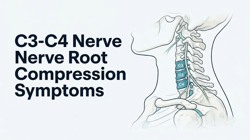
C3-C4 nerve root compression symptoms typically include pain at the base of the skull, lateral neck pain, shoulder cap discomfort, and in more severe cases, tingling or numbness across the upper chest and shoulder area, reflecting the specific nerve territories served by the C3 and C4 spinal nerve roots. Unlike compression at lower cervical levels such as C5-C6 or C6-C7, C3-C4 involvement does not typically radiate into the arm or fingers, which means many people experiencing this condition are confused about why their symptoms feel so different from the “classic” pinched nerve description they’ve read about.
According to the National Institute of Neurological Disorders and Stroke (NINDS), cervical radiculopathy affects an estimated 85 people per 100,000 in the general population annually, with the peak incidence occurring between ages 50 and 54. While C5-C6 and C6-C7 are the most frequently involved levels overall, C3-C4 disease becomes proportionally more common in older adults with multilevel cervical spondylosis, and its symptoms are among the most frequently misattributed to headache disorders, shoulder pathology, or temporomandibular joint problems.
This article covers every clinically recognized symptom of C3-C4 nerve root compression with the anatomical and physiological reasoning behind each one, explains how disc bulges differ from herniations at this level, identifies the critical distinction between radiculopathy and myelopathy at C3-C4, and clearly states which symptoms require emergency evaluation.
C3-C4 Nerve Root Compression Symptoms
C3-C4 nerve root compression symptoms are primarily felt in the neck, the base of the skull, the upper shoulder, and the area around the clavicle, and they do not typically extend into the arm or hand the way lower cervical nerve root compressions do.
The symptoms occur because the C3 and C4 nerve roots exit the cervical spine through small bony openings called neural foramina, and when these openings narrow from disc pathology or degenerative bone changes, the nerve root is squeezed. That compression triggers ectopic electrical firing along the axon, which the brain interprets as pain, burning, or unusual sensation in whatever body region the nerve normally supplies.
The full symptom picture at this level includes:
- Aching or burning pain at the base of the skull (occiput) and posterior scalp
- Lateral and posterior neck pain, often worse with rotation or lateral bending
- Pain or discomfort radiating to the upper shoulder and shoulder cap (deltoid region)
- Numbness or tingling across the clavicular zone and upper chest
- Cervicogenic headache, which originates in the neck but is perceived in the head
- Reduced range of neck motion, particularly rotation toward the affected side
- Tenderness over the posterior cervical musculature at the C3-C4 level
- In myelopathy cases: bilateral limb signs, gait changes, bladder dysfunction (discussed separately below)
For people over 60, symptoms may be bilateral rather than one-sided, because multilevel degenerative spondylosis can narrow the central canal and affect both C3 and C4 roots simultaneously. In younger individuals, a single-level disc herniation more commonly produces one-sided symptoms.
What Is C3-C4 Nerve Root Compression and How Does It Occur
C3-C4 nerve root compression occurs when the C3 or C4 spinal nerve root, or both, are mechanically impinged within the neural foramen or central spinal canal at the vertebral level between the third and fourth cervical vertebrae.
Think of the C3-C4 intervertebral disc as a shock-absorbing cushion seated between two bony cylinders. The disc has a tough outer shell called the annulus fibrosus and a gel-like inner core called the nucleus pulposus. When the annulus weakens from degenerative wear, repetitive strain, or acute trauma, the nucleus can push outward, reducing the space through which the adjacent nerve root travels. The nerve root doesn’t need to be fully crushed to produce symptoms. Even modest pressure on the dorsal root ganglion, the cluster of nerve cell bodies sitting just inside the foramen, is enough to trigger ectopic neural firing.
Several distinct mechanisms produce compression at C3-C4:
| Mechanism | What Physically Happens | Typical Onset Pattern |
|---|---|---|
| Acute disc herniation | Nucleus pulposus breaches annulus fibrosus | Sudden onset, often after trauma or strain |
| Degenerative disc bulge | Annulus fibrosus bulges without full breach | Gradual onset, worsens over weeks to months |
| Uncovertebral osteophyte | Bone spur from joint of Luschka narrows foramen | Chronic, progressive, often bilateral |
| Facet joint hypertrophy | Enlarged facet joint encroaches on foramen | Gradual, often with axial neck pain |
| Ligamentum flavum thickening | Folded ligament reduces canal space | Slow, insidious, more common in older adults |
The joints of Luschka (uncovertebral joints) sit just anterior to the neural foramen at every cervical level, and C3-C4 is no exception. Degenerative bony outgrowths from these joints are a particularly common source of foraminal narrowing at C3-C4 in adults over 50, and they explain why symptoms at this level often develop gradually rather than suddenly.
C3 Dermatome and C4 Dermatome Symptom Territories Explained
The C3 dermatome covers the posterior scalp, the area behind and below the ear, the angle of the jaw, and the lateral and posterior neck. The C4 dermatome covers the upper shoulder cap, the clavicular region, and the upper anterior chest just below the clavicle.
This geography is the reason C3-C4 nerve root compression produces such a distinctive symptom pattern. The pain and sensory changes don’t go into the arm. They stay in the neck, head, jaw area, shoulder top, and upper chest. Many people with confirmed C3-C4 compression spend months being evaluated for headache disorders, temporomandibular joint dysfunction, or shoulder rotator cuff problems before anyone images their cervical spine.
Understanding dermatome territories is like understanding a wiring diagram. Each nerve root is the electrical supply line for a specific skin zone. Damage to the supply line causes abnormal signals specifically in the zone it serves, not in zones served by other lines.
The clinical implication matters enormously for self-assessment:
- Pain behind the ear or at the base of the skull on one side: C3 territory
- Aching across the top of the shoulder (the “cap” of the shoulder, not the front or lateral arm): C4 territory
- Tingling just below the clavicle on the chest wall: C4 territory
- Pain or numbness in the forearm or hand: this is NOT C3 or C4 territory; these symptoms point to C5, C6, C7, or C8 involvement
According to the American Academy of Orthopaedic Surgeons, accurate dermatome localization is one of the primary tools clinicians use to identify which cervical level is responsible for a patient’s symptoms before imaging is ordered.
For individuals with diabetes mellitus, peripheral diabetic neuropathy can produce overlapping sensory changes in similar distribution zones, making clinical dermatome assessment less reliable and increasing the importance of electrodiagnostic testing.
C4 Nerve Root Compression Symptoms
C4 nerve root compression specifically produces pain, numbness, and weakness in the distribution of the C4 dermatome and myotome: the shoulder cap, the clavicular region, the upper anterior chest, and the upper back between the shoulder blades.
The C4 nerve root, after exiting through the C3-C4 neural foramen, contributes to the cervical plexus and sends sensory fibers to the clavicular skin zone and shoulder cap. Its motor fibers innervate the upper trapezius muscle and contribute to the phrenic nerve (along with C3 and C5). When C4 is compressed, the shoulder cap area aches or burns. Lifting the arm or rotating the neck toward the affected side typically worsens pain.
Specific C4 nerve root compression symptoms include:
- Aching, burning, or sharp pain across the top of the shoulder and shoulder blade
- Numbness or reduced sensation over the clavicular area and upper anterior chest
- Referred pain along the upper trapezius from the neck to the shoulder
- Possible weakness in the upper trapezius, noticeable as difficulty shrugging the affected shoulder against resistance
- Pain that worsens when tilting the head to the opposite side (stretching the compressed root)
- Pain relief when tilting the head toward the affected side (reducing root tension)
A 2021 study published in the Journal of Neurosurgery: Spine noted that C4 radiculopathy is frequently underdiagnosed because its symptoms overlap substantially with shoulder impingement syndrome, subacromial bursitis, and acromioclavicular joint pathology. The distinguishing feature is that C4 radiculopathy produces neck pain as its primary complaint, whereas rotator cuff pathology typically produces pain with specific shoulder movements rather than with neck position changes.
Women with fibromyalgia are particularly susceptible to symptom misattribution at this level, as the widespread musculoskeletal pain of fibromyalgia can obscure the directional, position-dependent quality that characterizes true radicular pain.
C3 Nerve Root Compression Symptoms
C3 nerve root compression produces pain and sensory changes at the base of the skull, the posterior scalp, the lateral and posterior neck, and sometimes around the angle of the jaw, all of which reflect the specific dermatome territory of the third cervical nerve root.
The C3 nerve root exits the spine through the neural foramen between C2 and C3 but is directly influenced by pathology at C3-C4, particularly when disc material or osteophytes compress the root from below or when foraminal stenosis at C3-C4 affects the root’s course. The dorsal root ganglion of C3 sits very close to the foramen, meaning even mild foraminal compromise can produce significant sensory symptoms.
Key C3 nerve root compression symptoms include:
- Pain at the base of the skull (occipital region), often described as a deep, aching pressure
- Scalp tenderness or hypersensitivity, particularly over the posterior scalp
- Pain radiating from the posterior neck toward the ear or jaw area
- Cervicogenic headache originating from cervical muscle tension and nerve root irritation
- Reduced sensation or tingling along the posterior and lateral neck
- Neck stiffness with painful restriction of rotation, particularly toward the ipsilateral (same) side
The C3 nerve root also contributes to the ansa cervicalis, the nerve loop that innervates several anterior neck muscles including the sternohyoid and omohyoid. In cases of significant C3 root involvement, some individuals report difficulty swallowing or a sensation of throat tightness, though this is uncommon and should prompt evaluation to exclude other causes.
In older adults with established cervical spondylosis, C3 symptoms can be bilateral and persistent rather than episodic, because multilevel degenerative change narrows the foramen on both sides simultaneously.
Key Takeaway: C3-C4 nerve compression symptoms stay in the neck, skull base, shoulder cap, and upper chest. Pain in the arm or hand means a different cervical level is involved.
C3-C4 Disc Bulge Symptoms
A C3-C4 disc bulge produces symptoms through a different mechanism than a full herniation, and understanding that difference changes what you’ll likely feel and how quickly symptoms progress.
In a disc bulge, the annulus fibrosus remains intact but the entire disc swells circumferentially outward, pressing against the posterior longitudinal ligament and narrowing the neural foramen or central canal. Because the outer disc wall is intact, the chemical irritant (the nucleus pulposus material) stays contained. The compression is mechanical rather than combined mechanical-and-chemical, which typically means a less intense, more aching symptom quality compared to an acute herniation.
C3-C4 disc bulge symptoms tend to be:
- A dull, aching neck pain that worsens after prolonged sitting or looking down at a screen
- Stiffness at the base of the neck and upper cervical region in the morning
- A feeling of pressure or tightness across the upper shoulders and lower neck
- Intermittent tingling at the shoulder cap or clavicular area, often worse by end of day
- Headaches starting at the base of the skull, particularly after extended neck flexion
A disc bulge at C3-C4 may produce no symptoms at all. Research published in the New England Journal of Medicine found that a notable proportion of asymptomatic individuals have disc bulges on MRI, meaning the presence of a bulge on imaging does not automatically explain a person’s symptoms. Clinical correlation, meaning matching the imaging finding to the specific nerve territory described by the patient, is required before attributing symptoms to the bulge.
People who perform repetitive overhead work or who spend extended hours with their neck in forward flexion (desk workers, surgeons, dentists) are at elevated risk for symptomatic C3-C4 disc bulge. Postmenopausal women experience accelerated disc dehydration due to estrogen loss, which increases disc bulge susceptibility at all cervical levels including C3-C4.
C3-C4 Disc Herniation Symptoms
A C3-C4 disc herniation produces symptoms that are typically sharper, more intense, and more precisely located than those of a disc bulge, because the breached nucleus pulposus directly contacts and chemically irritates the nerve root.
When the annulus fibrosus tears and the nucleus pulposus leaks into the neural foramen or spinal canal, two simultaneous processes inflame the nerve root. Mechanical pressure compresses the axon and dorsal root ganglion. At the same time, nucleus pulposus tissue releases phospholipase A2, TNF-alpha, and prostaglandin E2, which directly sensitize the nerve root’s firing threshold. Research published in the Spine journal has confirmed elevated phospholipase A2 activity in herniated disc tissue, linking chemical radiculitis to the intensity of radicular pain even when mechanical compression appears modest on imaging.
C3-C4 disc herniation symptoms include:
- Sharp or burning pain at the occiput, neck, or shoulder cap, often severe at onset
- Intense unilateral neck pain that changes character with head position
- Tingling or electric-shock sensations in the C3 or C4 dermatome territory
- Neck muscle spasm severe enough to restrict movement to a few degrees
- Pain worsening with neck extension or with the Spurling maneuver (turning and tilting the head toward the symptomatic side)
- Pain relief in some positions, particularly when placing the arm overhead (reducing nerve root tension)
- In posterior-central herniations: bilateral symptoms or early myelopathic signs
Unlike lower cervical herniations, a C3-C4 herniation that compresses the cord centrally rather than the foramen laterally can produce myelopathy rapidly. This is a key safety distinction. Paramedian and central disc herniations at C3-C4 require urgent specialist evaluation.
Younger adults (under 40) more often experience acute herniations from a single traumatic event. Adults over 50 more commonly develop herniations through a degenerative cascade in which progressive disc dehydration makes the annulus brittle and susceptible to tearing with everyday activity.
C3-C4 Occipital Headache and Neck Pain
Cervicogenic headache originating from C3-C4 nerve root irritation is felt as a dull, aching pressure at the base of the skull that spreads forward toward the forehead, temple, or behind one eye, and it is one of the most frequently misdiagnosed headache types in clinical practice.
The mechanism is not a mystery. The trigeminal nucleus caudalis in the brainstem receives convergent input from both the trigeminal nerve (supplying the face and forehead) and the upper cervical nerve roots (C1 through C3). This anatomical crosswiring means that irritation of the C3 nerve root from disc pathology at C3-C4 is perceived by the brain as pain in the forehead or behind the eye. This is referred pain, not migraine, not sinus pressure, and not tension headache in the conventional sense.
According to a 2019 study published in the Journal of Headache and Pain, cervicogenic headache accounts for approximately 4.1% of all headache presentations, but it is widely believed to be significantly underdiagnosed because its referred pain pattern closely mimics migraine and tension-type headache.
Distinguishing features of C3-C4-related cervicogenic headache:
- Headache consistently worsened by specific neck movements or sustained neck positions
- Headache that begins in the neck or occiput and spreads forward
- Neck stiffness and restricted range of motion present alongside the headache
- No aura, no photophobia or phonophobia as the primary feature (which would suggest migraine)
- Tender posterior cervical muscles at the C3-C4 level on palpation
- Headache reproduced by applying pressure to the ipsilateral posterior neck
Key Takeaway: If your headache consistently starts at the back of your neck and spreads forward, and worsens when you move your neck, C3-C4 nerve root irritation is a plausible anatomical explanation worth raising with your physician.
Individuals who have a prior diagnosis of migraine are not immune to developing overlapping cervicogenic headache. The two conditions can coexist, and treatment that addresses only the migraine component will leave the cervical contribution untreated.
C3 C4 Numbness and Tingling: Where It Spreads and Why
Numbness and tingling from C3-C4 nerve root compression appear in a highly specific location: the posterior scalp, posterior neck, upper shoulder cap, clavicular zone, and upper anterior chest, and these locations are fixed by the anatomy of the C3 and C4 dermatomes.
The reason the sensory change feels like tingling or numbness rather than sharp pain relates to the type of nerve fibers being affected. Large-diameter, rapidly conducting A-beta fibers carry light touch and vibration sensation. When these fibers are compressed, their signal is disrupted, producing numbness or a pins-and-needles quality rather than burning pain. In contrast, when smaller C-fibers (which carry pain and temperature signals) are the primary targets of the chemical and inflammatory component of disc herniation, the sensation is burning or aching rather than numb.
This distinction has a practical diagnostic implication:
| Sensation Type | Fiber Type Primarily Affected | Clinical Significance |
|---|---|---|
| Tingling, pins-and-needles | A-beta fibers (large myelinated) | Mechanical compression predominant |
| Burning, electric pain | C-fibers and A-delta fibers | Chemical/inflammatory radiculitis |
| True numbness (loss of feeling) | All sensory fibers, severe compression | More significant nerve involvement |
| Weakness, coordination loss | Motor fibers (alpha motor neuron) | Myotomal involvement, needs urgent evaluation |
People with long-standing type 2 diabetes mellitus have reduced A-beta fiber conduction speed as part of diabetic peripheral neuropathy, which can mask or modify the tingling quality of C3-C4 radiculopathy. A neurologist should assess whether symptoms in this population reflect cervical radiculopathy, diabetic neuropathy, or both, using electrodiagnostic testing.
C3-C4 Muscle Weakness and Affected Myotomes
Muscle weakness from C3-C4 nerve root compression is less immediately obvious than arm weakness from lower cervical levels, but it is real and clinically testable through specific resistance maneuvers that target the muscles supplied by C3 and C4 myotomes.
The C3 myotome primarily includes the sternocleidomastoid and the splenius capitis muscle group. The C4 myotome includes the upper trapezius and the levator scapulae, with shared contribution to the diaphragm through the phrenic nerve alongside C3 and C5. A clinician testing for C4 myotomal weakness will typically ask the patient to shrug the shoulder against downward resistance. Subtle asymmetry in shrug strength is a meaningful positive finding.
C3-C4 myotomal weakness may present as:
- Difficulty turning the head against resistance (sternocleidomastoid, C3)
- Reduced shoulder shrug strength on the affected side (upper trapezius, C4)
- Difficulty maintaining the head in neutral against gravity after prolonged activity (deep cervical flexors, C3-C4 contribution)
- A subtle droop or asymmetry of the shoulder girdle
- Fatigue in neck-stabilizing muscles faster than the opposite side
According to the North American Spine Society clinical guidelines for cervical radiculopathy, myotomal weakness that is progressive, meaning it worsens across serial examinations, is an indication for expedited imaging and specialist referral regardless of pain severity. Weakness without pain can occur in radiculopathy and is sometimes the first sign that nerve damage is accumulating.
Athletes who perform repetitive overhead activities such as swimming, tennis, or throwing sports may have pre-existing asymmetries in these muscle groups that complicate myotomal assessment. A physical medicine and rehabilitation specialist (physiatrist) or a sports medicine physician with spine expertise is best positioned to interpret clinical strength testing in this context.
C3-C4 Foraminal Stenosis Symptoms
C3-C4 foraminal stenosis produces symptoms identical to those of disc herniation at the same level, but the underlying cause is narrowing of the neural foramen by bony overgrowth rather than disc material, which affects the chronicity and treatability of the condition.
The neural foramen at C3-C4 is bounded anteriorly by the uncovertebral joint (joint of Luschka) and the posterior disc, posteriorly by the facet joint, and superiorly and inferiorly by the pedicles of C3 and C4. Degenerative arthritis in both the uncovertebral joint and the facet joint simultaneously produces osteophyte formation that gradually reduces the foramen’s diameter. Unlike a herniated disc, which may partly resorb over time, osteophytic stenosis is essentially permanent without intervention.
Foraminal stenosis symptoms at C3-C4 tend to be:
- Chronic rather than acute in onset, developing over months to years
- Consistently worse with neck extension, because extension reduces foraminal diameter by 20% to 30%
- Position-dependent, with relief found in slight neck flexion
- Associated with morning neck stiffness that gradually loosens through the day
- Intermittently worse during periods of increased inflammation (such as a viral illness or overuse)
To understand foraminal stenosis, think of the neural foramen as a garden hose. A healthy foramen is a wide hose with plenty of flow. Osteophytes work like someone slowly pinching the hose over years. The nerve root, like the water, is progressively restricted. When the pinch is mild, the nerve functions but complains. When the pinch is severe, function drops.
Individuals with ossification of the posterior longitudinal ligament (OPLL), a condition with higher prevalence in individuals of East Asian descent, face a particularly narrow margin at C3-C4 because the pre-existing canal narrowing from OPLL compounds foraminal stenosis significantly. Any new or worsening neurological symptom in someone known to have OPLL warrants prompt spine specialist evaluation.
Key Takeaway: Foraminal stenosis at C3-C4 causes the same symptoms as disc herniation but develops slowly and does not self-resolve, making specialist evaluation important for persistent or progressive cases.
Pinched Nerve C3 C4 Symptoms: Acute Versus Chronic Presentation
The pinched nerve at C3-C4 presents very differently depending on whether it is acute (sudden onset from disc herniation or trauma) or chronic (gradual onset from degenerative stenosis), and treating them as the same condition would miss important clinical differences.
In an acute presentation, the onset is typically identifiable. A specific movement, a car accident, a fall, or a period of heavy lifting precedes the sudden appearance of severe neck pain with referral into the C3 or C4 dermatome. The pain is often so intense that the person restricts all neck movement. Muscle spasm locks the cervical spine. This acute picture usually reflects sudden disc herniation with both mechanical compression and chemical radiculitis.
In a chronic presentation, the symptoms build gradually. The person recalls months of increasing neck stiffness, worsening headaches at the occiput, and progressive shoulder cap aching. There is rarely a single precipitating event. Symptom intensity varies with activity level, posture, and sleep position, but there is no period of complete resolution.
| Feature | Acute C3-C4 Presentation | Chronic C3-C4 Presentation |
|---|---|---|
| Onset | Sudden, often event-related | Gradual over weeks to months |
| Pain intensity | Severe, often immobilizing | Moderate, waxing and waning |
| Primary cause | Disc herniation | Degenerative stenosis, spondylosis |
| Muscle spasm | Pronounced | Mild to moderate |
| Neurological signs | May appear rapidly | Develop slowly |
| Spontaneous improvement | Possible within 6 to 12 weeks | Less likely without intervention |
| Urgency of evaluation | High, especially with motor deficit | Moderate, but progressive weakness warrants prompt review |
According to the Mayo Clinic, the majority of acute cervical radiculopathy episodes improve with conservative management within 4 to 6 weeks in patients without progressive neurological deficit. Chronic presentations, however, require structured clinical assessment to determine whether conservative care can halt progression or whether surgical decompression is needed.
Older adults with established multilevel spondylosis are more likely to present chronically. Younger adults under 40 presenting with acute-onset symptoms after a clear precipitating event are more likely to have an acute disc herniation that may respond well to conservative management.
C3-C4 Myelopathy Symptoms: When the Spinal Cord Is Involved
C3-C4 myelopathy occurs when pathology at the C3-C4 level compresses not just the exiting nerve root but the cervical spinal cord itself, producing a distinct and more serious symptom pattern that includes bilateral limb dysfunction, gait abnormalities, and bowel or bladder changes.
This is the distinction that no top-ranking competitor content addresses adequately. Radiculopathy means one nerve root is compressed. Myelopathy means the spinal cord is compressed. At C3-C4, this distinction is particularly consequential because the corticospinal tracts carrying motor commands to both arms and legs run through the cervical spinal cord at this level. Compression here can impair motor function below the lesion in all four limbs.
Cervical spondylotic myelopathy at C3-C4 produces:
- Bilateral hand clumsiness (difficulty buttoning shirts, writing, or using utensils)
- Weakness in both arms and legs, often noticed first as difficulty climbing stairs
- A wide-based, shuffling gait (the gait looks unsteady, similar to a balance disorder)
- Electric shock sensations down the spine when the neck is flexed (Lhermitte sign)
- Hyperreflexia (exaggerated deep tendon reflexes below the level of compression)
- A positive Hoffmann sign (flicking the middle finger nail causes involuntary thumb flexion, indicating upper motor neuron dysfunction)
- Bladder urgency, hesitancy, or incontinence
- Sexual dysfunction
- In severe cases: loss of independent ambulation
A 2022 study published in the Journal of Neurosurgery: Spine found that cervical spondylotic myelopathy is the most common cause of spinal cord dysfunction in adults over 55, with C3-C4 and C4-C5 levels implicated in a substantial proportion of multilevel cases. The study emphasized that myelopathy progresses in a stepwise fashion in most patients, meaning periods of stability are punctuated by sudden functional decline, and that early surgical decompression offers better functional outcomes than delayed surgery.
If you have any combination of bilateral hand clumsiness, gait instability, and urinary changes alongside neck symptoms, this is not a self-monitoring situation. See a neurologist or spine surgeon promptly.
C3-C4 Nerve Compression Breathing and Diaphragm Concerns
C3-C4 nerve compression can affect breathing in rare but serious cases because the phrenic nerve, which is the primary motor nerve controlling the diaphragm, originates from the C3, C4, and C5 nerve roots.
This is the symptom most completely absent from every competing article, and it is clinically relevant. The diaphragm is the primary muscle of breathing. Its full motor supply travels down through the phrenic nerve, which begins in the cervical plexus from contributions of C3, C4, and C5. Severe compression of the C4 root can reduce phrenic nerve motor output, which impairs diaphragm movement on the ipsilateral (same) side.
In everyday clinical practice, C3-C4 compression rarely produces overt breathing difficulty because the contralateral phrenic nerve and accessory respiratory muscles compensate. However, specific circumstances increase the risk:
- Bilateral C3-C4 nerve root compression affecting both C4 roots simultaneously
- A high central disc herniation or significant central canal stenosis that compromises cord function at this level
- Significant myelopathy at C3-C4 affecting the cord’s descending pathways to respiratory motor neurons
- Surgical or traumatic injury to the C3-C4 region (such as a fracture-dislocation)
Symptoms that may reflect phrenic nerve involvement at C3-C4 include:
- Unexpected shortness of breath that worsens when lying flat (orthopnea)
- Reduced breath sounds on one side of the chest
- Paradoxical abdominal movement during breathing (the abdomen moves inward rather than outward with inhalation)
- Rapid or labored breathing without a clear pulmonary cause
Call 911 or go to the nearest emergency room immediately if you have neck symptoms combined with new shortness of breath, particularly if it worsens when lying down. This symptom combination requires emergency evaluation. Do not attribute this to anxiety or assume it will resolve.
How Age, Sex, and Comorbidities Change C3-C4 Symptom Patterns
The way C3-C4 nerve root compression presents varies substantially by age, biological sex, and underlying health conditions, and standard symptom descriptions often fail to reflect this variation accurately.
Age-related variation: Adults under 40 most often present with acute C3-C4 disc herniation. Symptoms are intense, typically unilateral, and the onset is identifiable. Adults over 50 more commonly develop symptoms through the gradual degenerative cascade: disc dehydration leads to disc height loss, uncovertebral joint arthrosis, and foraminal narrowing. Symptoms are chronic, bilateral, and often described as “always there, just varying in intensity.” Adults over 70 with multilevel spondylosis may have C3-C4 myelopathy without prominent neck pain, because the cord compression develops so slowly that the body compensates until a threshold is crossed.
Biological sex variation: Research published in the European Spine Journal has found that cervical disc herniation presents at a younger age in males (peak in the 30s to 40s), whereas degenerative spondylotic disease affecting the same levels presents more commonly in postmenopausal females. Estrogen deficiency after menopause accelerates intervertebral disc dehydration and reduces disc proteoglycan content, increasing susceptibility to symptomatic foraminal narrowing at C3-C4. Females also report higher pain intensity scores for the same degree of nerve root compression, a finding attributed to differences in central sensitization rather than peripheral nerve differences.
Comorbidity variation:
- Rheumatoid arthritis produces cervical instability and pannus formation at the upper cervical spine, which can simulate or accelerate C3-C4 compressive pathology. These patients require MRI with flexion-extension views.
- Diabetes mellitus slows axonal recovery from compression, worsens neuropathic symptoms, and complicates electrodiagnostic interpretation.
- OPLL (ossification of the posterior longitudinal ligament) significantly narrows the available spinal canal space, making C3-C4 myelopathy far more likely with even modest disc bulging.
Key Takeaway: The same C3-C4 MRI finding produces different symptoms in a 35-year-old male, a 60-year-old postmenopausal woman, and an older adult with rheumatoid arthritis. The clinical picture must be matched to the individual, not the imaging alone.
How Is C3-C4 Nerve Root Compression Diagnosed
C3-C4 nerve root compression is diagnosed through a combination of clinical history, physical examination findings specific to the C3 and C4 nerve territories, and confirmatory imaging or electrodiagnostic testing.
The diagnostic process typically unfolds in this sequence:
- Clinical history: A physician maps the location, character, and triggers of symptoms and asks specifically whether neck position changes provoke or relieve pain. The pattern of headache location, shoulder cap pain, and clavicular tingling points toward C3-C4 involvement rather than lower cervical levels.
- Physical examination: The Spurling test (applying downward compression to the head while tilting it toward the symptomatic side) reproduces radicular pain in approximately 30% of confirmed C3-C4 radiculopathy cases per AAOS data. The Hoffmann sign and Babinski reflex are tested to screen for myelopathy. Manual muscle testing of the upper trapezius and levator scapulae targets C4 myotomal integrity.
- MRI of the cervical spine: The first-line imaging study. MRI without contrast demonstrates disc pathology, foraminal stenosis, osteophytes, and spinal cord signal changes. A T2-weighted sequence showing high signal intensity within the cervical cord at C3-C4 is a myelopathy warning sign requiring urgent review.
- CT scan or CT myelogram: Ordered when MRI is contraindicated (pacemaker, certain implants) or when bony detail is needed. CT myelogram directly visualizes the nerve root’s relationship to bony stenosis.
- Electromyography (EMG) and nerve conduction velocity (NCV) study: Ordered when clinical and imaging findings do not fully correlate. EMG can detect acute denervation or chronic reinnervation in muscles supplied by C3 or C4, confirming root-level dysfunction. It is also the primary tool for distinguishing cervical radiculopathy from peripheral entrapment neuropathy when symptoms are ambiguous.
- Diagnostic nerve root block: In selected cases, a targeted injection of local anesthetic around the C3 or C4 nerve root under fluoroscopic guidance confirms that the imaged level is the symptomatic level.
A board-certified orthopedic spine surgeon, a board-certified neurosurgeon with spine specialty, or a neurologist with expertise in cervical spine disease is the appropriate specialist for evaluating confirmed or suspected C3-C4 nerve root compression with neurological signs. A physiatrist (physical medicine and rehabilitation specialist) is appropriate for directing conservative management once a structural diagnosis is confirmed without urgent surgical indications.
Emergency Symptoms: When to Call 911 or Go to the ER
Certain symptoms associated with C3-C4 nerve root compression require immediate emergency evaluation. Do not wait to see if these resolve on their own.
Call 911 or go to the nearest emergency room immediately if you experience:
- Sudden onset of difficulty breathing or shortness of breath that worsens when lying flat, alongside neck pain (possible phrenic nerve involvement)
- Rapid onset of weakness or paralysis in both arms and both legs following a neck injury
- Loss of bladder or bowel control alongside neck pain and new limb weakness (possible acute cervical myelopathy)
- Electric shock-like pain down the spine triggered by neck movement (Lhermitte sign) appearing suddenly in the context of new neck trauma
- Sudden inability to walk normally or maintain balance combined with hand clumsiness and neck symptoms
- Neck pain following a high-speed car accident, fall from height, or direct neck trauma, even without immediate neurological symptoms (cervical fracture must be excluded before the neck is moved)
These presentations can indicate acute cervical spinal cord compression, cervical fracture-dislocation, or critical phrenic nerve dysfunction, and they require emergency medical assessment, not an urgent care appointment or a phone call.
Frequently Asked Questions About C3-C4 Nerve Root Compression Symptoms
What are the most common symptoms of C3-C4 nerve root compression?
The most common symptoms of c3-c4 nerve root compression include pain at the base of the skull, neck pain with restricted rotation, shoulder cap aching, and tingling or numbness across the clavicular region and upper chest.
These symptoms reflect the specific dermatome territories of the C3 and C4 nerve roots, which do not extend into the arm or hand.
Cervicogenic headache originating at the occiput and spreading forward is also a frequently reported symptom, often mistaken for tension-type or migraine headache.
Can C3-C4 nerve compression cause headaches?
Yes, C3-C4 nerve root irritation is a recognized cause of cervicogenic headache, which is felt as a unilateral pain starting at the back of the skull and spreading toward the forehead or behind one eye.
The mechanism involves convergent input from the upper cervical nerve roots and the trigeminal nerve in the brainstem, causing neck-originated pain to be perceived as a head pain.
A 2019 study in the Journal of Headache and Pain identified cervicogenic headache in approximately 4.1% of all headache presentations, with cervical spine pathology as the underlying cause.
How do I know if my C3-C4 disc bulge is causing my symptoms?
A C3-C4 disc bulge causes symptoms only when it compresses a nerve root or narrows the central canal sufficiently to irritate neural tissue.
The key indicator is whether your pain or sensory symptoms follow the C3 or C4 dermatome territory: skull base, posterior neck, shoulder cap, or clavicular zone.
A neurologist or spine specialist can confirm clinical correlation between the imaging finding and your symptoms through physical examination and, if needed, electrodiagnostic testing.
What is the difference between C3-C4 radiculopathy and C3-C4 myelopathy?
C3-C4 radiculopathy means one or both nerve roots are compressed, producing pain and sensory changes in the neck, shoulder cap, and upper chest on one side.
C3-C4 myelopathy means the spinal cord itself is compressed at this level, producing bilateral limb weakness, gait disturbance, hand clumsiness, and bladder or bowel changes.
Myelopathy is a more serious condition requiring urgent specialist evaluation, whereas mild radiculopathy without progressive motor deficit may be managed conservatively.
Can C3-C4 nerve compression cause arm or hand pain?
C3-C4 nerve root compression does not typically cause arm or hand pain, because the C3 and C4 dermatomes do not extend below the shoulder and clavicular region.
Arm pain, forearm tingling, or hand numbness point toward compression at C5-C6 (affecting the C6 root) or C6-C7 (affecting the C7 root).
If you have both C3-C4 findings on MRI and arm or hand symptoms, the arm symptoms likely originate from a different spinal level and require full cervical imaging review with a spine specialist.
When should I see a doctor for C3-C4 nerve root compression symptoms?
See a primary care physician promptly if you have persistent neck pain with skull base headaches, shoulder cap tingling, or upper chest numbness lasting more than two weeks without improvement.
See a neurologist, board-certified orthopedic spine surgeon, or neurosurgeon urgently if you develop progressive muscle weakness, bilateral limb symptoms, gait changes, bladder or bowel changes, or worsening symptoms despite conservative management.
Go to the emergency room immediately if you develop sudden difficulty breathing alongside neck symptoms, or if any neurological symptoms appear after a neck injury.
Putting This Together: What to Do With What You’ve Learned
The defining clinical feature of C3-C4 nerve root compression is that its symptoms stay in the neck, skull base, shoulder cap, and upper chest. If that pattern matches what you’re experiencing, the next concrete step is a visit to your primary care physician with a clear description of where the pain starts, where it spreads, and which neck positions make it better or worse. That information is what guides the decision to order imaging.
If you have been told you have a disc bulge or disc herniation at C3-C4 on imaging, the imaging alone does not explain your symptoms. Clinical correlation with your nerve territory is what makes the diagnosis meaningful. Push for that clinical conversation with a neurologist or spine specialist, not just a review of the MRI report.
The one thing to keep front of mind: progressive weakness, bilateral limb symptoms, gait changes, or bladder changes alongside neck symptoms are not “wait and see” findings. They warrant the same urgency as any other progressive neurological sign. Know the difference between a nerve root that is irritated and a spinal cord that is compressed, and let that distinction guide how fast you act