Jaw Infection After Root Canal Symptoms and Warning Signs
The symptoms of jaw infection after root canal treatment range from persistent throbbing jaw pain and visible swelling to fever, drainage, and difficulty opening your mouth. These are not the same as the normal soreness that follows any root canal procedure, and knowing the difference matters enormously for your safety.
According to the American Association of Endodontists (AAE), most root canal procedures have a high success rate, but periapical infections do develop in a subset of cases, either because residual bacteria survived the cleaning process, the tooth’s coronal seal failed, or a previously undetected canal was left untreated. The National Institute of Dental and Craniofacial Research (NIDCR) notes that odontogenic (tooth-origin) infections account for a substantial proportion of emergency department facial swelling presentations annually in the United States.
This article covers the full clinical picture of post-root canal jaw infection: every symptom explained by its physiological mechanism, a clear framework for distinguishing normal healing from genuine infection, who faces the highest risk, and the specific symptoms that require emergency care rather than a dentist appointment.
Jaw Infection After Root Canal: What Is Actually Happening
A jaw infection after root canal develops when bacteria colonize the periapical tissues, the area surrounding the tip of the tooth’s root inside the jawbone.
During a root canal, the dentist or endodontist removes the infected or inflamed dental pulp from the pulp chamber and root canals, then shapes, disinfects, and fills those canals with an inert material called gutta-percha. The goal is complete elimination of bacterial load inside the tooth’s root system. When that goal is not fully achieved, gram-negative anaerobic bacteria including Fusobacterium nucleatum, Porphyromonas gingivalis, and Treponema denticola can persist at or beyond the apical foramen, the small opening at the root tip where the tooth’s blood supply and nerves once entered.
These bacteria release lipopolysaccharides (LPS), which activate toll-like receptor 4 (TLR4) on local macrophages and mast cells. That activation triggers a cascade of inflammatory mediators including interleukin-1-beta (IL-1 beta), tumor necrosis factor-alpha (TNF-alpha), and prostaglandin E2 (PGE2). The result is increased vascular permeability in the periapical tissues (producing swelling), sensitization of local pain receptors by prostaglandins (producing throbbing pain), and neutrophil recruitment that forms pus as immune cells die fighting the infection.
Think of it like a slow pressure leak in a sealed pipe. The tooth’s interior is now sealed with gutta-percha, but bacteria at the tip of the root are generating inflammatory pressure in the surrounding bone. That pressure has no easy escape route, so it builds, spreads, and eventually forces its way through the path of least resistance: through the alveolar bone, along the periosteum, and into the soft tissues of the jaw and face.
Infection can also develop when the tooth’s permanent crown or filling placed after the root canal fails to seal properly, allowing oral bacteria to re-enter the treated canal system from above. This coronal microleakage scenario is one of the most common causes of late-onset post-root canal infection, sometimes appearing months to years after the original procedure.
Symptoms of Jaw Infection After Root Canal: Full Clinical Picture
The core symptoms of jaw infection after root canal include throbbing jaw pain, visible facial or jaw swelling, fever, a persistent bad taste or drainage, swollen lymph nodes under the jaw, and in severe cases difficulty opening the mouth or swallowing.
Here is the complete symptom inventory with the physiological mechanism behind each:
| Symptom | Physiological Mechanism | Clinical Significance |
|---|---|---|
| Throbbing jaw pain | Prostaglandin E2 sensitizes nociceptors at the periapex; increased tissue pressure amplifies pain signal | Moderate to severe; infection signal when worsening past 72 hours |
| Jaw or facial swelling | Increased vascular permeability from IL-1-beta and histamine drives fluid into periapical and soft tissues | Mild to severe; severe swelling is urgent |
| Fever (over 100.4°F / 38°C) | Systemic pyrogen release (IL-1-beta, TNF-alpha) resets the hypothalamic thermostat | Any fever after root canal warrants same-day dental contact |
| Bad taste or drainage | Pus from neutrophil accumulation breaks through bone or soft tissue and drains intraorally | Indicates abscess has decompressed, but infection still requires treatment |
| Swollen submandibular lymph nodes | Regional lymph nodes drain the infected area and enlarge as they filter bacteria and debris | Tender, enlarged nodes signal active immune response to infection |
| Trismus (restricted jaw opening) | Swelling or infection invades the masseter or medial pterygoid muscles, causing protective spasm | Indicates infection has spread to jaw muscles; requires urgent evaluation |
| Tooth mobility or extrusion | Periradicular bone resorption from osteoclast activation loosens the tooth’s bone anchoring | Signals advanced bone involvement |
| Sinus pressure or drainage | Maxillary posterior root apices sit immediately below the maxillary sinus floor; infection can penetrate sinus | Upper tooth root canals specifically; sinus congestion post-procedure can signal this |
Note for patients taking immunosuppressant medications, including corticosteroids for conditions like rheumatoid arthritis or organ transplant rejection: fever may be absent or blunted even in the presence of a serious infection. Relying on fever alone as the indicator of infection in this group is clinically unreliable.
Jaw Pain After Root Canal: Normal Soreness vs Infection Pain
Jaw pain after root canal is expected for the first 48 to 72 hours and reflects the normal inflammatory response to instrumentation of the root canal system, not infection.
During root canal treatment, files used to shape and clean the canals can extend slightly beyond the apical foramen, pushing debris and irrigation solution into the periapical space. This mechanical and chemical irritation triggers a localized inflammatory response that produces genuine soreness in the surrounding jawbone and periodontium. That response is physiologically identical in its early mechanism to infection, because both involve prostaglandin E2 sensitizing pain receptors. The critical difference is trajectory.
Normal post-procedure jaw pain follows a predictable downward curve. It peaks at around 24 to 48 hours, then steadily improves. By day three to five, most patients describe residual tenderness to biting pressure but no spontaneous throbbing pain at rest. A 2019 study published in the Journal of Endodontics found that approximately 40% of patients report moderate to severe pain in the first 24 hours after root canal treatment, with the majority reporting acceptable pain levels by day three.
Infection pain, by contrast, either fails to improve after 72 hours or actively worsens. It tends to be spontaneous, throbbing, and often described as pressure-building rather than ache-like. Biting on the tooth produces sharp, intense pain rather than mild tenderness. Pain at rest that wakes the patient from sleep is a particularly telling signal.
Differentiating the two:
- Normal soreness: Improving trajectory from 48 hours onward, mild to moderate, responds to over-the-counter ibuprofen or acetaminophen
- Infection pain: Worsening or plateau after 72 hours, spontaneous throbbing at rest, poor response to over-the-counter pain relief, pain waking you from sleep
Older adults over 65 sometimes report less intense pain even with active infection, because age-related changes in nerve sensitivity and immune response can blunt the nociceptive signal. If swelling or systemic symptoms appear without proportional pain in an older patient, take the swelling seriously regardless of the pain level.
Jaw Swelling After Root Canal: What It Means and When to Worry
Jaw swelling after root canal can represent either the tail end of normal post-procedural inflammation or the early sign of an abscess building pressure in the periapical bone.
Mild facial puffiness localized directly over the treated tooth area in the first 24 to 48 hours is within the expected healing range. The inflammatory cascade that cleans up debris from instrumentation increases vascular permeability, allowing protein-rich fluid to move into the surrounding soft tissue. That fluid accumulation produces puffiness. It typically resolves within 48 to 72 hours as the inflammatory phase gives way to the proliferative phase of healing.
Infection-driven swelling behaves differently. It appears or worsens after the first 48 hours rather than improving. It tends to be firmer, warmer to the touch, and more diffuse as the abscess spreads through the alveolar bone and into the soft tissues. When the infection pushes through the cortical plate of the mandible (lower jaw) or maxilla (upper jaw) and into the overlying soft tissue, the swelling becomes visible externally, sometimes dramatically so.
Infection spreading from a lower molar or premolar root canal can track into the submandibular space below the jaw. That is not merely a cosmetic problem. The submandibular space connects to the parapharyngeal space and then to the deeper neck fascial spaces. Swelling in that territory can compromise the airway.
Watch for these swelling patterns that require same-day or emergency evaluation:
- Swelling that crosses the jaw midline
- Swelling that extends below the jawbone toward the neck
- Swelling of the floor of the mouth
- Swelling that pushes the tongue upward or backward
- Swelling accompanied by difficulty swallowing or any change in voice quality
Pregnant patients, particularly in the second trimester, face additional complexity: dental infections during pregnancy carry potential risks to fetal outcomes, and some antibiotic options are restricted. A pregnant patient with post-root canal jaw swelling should contact both their endodontist or dentist and their obstetrician-gynecologist on the same day.
Key Takeaway: Normal jaw pain and mild swelling after a root canal improve within 48 to 72 hours. Any symptom that worsens after that window, particularly throbbing pain at rest, spreading swelling, or fever, signals infection rather than healing and requires same-day dental evaluation.
Normal Healing vs Infected Root Canal: How to Tell the Difference
Normal root canal healing and a developing jaw infection share overlapping symptoms in the first 48 hours, but their trajectories diverge clearly by day three.
The distinction comes down to five observable factors: direction of symptoms (improving vs. worsening), fever presence, swelling behavior, drainage, and functional limitation. Use this framework as a guide, not as a self-diagnostic tool. Only a dentist or endodontist can confirm the status of a treated tooth through clinical examination and radiography.
| Feature | Normal Healing | Infected Root Canal |
|---|---|---|
| Pain trajectory | Improving steadily from 24 to 72 hours | Plateau or worsening after 72 hours |
| Fever | None | Often present (above 100.4°F / 38°C) |
| Swelling | Mild puffiness, resolving by 48 to 72 hours | Increasing, firmer, may spread |
| Drainage or taste | None | Bad taste, salty/bitter drainage possible |
| Jaw opening | Mildly limited due to soreness, improving | Significantly restricted (trismus) or worsening |
| Lymph nodes | Not swollen or mildly reactive | Tender, enlarged under jaw or neck |
| Response to pain relievers | Good control with ibuprofen or acetaminophen | Poor or incomplete relief |
According to the Mayo Clinic, a tooth abscess does not resolve without treatment. Unlike some localized infections elsewhere in the body, a periapical dental abscess will not clear on its own. The bacteria involved are anaerobes that thrive in the low-oxygen environment of necrotic tissue and bone. Without drainage, antibiotic therapy, or retreatment of the root canal, the infection can persist for months, causing progressive bone loss, and eventually spread to adjacent structures.
Patients who have recently completed a course of antibiotics for another reason may have a masked infection: partial antibiotic suppression can temporarily reduce symptoms without eliminating the underlying bacterial source. This pattern makes the healing-vs-infection distinction harder to read and warrants extra caution.
Fever After Root Canal: When Temperature Signals Trouble
Any fever above 100.4 degrees Fahrenheit (38 degrees Celsius) occurring after a root canal procedure is a signal of systemic infection response and requires same-day contact with your dentist or endodontist.
Fever after a dental procedure is not a normal part of the healing process. The inflammatory response to routine root canal instrumentation does not produce systemic temperature elevation in immunologically healthy adults. Fever occurs when inflammatory pyrogens, specifically IL-1-beta and TNF-alpha, enter systemic circulation and act on the hypothalamus, resetting the body’s thermostat upward. For that to happen, the infection must be releasing enough bacterial products or cytokines to reach the bloodstream.
A temperature between 100.4°F and 102°F (38°C to 38.9°C) developing in the 48 to 72 hours after a root canal, accompanied by jaw pain or swelling, suggests an early systemic infectious response originating at the periapical site. That patient needs clinical evaluation and likely antibiotic therapy, determined by their treating provider.
Temperature above 102°F (38.9°C), particularly with chills, sweating, and worsening jaw or neck swelling, suggests more extensive infection involvement and warrants urgent or emergency evaluation. High fever combined with jaw swelling is not a situation to manage over the phone.
Call 911 or go to the emergency room immediately if fever accompanies any difficulty breathing, difficulty swallowing, or visible swelling of the throat or floor of the mouth.
Patients with Type 2 diabetes mellitus face a specific risk here. Research published in the Journal of Endodontics and consistent with NIDCR guidance confirms that diabetic patients have impaired neutrophil chemotaxis and phagocytic function, meaning their immune cells are slower to reach and neutralize the infection site. As a result, dental infections in diabetic patients can progress faster, cause more bone destruction, and spread more readily than in patients without diabetes. A diabetic patient who develops any fever after root canal should treat it as urgent, regardless of how low the temperature reading is.
Bad Taste in the Mouth and Drainage After Root Canal
A persistent bad, salty, or bitter taste in the mouth after root canal treatment, particularly one that returns after brushing, signals that pus is draining from an abscess near the treated tooth.
Pus is the byproduct of the immune response: neutrophils (white blood cells) migrate to the infection site and die after engulfing bacteria, releasing their cellular contents into the tissue. That accumulation of dead neutrophils, bacteria, cellular debris, and inflammatory fluid is what produces pus. When enough pressure builds in the periapical space, the abscess may erode through bone and periosteum and form a channel called a sinus tract (also called a parulis or dental fistula) that opens into the mouth, usually as a small pimple-like bump on the gum near the affected tooth.
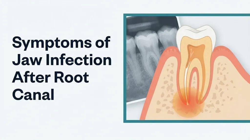
Drainage through a sinus tract temporarily relieves pressure, which is why some patients notice their pain level drops when the drainage starts. That reduction in pain does not mean the infection has resolved. The bacterial source inside the tooth or at the root apex is still present and will continue to fuel the process until treated.
The taste associated with intraoral abscess drainage is distinctive: most patients describe it as persistently foul, salty, or reminiscent of blood mixed with something metallic or bitter. It recurs no matter how frequently you rinse or brush, because the source is continuous.
Quick clinical tips for patients experiencing this symptom:
- Do not attempt to pop or squeeze the gum pimple (sinus tract). Disrupting it manually introduces new surface bacteria and does not address the root source.
- Warm salt water rinses can help keep the area clean while you arrange dental evaluation, but they are not a substitute for treatment.
- Note whether the bad taste is constant or intermittent. Intermittent drainage suggests a sinus tract that opens and closes. Constant drainage may indicate a larger or more actively draining abscess.
- Contact your endodontist or dentist the same day you notice this symptom. Drainage from a dental abscess is not a watch-and-wait scenario.
Key Takeaway: Drainage from a sinus tract (that small pimple on the gum near a root canal tooth) signals an active abscess that has broken through bone. The pain may temporarily ease, but the infection is not clearing on its own.
Lymph Node Swelling After Root Canal Infection
Tender, enlarged lymph nodes under the jaw or along the neck after a root canal signal that the regional immune system is actively responding to a dental infection and warrants dental evaluation.
The submandibular lymph nodes, located just beneath the lower jaw, are the primary drainage nodes for most of the teeth, gums, and oral floor. When bacteria or inflammatory debris from a periapical abscess travel through the lymphatic channels, these nodes enlarge and become tender as lymphocytes and macrophages work to filter and destroy the pathogens. The submental lymph nodes, positioned just below the chin, drain the central incisors and lower lip. The cervical lymph nodes along the neck become involved when infection spreads more extensively.
Lymph node swelling in the setting of post-root canal jaw infection is typically unilateral, meaning it appears on the same side as the treated tooth. The affected nodes are usually tender to the touch, firm but not hard, and movable under the skin. Nodes that are fixed (immovable), stony hard, or progressively enlarging over weeks without other signs of acute infection warrant evaluation for a different cause, which would be assessed by a primary care physician.
For context on what lymph node involvement indicates clinically: the degree of lymphadenopathy (lymph node enlargement) does not always correlate neatly with the severity of the dental infection. A 2021 review in the International Endodontic Journal noted that odontogenic infections can trigger regional lymphadenopathy even in cases where the abscess remains well-localized to the periapical bone.
Children under 12 years of age develop lymph node swelling with dental infections more readily than adults, and their nodes can become quite large relative to their facial size. Parents should not wait to see if enlarged neck nodes resolve in a child who has recently had dental work. A pediatric dentist or pediatric emergency medicine physician should evaluate any child with visible neck swelling, fever, and recent dental procedure history.
Root Canal Jaw Pain Weeks Later: What That Usually Means
Jaw pain that returns or persists weeks after a root canal procedure, past the point where normal healing should have resolved discomfort, is a strong clinical indicator that the treated tooth has an ongoing problem requiring professional evaluation.
There are several distinct scenarios that produce late-onset post-root canal jaw pain. First, a previously missed root canal. Posterior teeth (molars) often have three, four, or in some cases five root canals, and anatomical variations can make one canal difficult to locate and treat during the original procedure. An untreated canal harbors the original infected pulp tissue, which continues to fuel the periapical inflammatory process.
Second, a cracked root. The mechanical forces of root canal instrumentation and obturation can, in uncommon cases, create or propagate a vertical root fracture. A fractured root allows bacteria to migrate along the fracture line into the periodontal ligament and periapical bone, producing pain that mimics infection but does not respond to retreatment. Root fractures are diagnosed by a combination of clinical examination and, increasingly, cone beam computed tomography (CBCT), a three-dimensional dental imaging technique.
Third, coronal microleakage, as discussed earlier: if the permanent crown or filling placed after the root canal breaks down or was never placed, oral bacteria can re-colonize the root canal system from the tooth’s crown. This pathway produces infection symptoms that can appear months to years after the original procedure.
Fourth, periapical scar tissue or periapical cyst formation. A small percentage of periapical radiolucencies (dark areas on dental X-rays representing bone loss) are cysts rather than abscesses. Cysts contain a fluid-filled cavity lined with epithelial cells. Unlike a true abscess, a periapical cyst may not respond fully to root canal retreatment alone and can require surgical intervention (apicoectomy).
Patients who had their root canal performed more than 12 months prior and are experiencing new jaw pain should request a periapical radiograph and a clinical examination from their dentist or endodontist. Do not assume that late-onset pain is residual from the original procedure.
Root Canal Infection Spreading Symptoms
A root canal infection that spreads beyond the periapical bone enters the soft tissue fascial spaces of the face, jaw, and neck, producing symptoms that move from concerning to medically urgent.
Odontogenic infection spreads through the path of least resistance. From the periapical bone, infection can erode through the cortical bone plate and enter:
- The buccal space (producing visible cheek swelling)
- The submandibular space (swelling below the jawline, a sign that infection has left the oral cavity proper)
- The sublingual space (swelling of the floor of the mouth)
- The parapharyngeal space (neck swelling, throat fullness, changes in voice)
- The masticator space (involving the jaw muscles, producing trismus)
When infection spreads simultaneously into the submandibular, sublingual, and submental spaces bilaterally, it produces Ludwig’s angina, a rapidly progressive bilateral cellulitis that is a life-threatening airway emergency. Ludwig’s angina causes the tongue to be displaced upward and backward, which can obstruct the pharynx and prevent breathing.
Recognizing signs that a root canal infection is spreading:
- Swelling extending below the jawbone
- Difficulty or pain swallowing
- Muffled, “hot potato” voice quality
- Inability to fully open the mouth (trismus)
- Neck swelling or stiffness
- Feeling that something is closing in the throat
- Drooling because swallowing is impossible
- Fever above 102°F with any of the above
A 2022 analysis in Oral Surgery Oral Medicine Oral Pathology Oral Radiology identified lower molar teeth as the most common odontogenic source for infections spreading to the submandibular and sublingual spaces, because the root apices of lower second and third molars sit below the attachment of the mylohyoid muscle, placing them anatomically inside the submandibular rather than the sublingual space.
Key Takeaway: Swelling that moves below your jawbone toward your neck after a root canal is not a dental inconvenience. It is a sign that infection is spreading into fascial spaces that border your airway.
Root Canal Failure Signs Every Patient Should Know
Root canal failure occurs when the treated tooth does not heal as expected or develops new or recurrent infection, requiring retreatment or extraction.
The American Association of Endodontists states that root canal treatment has a long-term success rate of approximately 85% to 97% when performed by trained clinicians and when the tooth is properly restored afterward. Failure falls within the remaining percentage, and recognizing its signs early allows retreatment before bone loss becomes irreversible.
Signs that strongly suggest root canal failure:
- Persistent pain or tenderness on the treated tooth lasting beyond two to three weeks post-procedure
- Swelling or a recurring pimple (parulis/sinus tract) on the gum near the treated tooth that resolves and returns
- New or growing dark area (radiolucency) on a dental X-ray around the treated root tip, appearing on a follow-up radiograph
- Tooth that remains unusually sensitive to biting or chewing pressure weeks after treatment
- Tooth discoloration (gray or dark) that was not present before, which can indicate pulp tissue breakdown products staining the dentin
- Gum recession or bone loss adjacent to the treated tooth on clinical examination
Radiographic follow-up is critical for confirming healing. According to the AAE, a periapical radiograph should typically be taken at least six to twelve months after root canal treatment to assess whether the periapical radiolucency (if present before treatment) has decreased in size, indicating healing, or increased, indicating failure.
Patients treated by a general dentist rather than a board-certified endodontist may wish to seek an endodontic opinion for any persistent symptoms, as endodontists have specialized training in retreatment techniques, three-dimensional imaging interpretation, and surgical management of complex root anatomy.
Who Is at Higher Risk for Jaw Infection After Root Canal
Certain systemic health conditions and medications significantly increase the risk of post-root canal jaw infection, slower healing, or more severe infection behavior.
Type 2 diabetes mellitus carries the most well-documented risk. Research published in the Journal of Endodontics and supported by NIDCR findings consistently shows that hyperglycemia impairs neutrophil chemotaxis (the ability of immune cells to navigate to the infection site), phagocytosis (their ability to engulf and destroy bacteria), and the oxidative burst mechanism that kills bacteria once engulfed. Diabetic patients also heal more slowly at the periapical level, and their infections are more likely to spread to adjacent fascial spaces.
Immunocompromised individuals, including organ transplant recipients on tacrolimus or mycophenolate, cancer patients undergoing chemotherapy, and individuals with HIV/AIDS and low CD4 counts, have diminished cell-mediated and humoral immune responses. In these patients, an odontogenic infection that would remain well-localized in a healthy adult can spread more aggressively, produce fewer local symptoms (masking severity), and respond less predictably to standard antibiotic regimens.
Patients on bisphosphonate or antiresorptive therapy (including alendronate for osteoporosis, zoledronic acid for cancer-related bone conditions, or denosumab) face a specific risk: medication-related osteonecrosis of the jaw (MRONJ), a condition in which the jaw bone fails to heal after dental procedures. Any invasive dental work in this population, including root canal treatment, carries a measurable risk of triggering exposed, non-healing bone. These patients should inform both their dentist and the prescribing physician before any root canal procedure.
Patients with prosthetic heart valves or a history of infective endocarditis require special consideration. The American Heart Association states that certain high-risk cardiac patients may require prophylactic antibiotic coverage before invasive dental procedures, because dental manipulation can cause transient bacteremia. After a root canal, any fever in these patients should be evaluated urgently, as bacteremia-seeded endocarditis can have life-threatening consequences.
Periapical Abscess After Root Canal Explained
A periapical abscess after root canal is a localized collection of pus that forms at or near the tip of a treated tooth’s root, inside the jawbone, as a result of persistent or recurring bacterial infection.
The word “periapical” means around (peri) the apex (apical), the root tip. When bacteria inside a treated root canal system or at the apical foramen stimulate the immune response described earlier, neutrophils flood the site, engulf bacteria, and die. Their accumulated remains, mixed with bacterial byproducts and tissue fluid, form the pus collection that constitutes the abscess. As pus volume increases, pressure builds. That pressure is responsible for the characteristic intense, throbbing, often positional pain of an abscess (worse when lying down because increased blood pressure to the head amplifies tissue pressure).
The periapical abscess exists on a spectrum with other periapical pathology:
| Condition | Definition | Pain Level | Bone Involvement | Drainage |
|---|---|---|---|---|
| Symptomatic apical periodontitis | Inflammation of periradicular ligament around treated root | Moderate; pain on biting | Minimal | None |
| Periapical abscess (acute) | Active pus collection at root apex in bone | Severe, throbbing | Emerging bone loss | May develop sinus tract |
| Periapical abscess (chronic) | Long-standing abscess, often with established sinus tract | Mild to none (decompressed) | Progressive bone loss visible on X-ray | Intermittent sinus tract drainage |
| Periapical cyst | Epithelium-lined fluid sac at root apex | Usually asymptomatic | Defined radiolucency on X-ray | None without rupture |
Chronic periapical abscesses are particularly deceptive. Because they have established a drainage pathway (the sinus tract), pressure is continuously relieved, and the patient may have minimal or no pain. However, the ongoing infection is silently destroying the surrounding alveolar bone. The NIDCR notes that chronic untreated dental infections are a recognized cause of progressive jaw bone loss, which complicates future implant or restorative treatment.
Emergency Symptoms: When to Call 911 or Go to the ER
Certain symptoms associated with jaw infection after root canal require immediate emergency evaluation. Do not wait to see if these resolve on their own.
Call 911 or go to the nearest emergency room immediately if you experience:
- Swelling of the throat, floor of the mouth, or neck alongside dental jaw symptoms
- Difficulty swallowing or an inability to swallow saliva
- Difficulty breathing, shortness of breath, or a sense that your airway is narrowing
- Your tongue being pushed backward or upward by floor-of-mouth swelling
- Inability to open your mouth (trismus) combined with fever and spreading swelling
- Voice changes: muffled, “hot potato” quality voice suggesting pharyngeal swelling
- High fever (above 102°F / 38.9°C) with chills, confusion, or rapid heartbeat alongside jaw infection symptoms
- Rapid spread of facial, jaw, or neck swelling over hours
- Drooling because you cannot safely swallow
These presentations can indicate Ludwig’s angina (bilateral submandibular cellulitis with airway compromise), deep space neck infection, or early sepsis originating from an odontogenic source, all of which require emergency medical assessment, surgical airway management consideration, and intravenous antibiotic therapy. These are not conditions an urgent care clinic or dental office is equipped to manage safely. They are hospital emergency department and surgical situations.
If any of these symptoms appear, call 911. Do not drive yourself. Do not wait for a dentist appointment.
Key Takeaway: Ludwig’s angina and deep neck space infections from dental sources can close an airway within hours. If jaw swelling moves toward your throat or neck, or if swallowing becomes difficult, treat it as an airway emergency and call 911.
How a Root Canal Infection Is Diagnosed
A dentist or endodontist diagnoses a jaw infection after root canal through a combination of clinical examination, patient symptom history, and dental imaging.
The clinical examination begins with percussion testing: the clinician taps the treated tooth vertically and horizontally with a blunt instrument. Sharp pain on percussion, particularly vertical percussion, suggests periapical inflammation or abscess. Palpation of the gum and jaw bone around the tooth tip assesses for tenderness, fluctuance (a spongy quality indicating fluid collection beneath the tissue), and the presence of a sinus tract.
Dental X-rays, specifically periapical radiographs, show the treated root canals and the bone immediately surrounding the root tips. A dark shadow (radiolucency) at the periapex indicates bone loss from infection. If a radiolucency was visible before treatment, the follow-up film at 6 to 12 months should show it decreasing in size if healing is proceeding normally. Enlargement of an existing radiolucency or appearance of a new one confirms failure of healing.
For complex cases, including suspected missed canals, root fractures, or uncertain anatomy, cone beam computed tomography (CBCT) provides three-dimensional imaging that far surpasses the diagnostic resolution of conventional two-dimensional dental X-rays. The AAE has published clinical recommendations supporting CBCT use in cases of persistent periapical pathology where conventional radiographs are inconclusive.
Laboratory testing is not routinely needed for straightforward dental abscesses but becomes relevant when systemic involvement is suspected:
- Complete blood count (CBC): Elevated white blood cell count (leukocytosis) with a left shift (predominance of immature neutrophils) confirms a systemic infectious response.
- C-reactive protein (CRP) and erythrocyte sedimentation rate (ESR): Elevated in systemic infection; useful for tracking treatment response.
- Blood cultures: Ordered when bacteremia or sepsis is suspected, particularly in high-risk cardiac patients.
The provider best positioned to diagnose and manage a post-root canal jaw infection is a board-certified endodontist, a dental specialist with two to three years of postgraduate training beyond dental school specifically focused on root canal treatment, periapical pathology, and retreatment. If surgical management (apicoectomy) or fascial space drainage is needed, referral to an oral and maxillofacial surgeon is appropriate. When systemic infection signs suggest sepsis or airway threat, emergency medicine physicians and, when needed, an infectious disease specialist take lead.
Root Canal Retreatment and Next Steps After Jaw Infection
Root canal retreatment is the first-line definitive treatment for most cases of jaw infection following an initial root canal procedure, provided the tooth structure is restorable and no fracture is present.
During retreatment, the endodontist removes the existing gutta-percha filling material from the root canals, re-cleans and reshapes the canals to a greater depth and precision than the original procedure, and re-obturates the system. Any previously missed canals are identified and treated. According to the AAE, root canal retreatment has a reported success rate in the range of 75% to 85%, somewhat lower than initial treatment, reflecting the greater complexity of retreating a previously treated system.
After retreatment, the tooth requires a new permanent restoration (crown or buildup and crown) as soon as clinically appropriate. Leaving a treated tooth without permanent coronal coverage re-creates the microleakage risk that may have contributed to the original failure.
When retreatment is not feasible due to root fracture, inability to negotiate blocked canals, or insufficient remaining tooth structure, two options remain. The first is periapical surgery (apicoectomy): the oral surgeon makes a small incision in the gum, removes the root tip (apical 3 millimeters) where persistent infection or a small cyst resides, and places a small filling at the surgically resected root end. This approach is appropriate for localized periapical pathology that does not respond to orthograde (through-the-crown) retreatment. The second option is tooth extraction followed by bone grafting and dental implant placement once the infection has fully resolved.
Antibiotic therapy is an adjunct to, not a replacement for, dental treatment. The American Academy of Family Physicians and the AAE both emphasize that antibiotics alone cannot eliminate a periapical abscess because the infection resides in an avascular environment (inside necrotic root canal tissue and bone) where antibiotic penetration is limited. Antibiotics reduce systemic infection spread and buy time before definitive treatment but do not resolve the source.
Steps to take after being diagnosed with a post-root canal jaw infection:
- Contact your endodontist immediately upon receiving the diagnosis or upon experiencing the symptoms described in this article.
- Ask specifically whether retreatment, periapical surgery, or extraction is the recommended path, and what the rationale is.
- Disclose all systemic health conditions and current medications, including bisphosphonate use, immunosuppressants, and anticoagulants, before any procedure.
- Ask what imaging has been or will be used: periapical radiograph alone, or CBCT for three-dimensional anatomy assessment.
- Ask whether antibiotic therapy is appropriate for your situation and, if so, whether a culture and sensitivity test will be done to identify the specific bacteria involved, particularly if you have a history of antibiotic-resistant infections.
- Schedule your permanent crown or restoration immediately after retreatment rather than delaying, to protect the coronal seal.
- Attend all follow-up radiographic appointments at 6 and 12 months post-retreatment to verify healing.
Patients on blood-thinning medications, including warfarin, apixaban, or clopidogrel, should discuss timing and potential medication management with their prescribing physician before any dental surgical procedure, including apicoectomy or extraction.
Frequently Asked Questions About Jaw Infection After Root Canal
How do I know if my root canal is infected or just sore?
Normal root canal soreness improves steadily between 48 and 72 hours after the procedure, responding reasonably well to ibuprofen or acetaminophen.
If pain worsens after 72 hours, becomes spontaneous and throbbing at rest, is accompanied by fever or swelling, or fails to respond to over-the-counter pain relief, the tooth likely has an infection rather than expected post-procedural soreness.
Contact your dentist or endodontist the same day you notice any of these signs rather than waiting for a scheduled follow-up.
Can a jaw infection after root canal go away on its own?
A periapical abscess following root canal treatment will not resolve without dental intervention.
The Mayo Clinic and the American Association of Endodontists both confirm that dental abscesses require either drainage, retreatment, or extraction of the infected tooth to eliminate the bacterial source.
Without treatment, the infection persists, causes progressive jawbone destruction, and risks spreading into fascial spaces of the face and neck.
What does an infected root canal feel like in the jaw?
An infected root canal typically produces throbbing pain in the jaw that occurs spontaneously, including at rest and during the night.
The jaw feels tender and sometimes swollen near the treated tooth, biting produces sharp pain, and some patients notice a persistent foul or salty taste if the abscess is draining through a sinus tract.
In advanced cases, the jaw may feel stiff or difficult to open fully because infection has involved the surrounding muscles.
How long does jaw pain after a root canal last normally?
Normal jaw pain after root canal treatment peaks within 24 to 48 hours and then progressively decreases, with most patients experiencing minimal discomfort by day three to five.
A 2019 study in the Journal of Endodontics found approximately 40% of patients report moderate to severe pain in the first 24 hours, with significant improvement in the majority by day three.
Pain that persists beyond seven to ten days at a moderate intensity, or that worsens at any point after the initial 72-hour peak, warrants clinical evaluation to rule out infection.
Can a root canal infection spread to my brain or neck?
Yes. Severe odontogenic infections can spread through the fascial spaces of the jaw and neck, and in rare but documented cases can extend into the carotid sheath, the mediastinum, or the intracranial space.
Lower molar infections carry the highest risk of spreading into the submandibular and parapharyngeal spaces, which sit adjacent to the airway and major neck vessels.
Call 911 immediately if any root canal infection symptom is accompanied by neck swelling, difficulty swallowing, difficulty breathing, or a voice change, as these are signs of potentially life-threatening airway or deep space involvement.
What antibiotic is used for jaw infection after root canal?
The choice of antibiotic for a periapical jaw infection is determined by a dental or medical clinician based on allergy history, infection severity, and local resistance patterns, and should never be self-selected.
According to AAE clinical guidance and consistent with the NIDCR, amoxicillin has historically been the first-line choice for odontogenic infections in non-penicillin-allergic patients, with clindamycin used in penicillin-allergic individuals; however, antibiotic selection is increasingly guided by culture and sensitivity results because resistance patterns among common oral anaerobes are evolving.
Antibiotics are an adjunct to definitive dental treatment and will not cure a periapical abscess without retreatment, drainage, or extraction of the infected tooth.
Closing
The symptoms of jaw infection after root canal exist on a clear and clinically recognizable spectrum. Mild soreness in the first 48 to 72 hours is physiology at work. Worsening pain, fever, spreading swelling, drainage, and restricted jaw movement after that window are signals of pathology requiring professional intervention.
The single most practical thing to take from this article: do not wait past 72 hours to contact your endodontist or dentist if symptoms are worsening instead of improving. The periapical abscess does not self-resolve. Early treatment means retreatment of the canal or antibiotic management of a localized infection. Delayed treatment means fascial space involvement, progressive jawbone destruction, and in serious cases an airway emergency.
You now know what each symptom means mechanically, which populations face heightened risk, and exactly what threshold demands a trip to the emergency room rather than a dental office. Act on that knowledge.