12 Symptoms of Long COVID: Signs, Causes, and 2026 Care
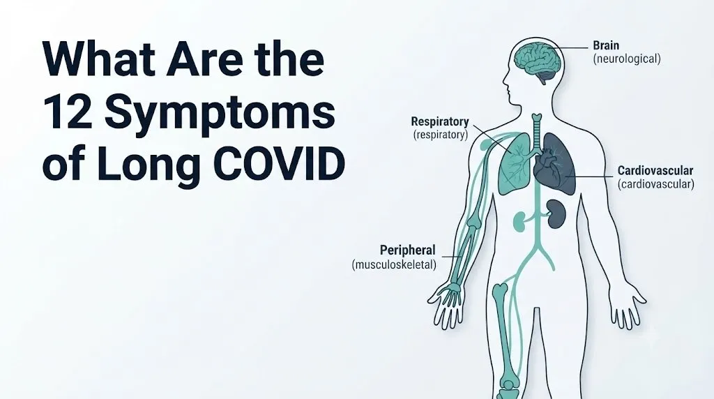
The 12 symptoms of long COVID include fatigue, brain fog, shortness of breath, post-exertional malaise, cardiovascular changes, neurological pain, autonomic dysfunction, digestive disturbance, smell and taste loss, sleep disruption, anxiety and depression, and persistent chest pain. These symptoms reflect real, measurable dysfunction across multiple organ systems, not a psychological response to illness.
The World Health Organization defines long COVID as a condition that occurs in individuals with a history of probable or confirmed SARS-CoV-2 infection, generally three months from the onset of infection, with symptoms lasting at least two months and not explained by an alternative diagnosis. According to the NIH RECOVER Initiative, approximately 10 to 30 percent of people who contracted COVID-19 have experienced symptoms lasting more than 12 weeks, affecting tens of millions of people globally.
This article covers each of the 12 recognized symptom categories in clinical detail, explains the specific physiological mechanism behind each one, addresses how symptoms differ by sex and age, and gives you a clear framework for deciding when to monitor at home, when to see a physician, and when the emergency room is the right call.
What Are the 12 Symptoms of Long COVID
The 12 core symptoms of long COVID, as documented in peer-reviewed clinical cohort studies, are: severe fatigue, post-exertional malaise, cognitive impairment (brain fog), shortness of breath, cardiovascular changes including palpitations and chest pain, neurological symptoms including neuropathic pain and headache, autonomic dysfunction including postural orthostatic tachycardia, gastrointestinal symptoms, loss of smell (anosmia) or taste (ageusia), sleep disturbance, anxiety and depression, and musculoskeletal pain.
A landmark 2021 patient survey published in The Lancet (Davis et al.) documented more than 200 symptoms across 10 organ systems in a cohort of 3,762 long COVID patients from 56 countries. The study found that 85.9 percent of respondents experienced fatigue, 81.2 percent experienced post-exertional malaise, and 67.8 percent experienced cognitive dysfunction, establishing these three as the most prevalent and functionally impairing symptoms in the long COVID clinical picture.
| Long COVID Symptom | Body System | Prevalence Estimate |
|---|---|---|
| Fatigue | Multi-system | 85% (Davis et al., The Lancet 2021) |
| Post-exertional malaise | Musculoskeletal/metabolic | 81% (Davis et al., The Lancet 2021) |
| Cognitive impairment (brain fog) | Central nervous system | 68% (Davis et al., The Lancet 2021) |
| Shortness of breath | Respiratory system | 54% (Al-Aly et al., JAMA 2021) |
| Palpitations/chest pain | Cardiovascular system | 30-50% (Al-Aly et al., JAMA 2021) |
| Neuropathic pain/headache | Peripheral nervous system | 44% (Davis et al., The Lancet 2021) |
| Autonomic dysfunction/POTS | Autonomic nervous system | 25-30% (clinical cohort estimates) |
| Digestive symptoms | Gastrointestinal system | 30% (Al-Aly et al., JAMA 2021) |
| Smell/taste loss | Olfactory/gustatory systems | 40% (clinical cohort estimates) |
| Sleep disturbance | Central nervous system | 72% (Davis et al., The Lancet 2021) |
| Anxiety/depression | Psychiatric/neurological | 38% (Taquet et al., The Lancet 2021) |
| Musculoskeletal pain | Musculoskeletal system | 44% (Davis et al., The Lancet 2021) |
Not everyone develops all 12 symptoms. Many people experience 3 to 5 symptoms that cluster around one or two dominant organ systems, while some develop a broader multi-system picture.
Long COVID Definition and Diagnostic Criteria
Long COVID, clinically designated post-acute sequelae of SARS-CoV-2 (PASC) and coded in the International Classification of Diseases as ICD-10: U09.9, is defined by the WHO as symptoms persisting or newly developing at least 3 months after initial SARS-CoV-2 infection, lasting at least 2 months, and not explained by an alternative diagnosis.
The NICE (National Institute for Health and Care Excellence) guidelines for post-COVID syndrome use a slightly different time threshold: symptoms continuing more than 12 weeks after acute infection that are not explained by an alternative diagnosis. Both definitions share the core principle that diagnosis is clinical, meaning no single laboratory test confirms long COVID. A physician establishes the diagnosis based on the infection history, symptom pattern, timeline, and the exclusion of other conditions that could explain the symptoms.
Critically, long COVID can develop after any severity of acute COVID-19 infection. A 2021 study published in Nature Medicine (Sudre et al.) found that long COVID occurs in individuals who had asymptomatic, mild, moderate, and severe acute illness, with some evidence that even people who did not require hospitalization develop persistent symptoms. This is one of the most clinically underappreciated facts: you do not need to have been hospitalized to develop significant, lasting post-COVID dysfunction.
Long COVID has no single disease mechanism. Current evidence supports at least four concurrent pathways: persistent viral antigen or intact virus in tissue reservoirs, ongoing immune dysregulation with autoantibody production, direct organ damage from the acute infection, and reactivation of latent viruses including Epstein-Barr virus triggered by the immune disruption of COVID-19.
Adolescents and younger adults develop long COVID at rates comparable to middle-aged adults. Children represent an additional population where long COVID recognition is actively developing, with the American Academy of Pediatrics noting that pediatric long COVID presents differently in school-age children, often manifesting as fatigue, headache, and difficulty concentrating rather than the cardiovascular and respiratory symptoms more common in adults.
Long COVID Fatigue and Post-Exertional Malaise
Long COVID fatigue is not ordinary tiredness. It is a form of pathological exhaustion produced by measurable dysfunction in cellular energy production, specifically impaired mitochondrial oxidative phosphorylation in skeletal muscle tissue, documented in research published in Nature Communications (2023).
Think of the mitochondria inside each muscle cell as tiny power plants converting fuel (glucose and fatty acids) into usable energy in the form of adenosine triphosphate (ATP). In long COVID fatigue, the electron transport chain within those mitochondria appears to function at reduced efficiency, producing less ATP per unit of metabolic fuel than a healthy mitochondrion would. The result is not just tiredness but a profound depletion of available cellular energy that is measurably different from the fatigue that follows ordinary overexertion.
Post-exertional malaise (PEM) is the specific clinical phenomenon where physical or cognitive exertion triggers a worsening of symptoms, typically 12 to 48 hours after the activity, that can last days to weeks. PEM is the hallmark feature that distinguishes long COVID fatigue from depression-related fatigue and from deconditioning. It also overlaps mechanistically with myalgic encephalomyelitis/chronic fatigue syndrome (ME/CFS), a condition with which long COVID shares multiple biological features according to research from the NIH.
PEM is clinically important because conventional wisdom about exercise for illness recovery is dangerous when applied to long COVID patients with PEM. Standard advice to “push through fatigue” or gradually increase exercise intensity can trigger PEM crashes that set a patient back substantially.
To track and describe fatigue and PEM to a primary care physician or post-COVID care clinic:
- Rate baseline fatigue each morning on a 1 to 10 scale before any activity
- Log every activity attempted and its duration and intensity
- Record any worsening of fatigue, pain, or cognitive symptoms in the 12 to 48 hours following activity
- Note whether the worsening resolves within 24 hours (less concerning) or extends beyond 48 hours (consistent with PEM)
- Bring this log to your appointment so the physician can distinguish PEM from general deconditioning
Women and individuals with pre-existing autoimmune conditions report PEM at higher rates in long COVID cohorts, though the mechanistic reason for this sex difference is not yet fully established in the literature.
Long COVID Brain Fog Symptoms
Long COVID brain fog is a clinically documented form of cognitive impairment caused by neuroinflammation, specifically the activation of microglial cells in the brain, combined with disruption of the blood-brain barrier that allows inflammatory cytokines including interleukin-6 (IL-6) and tumor necrosis factor-alpha (TNF-alpha) to enter the central nervous system.
Microglia are the brain’s resident immune cells. Under normal conditions they perform surveillance and cleanup functions. When systemic inflammation from the SARS-CoV-2 immune response triggers microglial activation, these cells shift to a pro-inflammatory state and release cytokines that impair synaptic transmission. The prefrontal cortex, which governs working memory, attention, and executive function, is particularly vulnerable to this inflammatory disruption. That is exactly why brain fog patients describe forgetting words mid-sentence, losing their train of thought, and struggling with tasks that previously required no effort.
A 2021 study published in The Lancet Psychiatry (Taquet et al.) of 1.2 million patients found that the incidence of neurological or psychiatric diagnoses in the six months following COVID-19 infection was 33.62 percent, higher than in matched cohorts with influenza or other respiratory infections. Brain imaging studies published in Nature (2022) found structural changes in gray matter and olfactory cortex volume in individuals post-COVID compared to matched controls, providing anatomical evidence for neurological change beyond subjective symptom reports.
Brain fog in long COVID is distinct from depression-related cognitive impairment, though the two can co-occur. Brain fog in long COVID is more likely to fluctuate with physical exertion (worsening after PEM triggers), whereas depression-related cognitive slowing tends to be more persistent and accompanied by prominent mood changes.
Quick Tip:
- Keep a daily symptom and cognitive difficulty log: this serves as objective documentation for a neurologist or primary care physician
- Avoid pushing through brain fog with high cognitive effort tasks; this can trigger PEM in the same way physical exertion does
- Older adults (over 65) with pre-existing mild cognitive impairment may experience more pronounced brain fog that is harder to attribute to long COVID alone, making neurologist evaluation particularly important in this group
Key Takeaway: Long COVID fatigue and brain fog are caused by measurable biological dysfunction including mitochondrial impairment and microglial neuroinflammation, not deconditioning or psychological distress, and pushing through both with exercise can worsen outcomes in patients with post-exertional malaise.
Long COVID Shortness of Breath and Respiratory Symptoms
Long COVID shortness of breath reflects persistent abnormalities in pulmonary gas exchange, specifically reduced diffusing capacity of the lungs for carbon monoxide (DLCO), even in patients whose chest X-rays appear normal.
The alveolar epithelium, the thin layer of cells lining the 300 million air sacs in each lung, is one of SARS-CoV-2’s primary infection targets. Damage to type II pneumocytes (the alveolar cells responsible for producing surfactant and participating in gas exchange) results in impaired oxygen transfer across the alveolar-capillary membrane. Even partial or patchy damage to this membrane surface area reduces the lungs’ ability to transfer oxygen into the bloodstream efficiently during exertion, producing breathlessness at activity levels that previously caused no difficulty.
According to a large veterans cohort study published in JAMA (Al-Aly et al., 2021) of more than 73,000 post-COVID patients compared to 5.1 million controls, individuals with COVID-19 had a 1.49-fold higher risk of incident dyspnea (shortness of breath) at one year post-infection. Pulmonary function abnormalities, particularly reduced DLCO, have been documented in long COVID patients who had only mild acute illness and no radiological evidence of pneumonia during their acute phase.
| Respiratory Symptom | Mechanism | Evaluation Needed |
|---|---|---|
| Breathlessness at rest | Alveolar damage or autonomic dysfunction | Urgent: same-day primary care or pulmonology |
| Breathlessness on exertion only | Reduced DLCO, deconditioning, or PEM | Scheduled primary care evaluation |
| Persistent cough | Airway inflammation, post-viral airway hyperreactivity | Scheduled primary care evaluation |
| Chest tightness with breathing | Pleural inflammation or cardiac involvement | Same-day evaluation |
| Breathlessness with leg swelling | Pulmonary embolism risk | Emergency room immediately |
People with pre-existing asthma or chronic obstructive pulmonary disease are at higher risk for prolonged respiratory symptoms because their alveolar reserve is already reduced before COVID-19 infection compounds the deficit. A referral to a pulmonologist for full pulmonary function testing including DLCO is appropriate when breathlessness limits normal daily activities at four weeks or more post-infection.
Long COVID Heart Symptoms and Cardiovascular Effects
Long COVID heart symptoms are produced by multiple mechanisms: direct viral damage to myocardial cells (the heart muscle), inflammatory injury to the pericardium (the sac surrounding the heart), disruption of the sinoatrial node (the heart’s electrical pacemaker), and autoantibody-mediated dysfunction of cardiac autonomic receptors.
Palpitations, the subjective sensation of a rapid, pounding, or irregular heartbeat, are among the most reported cardiovascular symptoms in long COVID cohorts. The mechanistic pathway differs from person to person: some patients develop true cardiac arrhythmias detectable on a cardiac monitor, while others experience tachycardia (elevated heart rate) driven by autonomic dysfunction rather than intrinsic cardiac disease. Distinguishing between these two causes matters because the diagnostic approach and management differ.
Research published in Nature Medicine (Xie and Al-Aly, 2022) using the same veterans database found that at one year post-infection, COVID-19 survivors had a 72 percent higher risk of heart failure, a 63 percent higher risk of atrial fibrillation, and a 52 percent higher risk of stroke compared to uninfected controls. These elevated risks were present even in patients who had not been hospitalized during acute infection, though the absolute risk remained higher in those with severe acute illness.
Chest pain in long COVID requires individualized evaluation. Pleuritic chest pain (sharp pain that worsens with deep breathing) can indicate pericarditis, a documented complication of COVID-19. A cardiologist referral with echocardiogram and potentially cardiac MRI is appropriate for any persistent chest pain, unexplained palpitations lasting more than a few days, or documented resting tachycardia (heart rate above 100 beats per minute consistently).
Men with long COVID and pre-existing cardiovascular risk factors (hypertension, diabetes, hypercholesterolemia) face disproportionately elevated risk for adverse cardiac events in the post-acute phase. Primary care physicians managing male long COVID patients with cardiovascular risk factors should consider early cardiology referral rather than watchful waiting.
Long COVID Neurological Symptoms
Long COVID neurological symptoms include neuropathic pain, sensory paresthesias (tingling, burning, or electric shock sensations), persistent headache, tinnitus (ringing in the ears), vision changes, and in some patients, cognitive decline that extends beyond brain fog into measurable deficits on neuropsychological testing.
The peripheral neurological symptoms, particularly the tingling, burning, and pain in the hands, feet, and face, are increasingly understood to involve small fiber neuropathy (SFN), damage to the smallest nerve fibers in the peripheral nervous system. A 2021 study published in the Annals of Neurology found reduced intraepidermal nerve fiber density on skin punch biopsy in a subset of long COVID patients with neuropathic pain, providing objective pathological evidence for SFN rather than functional or psychological symptom generation.
Small fiber nerves serve two functions: sensory transmission (carrying pain, temperature, and touch signals) and autonomic regulation (controlling heart rate, blood pressure, sweat gland activity, and gastrointestinal motility). This explains why SFN in long COVID can simultaneously produce neuropathic pain AND autonomic dysfunction, making it a unifying mechanism for several seemingly unrelated long COVID symptoms in the same patient.
Persistent headache in long COVID occurs in approximately 44 percent of patients by some estimates and is believed to involve intracranial pressure changes, cerebrovascular reactivity abnormalities, and direct neuroinflammatory effects on the meningeal pain-sensing fibers. Unlike tension headache, long COVID headache frequently does not respond to standard analgesics and may worsen with exertion, a pattern that should be reported specifically to a neurologist.
Patients with pre-existing migraine disorder report that COVID-19 infection significantly worsened their migraine frequency and severity, with some developing new daily persistent headache that was not present before infection. A neurologist evaluation is appropriate for new persistent headache, paresthesias, tinnitus, or vision changes that have lasted more than 4 weeks post-infection.
Key Takeaway: Long COVID neurological symptoms including paresthesias and neuropathic pain are increasingly documented as small fiber neuropathy, a measurable nerve fiber abnormality confirmed on skin punch biopsy, not a functional or psychosomatic symptom.
Long COVID Autonomic Dysfunction and POTS
Postural orthostatic tachycardia syndrome (POTS) is a recognized and well-documented long COVID manifestation in which the autonomic nervous system fails to properly regulate heart rate in response to positional changes, causing heart rate to increase by 30 or more beats per minute within 10 minutes of standing.
Think of the autonomic nervous system as the body’s automatic control panel, managing heart rate, blood pressure, breathing rate, and digestive activity without conscious effort. In POTS, the section of this control panel that coordinates blood redistribution when you stand up malfunctions. When you stand, blood normally pools in the legs briefly before the autonomic system compensates by increasing vascular tone and heart rate just slightly. In POTS, that compensation is excessive: the heart rate surges dramatically to maintain cerebral blood flow against gravity, producing palpitations, dizziness, lightheadedness, and sometimes syncope (fainting).
The mechanism in long COVID-associated POTS appears to involve multiple pathways: autoantibodies against autonomic receptors (particularly alpha-1 adrenergic and beta-2 adrenergic receptors), damage to the small fiber nerves that regulate vascular tone in the peripheral vasculature, and possibly direct injury to the vagus nerve, which provides the primary parasympathetic brake on heart rate. Research from the NIH RECOVER Initiative has identified autonomic dysfunction as one of the most functionally impairing long COVID phenotypes.
POTS is diagnosed by a tilt table test administered by an autonomic specialist or cardiologist, or by a simplified active stand test where heart rate is measured supine and then standing at 1, 3, 5, and 10 minutes. An increase of 30 or more beats per minute (or 40 in adolescents) is diagnostic. Because POTS is disproportionately common in younger women (ages 15 to 50) generally, the long COVID variant also skews toward this demographic, though it occurs in men and older adults as well.
Long COVID Digestive Symptoms
Long COVID digestive symptoms, occurring in approximately 30 percent of long COVID patients according to the JAMA veterans cohort study, include nausea, abdominal pain, diarrhea, constipation, acid reflux, and appetite loss. These symptoms are produced by disruption of the enteric nervous system and gut microbiome dysbiosis following acute SARS-CoV-2 infection.
The enteric nervous system, a network of approximately 500 million neurons embedded in the gastrointestinal tract wall, independently regulates gut motility, secretion, and blood flow. SARS-CoV-2 infects the gut via ACE2 receptors expressed on intestinal epithelial cells. Viral infection and the subsequent inflammatory response disrupt the enteric neural network and damage enterochromaffin cells, which are the gut’s primary producers of serotonin. Approximately 90 percent of the body’s serotonin is produced in the gut, where it regulates intestinal motility. Damage to enterochromaffin cells produces disordered motility: the gut moves too fast (diarrhea) or too slow (constipation), often alternating between both.
Gut microbiome composition is measurably altered following SARS-CoV-2 infection, with reduced diversity and depletion of beneficial bacterial species including Lactobacillus and Bifidobacterium documented in multiple studies. A 2021 study published in Gut (Yeoh et al.) found that microbiome composition at the time of hospital admission predicted whether patients developed long COVID symptoms, raising the possibility that gut dysbiosis is not just a consequence but a contributing mechanism.
Nausea and appetite loss that are severe enough to cause unintentional weight loss of more than 5 percent of body weight within a month, or digestive symptoms accompanied by blood in the stool, require prompt evaluation by a gastroenterologist to exclude inflammatory bowel disease, gastrointestinal bleeding, or other structural causes that may have been unmasked or triggered by the infection.
Long COVID Smell and Taste Loss
Long COVID anosmia (complete smell loss) and parosmia (distorted smell perception, often described as pleasant smells becoming foul or chemical) are caused by damage to non-neuronal sustentacular cells in the olfactory epithelium, with secondary effects on the olfactory sensory neurons they support.
The olfactory epithelium, located in the upper nasal cavity, contains the specialized neurons (olfactory sensory neurons) that carry smell signals directly to the olfactory bulb in the brain via the olfactory nerve (cranial nerve I). SARS-CoV-2 does not appear to directly infect olfactory sensory neurons but does infect the sustentacular cells that support and maintain them. When sustentacular cells die or become dysfunctional, the olfactory sensory neurons they support lose metabolic support and become temporarily unable to fire correctly.
This mechanism explains why most people with COVID-related smell loss recover it: the sustentacular cells regenerate and olfactory neurons regain function. However, a clinically documented subset of long COVID patients develops parosmia, a qualitative smell distortion that arises when recovering olfactory neurons reconnect in disorganized patterns, sending incorrect signals to the olfactory cortex. Foods that previously smelled appealing begin to smell like rotting matter, sulfur, or chemicals.
A 2021 study published in JAMA Otolaryngology found that among patients with COVID-19-related smell loss, 63 percent had not fully recovered smell function at 12 months. Persistent anosmia or parosmia at three months post-infection warrants referral to an otolaryngologist (ear, nose, and throat specialist) for evaluation and consideration of olfactory training, a structured sensory rehabilitation protocol with emerging supportive evidence.
Key Takeaway: Long COVID anosmia and parosmia are caused by sustentacular cell damage in the olfactory epithelium rather than direct olfactory nerve infection, which explains both the partial recovery most patients experience and the distorted smell sensations that develop during regeneration.
Long COVID Mental Health Symptoms
Long COVID mental health symptoms, including anxiety, depression, post-traumatic stress responses, and emotional dysregulation, arise from a combination of biological mechanisms and the psychosocial impact of prolonged illness. The biological component is specifically important: neuroinflammation in long COVID directly disrupts the limbic system, including the amygdala and hippocampus, which regulate emotional processing and stress response.
The hypothalamic-pituitary-adrenal (HPA) axis, the body’s primary cortisol stress-response system, shows dysregulation in a subset of long COVID patients, as documented in several observational studies. Abnormal cortisol production or rhythm disrupts sleep, amplifies anxiety, and reduces emotional resilience independently of the patient’s psychological response to illness. This means that long COVID anxiety and depression in some patients have a direct neuroendocrine substrate, not just a psychosocial one.
A study published in The Lancet Psychiatry (Taquet et al., 2021) found that 23.18 percent of COVID-19 patients received a psychiatric diagnosis for the first time in the six months following their infection, compared to significantly lower rates in matched control populations with other respiratory infections. Depression and anxiety were the most common new diagnoses.
If you are in crisis or experiencing thoughts of self-harm or suicide, contact the 988 Suicide and Crisis Lifeline by calling or texting 988 at any time. This service is free, confidential, and available 24 hours a day, 7 days a week.
Women with long COVID report higher rates of anxiety and depression than men in most cohort studies, though some researchers propose this reflects reporting differences as well as biological susceptibility. Regardless of biological sex, any long COVID patient experiencing persistent anxiety, depressive symptoms, intrusive thoughts, or emotional dysregulation that interferes with daily function warrants evaluation by a psychiatrist or licensed clinical psychologist, both of whom can distinguish neuroinflammation-mediated mood changes from primary psychiatric disorder and provide appropriate evidence-based treatment.
Long COVID Mast Cell Activation Syndrome and MCAS Overlap
Mast cell activation syndrome (MCAS) is a condition in which mast cells (immune cells found in connective tissue throughout the body, particularly in skin, lungs, and gastrointestinal tissue) release excessive amounts of inflammatory mediators including histamine and tryptase in response to triggers that would not normally cause a reaction.
A growing body of clinical observation and emerging research suggests that some long COVID patients develop or unmask MCAS following SARS-CoV-2 infection. The proposed mechanism involves viral activation of mast cells that then become hypersensitive: rather than degranulating only during genuine immune threats, they release histamine, prostaglandins, and other inflammatory mediators in response to ordinary stimuli including foods, odors, temperature changes, and emotional stress.
The clinical picture of MCAS in long COVID includes flushing, hives or skin rashes that come and go unpredictably, gastrointestinal symptoms triggered by specific foods, brain fog worsening with dietary triggers, palpitations, and anaphylaxis-like episodes. These symptoms can be mistaken for anxiety, irritable bowel syndrome, or food intolerance without recognizing the mast cell mechanism underlying them.
Elevated mast cell tryptase in serum, elevated 24-hour urine histamine, and response to antihistamine medications are key diagnostic markers, according to clinical specialists in immunology. An allergist-immunologist or specialist familiar with mast cell disorders is the appropriate referral when this symptom constellation is present.
People with pre-existing conditions that prime mast cell hyperreactivity, including existing allergic conditions, eosinophilic disorders, or connective tissue disorders such as hypermobile Ehlers-Danlos syndrome, appear at elevated risk for developing MCAS features following COVID-19, according to clinical observation in post-COVID specialty clinics.
Long COVID in Women Versus Men
Women are diagnosed with long COVID at approximately twice the rate of men in most epidemiological cohorts, a disparity that reflects both biological mechanisms and potential differences in health-seeking behavior, though the biological component is increasingly supported by evidence.
The immune system responds differently across biological sexes, driven substantially by estrogen. Estrogen upregulates the production of type I interferons, the immune proteins responsible for the initial antiviral response. This stronger initial immune response, while protective in acute viral illness, also means women’s immune systems are more likely to generate autoantibodies and sustain a prolonged inflammatory response after the virus is cleared. Autoimmunity broadly affects women at rates approximately 4 to 1 compared to men, and the autoimmune-like features of long COVID follow this same pattern.
Women with long COVID are disproportionately represented among patients with POTS, MCAS, and multi-system symptom presentations, while men with long COVID show higher rates of cardiovascular complications and higher rates of death during the acute phase. This sex-differentiated risk profile means the clinical picture of long COVID that a woman should expect to discuss with her physician differs meaningfully from what a man should expect.
A 2021 study published in Nature Medicine (Sudre et al.) found that female sex was one of the strongest predictors of developing long COVID at 28 days after infection. Reproductive-age women may also notice interactions between long COVID symptoms and their menstrual cycle, with symptom exacerbation during specific cycle phases, a pattern potentially linked to cyclical estrogen fluctuations modifying immune activity and mast cell reactivity.
Women experiencing these symptoms after COVID-19 should specifically ask their primary care physician or an obstetrician-gynecologist whether menstrual-cycle-related symptom tracking is appropriate in their case, as this information can guide specialist referral and management planning.
Key Takeaway: Women develop long COVID at approximately twice the rate of men and are disproportionately affected by POTS, MCAS, and autoantibody-driven multi-system symptoms, a pattern rooted in estrogen-mediated immune upregulation rather than psychosocial factors.
How Long Does Long COVID Last
Long COVID duration varies widely: some patients recover fully within 3 to 6 months, others experience persistent or cycling symptoms for years, and a subset develop chronic conditions that persist indefinitely without remission.
According to data from the NIH RECOVER Initiative, approximately 85 percent of long COVID patients report some symptom improvement over the first year. However, improvement is not the same as resolution: many patients improve from severe functional impairment to moderate impairment without returning to their pre-COVID baseline. A 2023 analysis of NIH RECOVER cohort data found that certain symptom clusters, particularly post-exertional malaise, autonomic dysfunction, and cognitive impairment, are more likely to persist beyond 12 months than respiratory symptoms, which tend to improve more predictably.
The natural history of long COVID is also non-linear. Patients commonly describe periods of relative improvement followed by symptom “crashes” triggered by overexertion, illness, or stress. This relapsing pattern is consistent with the ME/CFS model, with which long COVID shares substantial biological overlap. Patients who follow a structured pacing strategy (matching activity level to current energy envelope rather than attempting gradual escalation) tend to have fewer and less severe crash episodes, though the evidence base for specific management strategies in long COVID remains an active area of research.
Factors associated with longer duration and poorer recovery include: older age at time of infection, female sex, pre-existing autoimmune or chronic inflammatory conditions, greater number of symptoms at four weeks post-infection, and presence of PEM as a dominant symptom. The CDC reports that prior vaccination reduces the risk of developing long COVID, and emerging evidence suggests some vaccinated individuals who develop long COVID may have a shorter or milder course, though this finding requires larger prospective studies to confirm.
Long COVID Diagnosis Criteria and Testing
Long COVID is diagnosed clinically, meaning no single blood test, imaging study, or laboratory result confirms or rules it out. The diagnostic process involves a thorough history, physical examination, exclusion of alternative diagnoses, and targeted testing based on the patient’s specific symptom profile.
To prepare for a first appointment with a primary care physician or post-COVID care clinic:
- Write out your complete symptom list, including symptoms you are uncertain are related, with onset dates relative to your COVID-19 infection
- Rate each symptom’s current severity on a 1 to 10 scale and note whether it is improving, stable, or worsening
- Document your COVID-19 infection timeline: date of first symptoms, test confirmation if available, severity of acute illness, and any treatments received
- Note any activities, foods, times of day, or events that reliably worsen or improve specific symptoms
- List all medications, supplements, and prior relevant medical history, particularly any autoimmune, cardiovascular, or neurological conditions
Common initial laboratory tests ordered by a physician for suspected long COVID include: complete blood count (CBC), C-reactive protein (CRP), erythrocyte sedimentation rate (ESR), ferritin, thyroid-stimulating hormone (TSH), D-dimer, antinuclear antibody (ANA) panel, and metabolic panel. These tests cannot diagnose long COVID but can identify treatable contributing conditions and rule out other explanations for symptoms.
Specialized testing depends on the symptom cluster: pulmonary function testing including DLCO for respiratory symptoms, echocardiogram and Holter monitor for cardiac symptoms, tilt table test for suspected POTS, skin punch biopsy for suspected small fiber neuropathy, and neuropsychological testing for cognitive impairment. These specialized tests are typically ordered by the relevant specialist after referral from primary care.
Emergency Symptoms: When to Call 911 or Go to the ER
Certain symptoms associated with long COVID require immediate emergency evaluation. Do not wait to see if these resolve on their own.
Call 911 or go to the nearest emergency room immediately if you experience:
- Sudden severe chest pain or chest pressure, particularly if it radiates to your jaw, left arm, or back, because this can indicate myocardial infarction (heart attack) or aortic dissection
- Severe shortness of breath at rest or breathlessness that prevents completing a sentence, which can indicate pulmonary embolism, acute heart failure, or severe respiratory decompensation
- Sudden one-sided weakness, facial drooping, slurred speech, sudden vision loss, or severe sudden headache unlike any previous headache, because these are stroke warning signs requiring emergency intervention within minutes
- Syncope (loss of consciousness or fainting) that is not immediately explained by positional change, because this can indicate dangerous cardiac arrhythmia or hemodynamic instability
- Sudden pleuritic chest pain (sharp pain with breathing) combined with rapid heart rate and shortness of breath, which together represent a clinical picture consistent with pulmonary embolism, a life-threatening clot in the pulmonary vasculature
- Palpitations with documented heart rate above 150 beats per minute at rest that do not resolve within minutes, because this can indicate a cardiac arrhythmia requiring emergency evaluation
- High fever (above 103°F / 39.4°C) with new confusion, stiff neck, or severe headache, which can indicate meningitis or encephalitis
These presentations can indicate pulmonary embolism, myocardial infarction, stroke, or dangerous cardiac arrhythmia and require emergency medical assessment. They cannot be managed at an urgent care appointment, a telehealth call, or a primary care clinic.
Frequently Asked Questions About Long COVID Symptoms
What are the most common symptoms of long COVID?
The most common long COVID symptoms are fatigue, post-exertional malaise, and cognitive impairment (brain fog), affecting approximately 85, 81, and 68 percent of long COVID patients respectively, according to the Davis et al. 2021 study published in The Lancet.
Shortness of breath, sleep disturbance, palpitations, and musculoskeletal pain are also among the most frequently reported symptoms across multiple large cohort studies.
These symptoms can occur after mild, moderate, or severe acute COVID-19 infection and are not limited to people who were hospitalized.
How do I know if what I have is long COVID or something else?
Long COVID is a clinical diagnosis made by excluding other explanations for persistent symptoms after a confirmed or probable SARS-CoV-2 infection, not by a single confirmatory test.
A primary care physician evaluating suspected long COVID will typically order laboratory work to rule out thyroid dysfunction, anemia, autoimmune conditions, and other treatable causes of similar symptoms.
If your symptoms began after COVID-19 infection, persist for more than 4 weeks, and are not explained by another diagnosis after evaluation, long COVID becomes the working clinical diagnosis under both the WHO and NICE definitions.
Can long COVID symptoms appear after a mild COVID infection?
Yes. Long COVID develops after mild, asymptomatic, moderate, and severe acute COVID-19 infections at documented rates across multiple cohort studies.
The 2021 Nature Medicine study by Sudre et al. found that long COVID was not restricted to patients who had severe acute illness, with many cases arising after infections that did not require medical care.
The absence of hospitalization during your acute illness does not reduce the likelihood of long COVID and should not be used as a reason to dismiss persistent symptoms.
What does post-exertional malaise feel like in long COVID?
Post-exertional malaise in long COVID feels like a profound crash of energy, pain, and cognitive function that typically begins 12 to 48 hours after physical or mental exertion and lasts days to weeks.
It is distinct from normal exercise fatigue, which resolves with rest within hours. PEM does not improve proportionally with rest.
Patients commonly describe it as feeling as though a severe illness has returned: heavy limbs, inability to concentrate, flu-like body aches, and complete depletion of the ability to function normally.
How long do long COVID symptoms typically last?
Long COVID symptoms last anywhere from 3 months to multiple years depending on the individual, with most patients showing some improvement within the first year according to NIH RECOVER Initiative data.
Certain symptom clusters, particularly post-exertional malaise, autonomic dysfunction, and cognitive impairment, are more likely to persist beyond 12 months than respiratory symptoms, which tend to improve earlier.
A subset of long COVID patients develop chronic conditions that persist indefinitely, a pattern similar to ME/CFS, which shares substantial biological and clinical overlap with long COVID.
When should I go to the emergency room for long COVID symptoms?
Go to the emergency room immediately for new chest pain with radiation to the jaw or arm, severe breathlessness at rest, sudden neurological changes including weakness or slurred speech, or loss of consciousness.
These presentations can indicate heart attack, pulmonary embolism, stroke, or dangerous cardiac arrhythmia, all of which require emergency evaluation within minutes, not hours.
Do not manage these presentations with a phone call to your doctor or an urgent care visit.
The Body Is Not Overreacting
Long COVID is not vague, not poorly defined, and not without biological substrate. The mechanisms behind each symptom cluster are documented in peer-reviewed research from some of the world’s leading medical journals, and the physiological changes are measurable. That clarity matters when you are trying to make decisions about your care.
The most important next step if you suspect long COVID is a structured conversation with a primary care physician who can take a complete post-infection history, order appropriate baseline laboratory work, and make targeted specialist referrals based on your dominant symptom clusters. Bring a written symptom log with dates, severity ratings, and activity correlations to that appointment. That documentation accelerates both diagnosis and referral.
You deserve care that takes the biology of your symptoms seriously. Armed with the clinical specifics in this article, you can have that conversation with precision