Broken Ankle vs Sprain: Symptoms and How to Tell Apart
Distinguishing broken ankle symptoms vs sprain comes down to three key clinical signals: the precise location of your pain, whether you can bear weight, and where the tenderness sits when the bone or ligament is pressed directly. A fracture produces point tenderness over the bony prominences of the ankle; a sprain produces tenderness primarily over the soft tissue ligaments on the outer or inner side of the joint.
The stakes of getting this wrong are real. According to the American Academy of Orthopaedic Surgeons, ankle injuries account for approximately 1 million emergency room visits in the United States every year, and a meaningful proportion of fractures are initially misidentified as sprains and sent home without imaging. An untreated fracture that bears weight can displace, requiring surgical repair that might otherwise have been avoided.
This article covers the anatomy of both injuries, their overlapping symptoms, the clinical signs that most reliably distinguish one from the other, the validated Ottawa Ankle Rules for deciding whether you need an X-ray, and the specific presentations that require emergency evaluation right now.
Broken Ankle Symptoms vs Sprain: The Core Comparison
The most reliable clinical distinction between broken ankle symptoms and a sprained ankle is the location of point tenderness: fractures produce tenderness directly over bone, while sprains produce tenderness over the ligaments that run between bones. Both injuries cause pain, swelling, and bruising, which is why visual inspection alone cannot reliably separate them.
Understanding this distinction requires knowing what each injury actually damages. A sprain tears or overstretches one or more ankle ligaments. A fracture breaks one or more of the bony structures that form the ankle mortise. The pain mechanisms are physiologically different: ligament pain comes from nociceptors within the torn ligament fibers and the inflammatory response in surrounding soft tissue; fracture pain comes from highly innervated periosteum that is disrupted when bone cortex breaks.
Periosteal pain has a distinctive character. It tends to be sharper, more precisely localized, and intensely sensitive to direct pressure over the bone itself. Ligamentous pain spreads more diffusely into the surrounding soft tissue and is less precisely localized to a single anatomical point.
Here is the core comparison at a glance:
| Feature | Ankle Sprain | Broken Ankle (Fracture) |
|---|---|---|
| Primary tissue injured | Ligaments (ATFL, CFL, deltoid) | Bone (lateral, medial, or posterior malleolus) |
| Pain location | Over ligament, lateral or medial soft tissue | Over bony prominence (malleolus) |
| Point tenderness | Soft tissue, 1-2 cm below malleolus tip | Directly on malleolus or bone surface |
| Swelling | Lateral or medial ankle, soft tissue | Over fracture site, often broader |
| Bruising | Lateral ankle extending toward sole | Can extend further, appear more rapidly |
| Weight bearing | Often possible (Grade 1-2) | Often impossible; 4 steps is Ottawa threshold |
| Deformity | Rare unless severe Grade 3 | Can be present with displacement |
| Sensation change | Rare | Possible if nerve involved |
Individual variation note: Children with open growth plates (typically under age 14 to 16) may present with all the same symptoms as a lateral ankle sprain but actually have a physeal (growth plate) fracture at the distal fibula. The growth plate is biomechanically weaker than the ankle ligaments in skeletally immature patients, making fracture more likely than pure ligament tear even with the same inversion force that would cause a sprain in an adult.
Ankle Anatomy: What Structures Are Injured in Each Condition
The ankle joint (talocrural joint) is formed by three bones, the tibia, fibula, and talus, held in precise alignment by ligament complexes on the inner and outer sides and a bony mortise structure that allows only dorsiflexion and plantarflexion movements. Injury to any one of these structures produces overlapping symptoms, which is why anatomy is the starting point for understanding the distinction.
The lateral ankle ligament complex consists of three named structures: the anterior talofibular ligament (ATFL), which runs from the front of the lateral malleolus to the talus and is the most commonly torn structure in ankle sprains; the calcaneofibular ligament (CFL), which runs from the lateral malleolus downward to the calcaneus; and the posterior talofibular ligament (PTFL), the strongest of the three, which is torn only in severe, high-energy sprains. The ATFL is injured in approximately 85 percent of lateral ankle sprains, according to clinical observations documented in the Journal of Bone and Joint Surgery.
On the inner side, the deltoid ligament complex protects against eversion forces. It is a broad, fan-shaped structure connecting the medial malleolus to the talus, calcaneus, and navicular. Isolated deltoid sprains are less common than lateral ankle sprains.
The bony structures most often fractured include the lateral malleolus (the rounded distal end of the fibula), the medial malleolus (the medial projection of the distal tibia), and the posterior malleolus (the posterior distal tibial rim). Fractures involving two of these structures are called bimalleolar fractures; those involving all three are trimalleolar fractures.
Individual variation note: Pregnant women experience increased ligamentous laxity throughout their joints due to the hormone relaxin, which relaxes smooth muscle and connective tissue to prepare the pelvis for delivery. This affects ankle ligament tension, theoretically increasing susceptibility to lateral ankle sprains during pregnancy.
Sprained Ankle Symptoms: What a Ligament Injury Feels Like
A sprained ankle produces immediate pain over the outer (or, less commonly, inner) ankle at the site of the torn ligament, followed by swelling that develops within minutes to hours and bruising that typically appears within 24 to 48 hours. The severity of these symptoms depends on which grade of sprain has occurred.
Ankle sprains are classified by grade based on the degree of ligament disruption. Grade 1 involves microscopic tears of the ligament fibers with mild pain, minimal swelling, and preserved weight-bearing ability. Grade 2 involves partial tearing with moderate pain, visible swelling, and reduced weight-bearing capacity. Grade 3 involves complete ligament rupture with severe pain at the moment of injury followed by paradoxical pain reduction (because the nerve fibers in the ruptured ligament are also disrupted), marked swelling, extensive bruising, and often complete inability to bear weight.
The mechanism of swelling in a sprain is well-understood. Torn capillaries within the ligament and surrounding soft tissue release blood into the tissue space. Simultaneously, inflammatory mediators including prostaglandins E2 and I2, histamine from mast cells, and bradykinin from plasma cause local vasodilation and increased capillary permeability. Plasma proteins and fluid leak into the interstitial space, producing the characteristic soft, pitting edema around the lateral malleolus.
Symptom checklist for a lateral ankle sprain:
- Immediate pain over the front-outer ankle, at the ATFL location
- Swelling that develops within minutes and peaks over 2 to 3 hours
- Bruising appearing 12 to 48 hours after injury, typically on the outer ankle and sometimes extending toward the arch or sole
- Tenderness when the soft tissue anterior and inferior to the lateral malleolus tip is pressed (not the bone itself)
- Sense of ankle instability or “giving way”
- Difficulty bearing full weight (Grade 2 and 3)
Individual variation note: Athletes who have sustained prior lateral ankle sprains may have residual proprioceptive deficits from previous ligament injuries, making them more susceptible to recurrent sprains and sometimes presenting with chronic instability symptoms on top of a new acute injury. A sports medicine physician can evaluate for chronic lateral ankle instability separately from the acute sprain assessment.
Key Takeaway: A sprained ankle’s tenderness sits in the soft tissue anterior and inferior to the lateral malleolus tip, not directly on the bone itself. If pressing the bone hurts more than pressing the soft tissue next to it, that pattern points toward fracture.
Broken Ankle Symptoms: What a Fracture Feels Like
A broken ankle (ankle fracture) typically produces immediate, severe pain directly over one of the malleoli or along the fibula shaft, often with a sensation described as a crack or pop at the moment of injury, followed by rapid swelling and an inability to bear weight that persists beyond the first few minutes. The intensity of pain can vary, and some fractures produce less pain than expected, which is why clinical testing matters more than pain score alone.
The lateral malleolus fracture is the most common single-bone ankle fracture, resulting from the fibula breaking at or near its distal tip when the ankle twists beyond its mechanical tolerance. Medial malleolus fractures typically occur with eversion or external rotation forces and are often part of a more complex bimalleolar or trimalleolar injury pattern. The Weber classification system (Weber A, B, and C) categorizes fibular fractures based on their relationship to the level of the distal tibiofibular syndesmosis, which determines ankle stability and surgical versus non-surgical management.
Think of the ankle mortise like a precisely fitted door frame. The lateral and medial malleoli form the sides of the frame, the tibial plafond forms the ceiling, and the talus sits inside as the door. When one side of the frame breaks (a single malleolus fracture), the door may still track correctly. When two sides break (bimalleolar), the frame becomes unstable and the door misaligns. That misalignment is exactly what surgeons are trying to prevent when they decide whether a fracture needs fixation.
Key symptoms that suggest fracture over sprain:
- Severe, immediate pain directly over the lateral malleolus, medial malleolus, or fibula shaft
- Audible or felt cracking or snapping at the moment of injury
- Inability to bear any weight in the first minutes after injury, persisting beyond five minutes
- Visible deformity or abnormal ankle contour (suggests displacement)
- Bony crepitus (a grating sensation) with any movement attempt
- Point tenderness directly on the malleolus tip or along the fibula bone
Individual variation note: Older adults with osteoporosis may sustain ankle fractures from low-energy mechanisms such as simply stepping off a curb or rotating suddenly, with pain intensity that feels similar to a moderate sprain rather than a severe fracture. The National Institute of Arthritis and Musculoskeletal and Skin Diseases (NIAMS) recommends that any ankle injury in a postmenopausal woman over age 50 with known or suspected low bone density be evaluated with imaging regardless of pain severity or weight-bearing ability.
Swelling and Bruising: Why Both Injuries Look Similar
Both ankle sprains and fractures produce swelling and bruising through the same fundamental mechanism of vascular disruption and inflammatory mediator release, which is why the appearance of the ankle cannot reliably distinguish the two injuries and imaging is often required. The differences in bruising pattern and timeline can offer supporting clues but are not definitive.
Swelling develops in both injuries through nearly identical physiological pathways. Torn blood vessels within the damaged tissue (whether ligament or periosteum) leak blood into the surrounding tissue. Histamine and bradykinin trigger vasodilation and increased capillary permeability within minutes. The resulting edema forms the characteristic swollen contour around the ankle. In both injury types, swelling peaks within 24 to 72 hours.
Bruising (ecchymosis) tends to develop more rapidly and extensively with fractures than with Grade 1 or 2 sprains, because fractures disrupt the periosteal blood supply and the more vascular medullary bone tissue. In fractures, bruising can be visible within the first hour. In moderate sprains, bruising may not appear for 12 to 24 hours and is typically confined to the outer ankle. In severe Grade 3 sprains or fractures, bruising can spread across the entire foot, including the sole, within 24 to 48 hours.
According to the American College of Emergency Physicians, the extent and location of bruising is not a reliable standalone indicator of fracture versus sprain in acute presentations because both injury types can produce extensive bruising, and patient factors such as anticoagulant use, aspirin therapy, and blood pressure medication can dramatically increase bruising in either injury type.
Individual variation note: Patients taking anticoagulants (warfarin, rivaroxaban, apixaban) or antiplatelet agents develop disproportionately extensive bruising after any ankle injury, which can make the visual assessment of injury severity unreliable. These patients should have a lower threshold for imaging even when the clinical picture otherwise seems mild.
Pain Location: The Most Reliable Clinical Way to Tell Them Apart
Pain location, specifically whether tenderness sits directly on the bony prominence of the malleolus versus in the soft tissue just anterior and inferior to it, is the single most clinically reliable physical examination finding for distinguishing a broken ankle from a sprained ankle before imaging is available. This distinction is what the Ottawa Ankle Rules formalize into a structured decision tool.
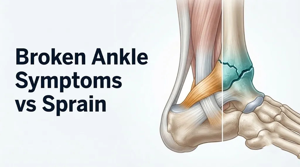
The tip of the lateral malleolus is the most palpable bony landmark on the outer ankle. Directly pressing on this bony tip and reproducing the patient’s worst pain is called a positive posterior edge malleolar zone tenderness finding. This is one of the two primary Ottawa Ankle Rule criteria for fracture.
The ATFL, by contrast, runs from the anterior edge of the lateral malleolus (not the tip) forward and downward toward the talus. Pressing 1 to 2 centimeters anterior and inferior to the malleolus tip, in the soft tissue between the fibula and the talus, identifies ATFL tenderness without pressing bone. When this specific soft tissue point is the primary pain location and the malleolus tip itself is not tender, a sprain is considerably more likely than a fracture.
To locate and assess tenderness accurately before seeing a provider:
- Sit down and place the injured ankle where you can reach it comfortably.
- Find the lateral malleolus: it is the rounded bony bump on the outer side of your ankle.
- Press firmly on the tip of that bump and the 6 centimeters of fibula directly above it. Rate the pain from 1 to 10.
- Move your finger 1 to 2 centimeters forward and downward from the malleolus tip into the soft tissue. Press firmly. Rate the pain again.
- If the bony tip produced significantly more pain than the soft tissue anterior to it, that pattern suggests bone involvement and warrants imaging. If the soft tissue point is more tender than the bone, ligament injury is more likely.
Individual variation note: People with significant ankle swelling may find all palpation acutely painful, making this soft-tissue vs. bone distinction harder to perform accurately. In highly swollen ankles within the first 2 to 4 hours, the Ottawa Ankle Rules applied by a trained clinician are more reliable than self-palpation.
Key Takeaway: Press the tip of your lateral malleolus and the fibula above it. If that bony prominence is the most painful point under direct pressure, get an X-ray. If the soft tissue anterior and inferior to the tip hurts more, a sprain is more likely but not confirmed.
Can You Walk on a Broken Ankle vs a Sprained Ankle?
The ability to take four or more steps immediately after an ankle injury does not rule out a fracture, but the inability to bear any weight for four steps is one of the two primary Ottawa Ankle Rule criteria indicating that X-ray is required. Weight-bearing ability is a useful clinical signal, not a definitive test.
Many people can limp on a non-displaced fracture. Lateral malleolus fractures that remain anatomically aligned (non-displaced) can still allow partial or even full weight bearing in the first minutes after injury, particularly if the ankle mortise is stable. Conversely, complete Grade 3 ankle sprains with full ligament rupture often produce initial inability to bear weight because of severe pain and joint instability, even without any bone injury.
The original Ottawa Ankle Rules, validated by Stiell and colleagues in a landmark 1992 study published in the Annals of Emergency Medicine, established that inability to bear weight for four steps both immediately after injury and in the clinical setting is associated with a high probability of fracture that warrants radiographic evaluation. The subsequent 1993 meta-analysis confirmed sensitivities of 96 to 99 percent for fracture detection when Ottawa criteria are applied.
Weight-bearing quality also matters, not just whether you can take four steps:
- Full weight bearing with minimal pain: more likely Grade 1 or 2 sprain
- Limping but can complete four steps: Ottawa Rule criterion not met, but fracture still possible
- Cannot complete four steps due to pain: Ottawa Rule criterion met; X-ray indicated
- Cannot bear any weight and ankle looks deformed: emergency evaluation required immediately
Individual variation note: Children often refuse to weight bear after any ankle injury due to pain and fear, even with minor sprains, making weight-bearing assessment less reliable in pediatric patients than in adults. A pediatric orthopedic clinician or emergency medicine physician should assess any ankle injury in a child under age 14 who will not bear weight, as growth plate fractures require specific imaging assessment.
High Ankle Sprain vs Fracture: A Distinct and Often Missed Injury
A high ankle sprain injures the distal tibiofibular syndesmosis rather than the lateral ligament complex, produces pain above the ankle rather than below it, and is frequently misidentified as either a simple lateral sprain or a fracture because its location and mechanism differ from both. This is the ankle injury that clinicians most often see sent home with the wrong diagnosis.
The distal tibiofibular syndesmosis is the fibrous joint that holds the tibia and fibula together just above the ankle joint. It consists of the anterior inferior tibiofibular ligament (AITFL), the posterior inferior tibiofibular ligament, the interosseous ligament, and the interosseous membrane. These structures maintain the precise spacing between the tibia and fibula that forms the ankle mortise socket. When they are torn, the mortise can widen, destabilizing the ankle joint even without a fracture.
High ankle sprains typically occur through external rotation forces applied to the ankle, most commonly in contact sports where the foot is planted and the body rotates over it. American football, hockey, and rugby players sustain these injuries at considerably higher rates than sports with primarily inversion-based ankle loading.
Distinguishing features of a high ankle sprain compared to a lateral sprain or fracture:
- Pain location is above the ankle joint, between the tibia and fibula, not at the malleolus tip or below it
- The squeeze test (compressing the tibia and fibula together mid-shaft, above the ankle) reproduces pain at the syndesmosis
- External rotation stress test produces pain at the syndesmosis when the ankle is externally rotated
- Tenderness tracks along the anterior tibiofibular interval, not the lateral malleolus
- Recovery typically takes 6 to 12 weeks for stable syndesmosis injuries, compared to 3 to 6 weeks for most lateral ankle sprains
Individual variation note: High ankle sprains are diagnosed at a rate approximately three to four times higher in American football players than in the general athletic population, according to observations in the sports medicine literature. Any athlete in a collision sport who presents with ankle pain above the malleolus level after a rotation mechanism should be evaluated by a sports medicine physician specifically for syndesmosis integrity.
Stress Fracture of the Ankle vs a Chronic Sprain
A stress fracture of the distal fibula or other ankle bones presents with gradually worsening ankle pain during activity that improves with rest, and it is frequently misdiagnosed as a chronic or recurrent ankle sprain because it lacks the dramatic acute onset of a traumatic fracture. This is the ankle injury pattern most likely to be missed in active people who train through pain.
Stress fractures develop when repetitive loading exceeds the bone’s capacity for remodeling between exercise sessions. The distal fibula is the most common ankle stress fracture site in runners and jumping athletes, followed by the medial malleolus. Unlike traumatic fractures, there is no single injury event. Pain builds progressively over days to weeks.
The clinical picture differs from both acute sprains and traumatic fractures in several ways. Swelling is minimal or absent early. Bruising is typically absent. Pain is reproducible with activity and relieved by rest. Point tenderness is present directly over the fibula shaft or malleolus when pressed, which distinguishes it from a soft tissue lateral ankle sprain.
Plain X-rays miss approximately 50 percent of stress fractures in the first two weeks because the fracture line is too fine to appear on standard radiography. According to the Mayo Clinic, MRI is the preferred imaging modality for suspected stress fractures because it detects bone marrow edema (the earliest change) before the fracture line becomes visible on X-ray or CT.
Individual variation note: Female athletes with the clinical triad of low energy availability, menstrual dysfunction, and low bone density (formerly called the female athlete triad, now classified within the broader Relative Energy Deficiency in Sport framework) face substantially elevated stress fracture risk at multiple sites including the ankle. A sports medicine physician evaluating a stress fracture in a young female athlete should screen for this syndrome specifically.
Key Takeaway: Ankle pain that builds gradually with activity and improves with rest, with point tenderness over the fibula or malleolus but minimal swelling, points toward stress fracture rather than sprain, and requires MRI rather than X-ray for accurate diagnosis in the first two weeks.
Ottawa Ankle Rules: The Evidence-Based Standard for X-Ray Decisions
The Ottawa Ankle Rules are a validated clinical decision tool developed to identify which ankle injuries require X-ray imaging, with a sensitivity of 96 to 99 percent for detecting clinically important ankle fractures, meaning they reliably identify nearly all fractures when properly applied. These rules are used in emergency departments globally as the standard of care for ankle injury triage.
The original Ottawa Ankle Rules were developed and validated by Stiell and colleagues in the Annals of Emergency Medicine in 1992, then subsequently confirmed in multiple prospective multicenter studies. They specify two anatomical zones: the malleolar zone and the midfoot zone.
An ankle X-ray is required if the patient has pain in the malleolar zone AND either of these findings:
- Bone tenderness along the distal 6 centimeters of the posterior edge of the fibula or the tip of the lateral malleolus, OR
- Bone tenderness along the distal 6 centimeters of the posterior edge of the tibia or the tip of the medial malleolus, OR
- Inability to bear weight both immediately after the injury and in the clinical setting (four steps)
A foot X-ray is required if there is midfoot pain AND either of these findings:
- Bone tenderness at the base of the fifth metatarsal, OR
- Bone tenderness at the navicular bone
The rules have a specificity of approximately 30 to 40 percent, meaning they correctly identify many injuries that do not require X-ray, reducing unnecessary imaging without missing fractures. They are not designed for self-application but provide a clear framework for understanding when imaging is clinically warranted.
Individual variation note: The Ottawa Ankle Rules were developed and validated in adults. Their application in children is debated because standard criteria may underperform in detecting Salter-Harris growth plate fractures, which do not always produce exactly the same tenderness pattern as adult malleolar fractures. Most pediatric emergency guidelines recommend lower thresholds for X-ray in children under age 12 with ankle injuries.
How Broken Ankle and Sprained Ankle Are Diagnosed
Ankle fractures are confirmed by plain X-ray (radiography) in most cases, while ankle sprains are diagnosed clinically by physical examination and, when ligament integrity needs precise assessment, by MRI. CT and bone scan are used in specific circumstances where X-ray is insufficient.
The standard diagnostic process for an ankle injury presenting to an emergency or urgent care setting follows a logical sequence:
- Clinical history: mechanism of injury, ability to bear weight immediately after, prior ankle injuries, medications (anticoagulants, steroids), and medical history (osteoporosis, diabetes affecting bone quality)
- Physical examination: Ottawa Ankle Rules assessment, palpation of malleoli, midfoot, and ligament locations, assessment of neurovascular status (sensation, capillary refill, pulse)
- X-ray (three views: anteroposterior, lateral, and mortise view): detects most traumatic fractures. The mortise view (slightly internally rotated) specifically evaluates the ankle mortise for displacement and widening
- MRI: ordered when ligament integrity, osteochondral lesions, or stress fracture is suspected and X-ray is negative; provides direct visualization of the ATFL, CFL, and syndesmosis
- CT scan: used to characterize complex fracture patterns pre-operatively and to assess articular surface involvement in plafond fractures
- Stress X-rays: manual or gravity stress applied during imaging to assess mortise widening and syndesmosis stability
| Imaging Test | What It Detects | When Ordered |
|---|---|---|
| Plain X-ray (3 views) | Traumatic bone fracture | Ottawa criteria met; standard first-line |
| MRI | Ligament tears, osteochondral injury, stress fracture | X-ray negative; chronic pain; stress fracture suspicion |
| CT scan | Complex fracture anatomy | Pre-operative planning; inconclusive X-ray |
| Bone scan | Stress fracture, early bone stress | Unavailable MRI; nuclear medicine access |
| Ultrasound | ATFL integrity, tendon injury | Bedside; operator-dependent |
Individual variation note: People with diabetes and peripheral neuropathy may have impaired pain sensation that masks the typical tenderness patterns used in clinical assessment. This can lead to underestimation of injury severity and delayed diagnosis, including for Charcot arthropathy, a bone and joint destruction process unique to neuropathic patients that can mimic an ankle sprain presentation initially.
Individual Variation: Children, Older Adults, and Other Populations
The distinction between a broken ankle and a sprained ankle is clinically harder to make in children under age 14, postmenopausal women with low bone density, and patients on anticoagulants, because in each of these groups the standard clinical signs are either less reliable or the injury pattern itself differs from the typical adult presentation. These groups warrant lower thresholds for imaging and specialist evaluation.
Children present a specific challenge because their physis (growth plate) at the distal fibula is the biomechanically weakest structure in the ankle complex before skeletal maturity. An inversion force that would tear the ATFL in an adult will instead fracture through the growth plate cartilage in a skeletally immature child. These physeal fractures are classified using the Salter-Harris system (Types I through V based on fracture pattern through the growth plate), and a Type I Salter-Harris fracture (through the physis only) may show no abnormality on plain X-ray despite a true fracture being present.
Older adults face the opposite problem. Reduced bone density means fractures can occur with forces that would produce only a sprain in younger adults. The NIAMS notes that osteoporosis affects approximately 10 million Americans, with postmenopausal women accounting for the majority. In this population, any ankle injury warrants X-ray evaluation regardless of whether Ottawa criteria appear met.
Risk factors that change the clinical picture:
- Children under 14: higher fracture risk with inversion forces; X-ray threshold lower
- Postmenopausal women: fractures with lower-energy trauma; never assume sprain without imaging
- Patients on corticosteroids: long-term steroid use reduces bone density, elevating fracture risk
- Athletes with high training loads: stress fracture must be included in the differential
- Patients on anticoagulants: bruising extent unreliable; clinical assessment validity reduced
Individual variation note: Adolescent female gymnasts have among the highest documented rates of distal fibula stress fractures in any athletic population, linked to high training volume combined with the bone density and hormonal factors associated with intensive training and sometimes low energy availability in this group.
Severity Grading: Broken Ankle Symptoms vs Sprain Severity Levels
Ankle sprains are graded by ligament disruption (Grade 1 to 3), while ankle fractures are classified by their anatomical pattern and stability, with the clinical severity of symptoms overlapping considerably between a severe Grade 3 sprain and a non-displaced single malleolus fracture. This overlap in symptom severity is the core reason clinical examination and imaging are both needed.
| Severity Level | Ankle Sprain | Ankle Fracture |
|---|---|---|
| Mild | Grade 1: minor ATFL stretch, weight bearing possible, minimal swelling | Non-displaced lateral malleolus fracture (stable); may weight bear with pain |
| Moderate | Grade 2: partial ATFL tear, reduced weight bearing, visible swelling and bruising | Displaced single malleolus fracture; weight bearing painful or impossible |
| Severe | Grade 3: complete ATFL (and often CFL) rupture, unable to weight bear, marked instability | Bimalleolar or trimalleolar fracture; joint instability, severe pain, often unable to weight bear |
| Emergency | Rare (ligament avulsion with bony fragment) | Open fracture, trimalleolar fracture with dislocation, neurovascular compromise |
A severe Grade 3 ankle sprain and a non-displaced lateral malleolus fracture can produce almost identical symptoms at clinical presentation. Both may produce inability to bear weight, marked swelling, diffuse bruising, and severe pain. Distinguishing them reliably requires X-ray. This is precisely the clinical scenario the Ottawa Ankle Rules were designed to triage.
The clinical consequences of misclassification differ by direction. Treating a fracture as a sprain risks malunion (healing in abnormal position), chronic instability, post-traumatic arthritis, and potential need for eventual surgical correction. Treating a Grade 3 sprain as conservatively as a Grade 1 risks chronic lateral ankle instability and recurrent injury.
Individual variation note: Elite athletes with prior ankle injuries may have baseline chronic lateral ankle instability that produces symptoms indistinguishable from an acute Grade 2 or 3 sprain on the day of injury. A sports medicine physician familiar with the athlete’s injury history can provide more accurate severity grading by accounting for pre-existing laxity during examination.
Key Takeaway: A severe Grade 3 ankle sprain and a stable single malleolus fracture can look and feel nearly identical at presentation. Only X-ray reliably differentiates them, which is why Ottawa criteria should prompt imaging rather than watchful waiting.
When to Go to the Emergency Room for an Ankle Injury
Certain presentations of ankle injury require immediate emergency evaluation. Do not wait to see if these resolve on their own.
Go to the nearest emergency room immediately or call 911 if you have any of the following:
- Visible deformity or abnormal ankle shape, suggesting bone displacement, dislocation, or fracture with displacement
- Bone visible through the skin (open fracture): this is a surgical emergency requiring immediate operative intervention and carries serious infection risk
- Numbness, tingling, or complete loss of sensation in the foot, suggesting injury to the peroneal nerve or posterior tibial nerve
- Pallor, coldness, or blue discoloration of the foot, suggesting vascular compromise to the foot from injury to the anterior tibial artery or peroneal artery
- Complete inability to move the foot or toes, beyond what pain alone would explain
- Ankle dislocation: the talus displaced from the ankle mortise, producing a grossly abnormal ankle contour
- Ankle injury in a child under age 5 or in any child with severe deformity or complete non-weight bearing
- Ankle injury following high-energy trauma such as a motor vehicle collision, fall from height, or sports collision with significant force
These presentations can indicate open fracture, ankle dislocation, vascular injury, or nerve laceration, all of which require emergency surgical or vascular assessment. An urgent care clinic or telephone advice is not appropriate for any of these presentations.
For presentations where Ottawa criteria are met but no emergency signs are present (inability to bear weight, malleolar bone tenderness), an emergency room or urgent care center with X-ray capability is appropriate for same-day evaluation.
Treatment Differences Between a Broken Ankle and a Sprain
The treatment of a sprained ankle focuses on protecting the ligament while it heals and restoring neuromuscular function, while the treatment of a broken ankle prioritizes restoring and maintaining the precise anatomical alignment of the ankle mortise, sometimes requiring surgical fixation to achieve this. The distinction between these treatment goals is why accurate diagnosis matters so much.
Grade 1 and 2 ankle sprains are typically managed with the RICE protocol (rest, ice, compression, elevation) in the acute phase, followed by a graded return to weight bearing, ankle bracing or taping for joint support, and a structured rehabilitation program targeting peroneal muscle strength and proprioceptive retraining. Grade 3 sprains may require a brief period in a protective walking boot or cast, followed by a supervised physical therapy program.
Ankle fractures require immobilization sufficient to maintain bone alignment during healing. Stable, non-displaced fractures can be managed in a non-weight bearing cast or walking boot depending on fracture type and stability. Unstable fractures (bimalleolar, trimalleolar, or fibular fractures with syndesmotic disruption) typically require open reduction and internal fixation (ORIF), a surgical procedure where plates and screws are used to hold the fracture in anatomical position during healing.
High-ankle sprains with syndesmosis disruption represent a specific management challenge. Stable syndesmotic injuries can be managed non-surgically in a boot with protected weight bearing. Unstable syndesmotic tears may require a syndesmosis screw or tightrope fixation to restore the mortise width.
Individual variation note: People with diabetes, peripheral vascular disease, or peripheral neuropathy who sustain ankle fractures face elevated risks of wound complications and delayed bone healing after surgical fixation. A board-certified orthopedic surgeon should evaluate these patients for the specific risks of surgical versus non-surgical management, factoring in vascular status and glycemic control.
Recovery Timelines: Sprained Ankle vs Broken Ankle
Recovery from a sprained ankle ranges from one to two weeks for Grade 1 injuries to three to six months for complete Grade 3 ruptures requiring structured rehabilitation, while recovery from a broken ankle ranges from six weeks for stable non-displaced fractures to six months or longer for complex multi-malleolar fractures requiring surgical fixation. These timelines assume appropriate treatment and compliance with rehabilitation.
Return to full function, including sport and demanding physical activity, often takes longer than bone or ligament healing alone, because neuromuscular function and proprioception require specific rehabilitation to restore. Research published in the British Journal of Sports Medicine has found that lateral ankle sprains, despite appearing straightforward, have re-injury rates of approximately 70 percent within the first 12 months without appropriate rehabilitation targeting peroneal muscle strength and ankle proprioception.
| Injury Type | Initial Weight Bearing | Typical Healing Time | Return to Sport |
|---|---|---|---|
| Grade 1 lateral sprain | Immediate, with discomfort | 1 to 3 weeks | 1 to 2 weeks with bracing |
| Grade 2 lateral sprain | Partial, with brace/boot | 3 to 6 weeks | 4 to 8 weeks with rehabilitation |
| Grade 3 lateral sprain | Non-weight bearing initially | 6 to 12 weeks | 3 to 6 months with rehabilitation |
| High ankle sprain (stable) | Restricted, boot | 6 to 12 weeks | 3 to 4 months minimum |
| Non-displaced lateral malleolus fracture | Boot, restricted | 6 to 8 weeks | 3 to 4 months |
| Bimalleolar fracture (surgical) | Non-weight bearing 6 weeks | 3 to 4 months bone healing | 4 to 6 months or longer |
| Trimalleolar fracture (surgical) | Non-weight bearing 6 to 8 weeks | 4 to 6 months | 6 to 12 months |
Individual variation note: Adults over age 60 with ankle fractures requiring surgical fixation face longer bone healing timelines due to reduced osteoblast activity and bone remodeling rates, longer rehabilitation courses due to pre-existing muscle weakness and balance deficits, and higher rates of post-operative complications including wound healing delay and infection, particularly if diabetes or peripheral vascular disease is present.
Frequently Asked Questions About Broken Ankle Symptoms vs Sprain
Can you walk on a broken ankle or only a sprained ankle?
You can sometimes walk on a broken ankle, particularly if the fracture is non-displaced and the ankle mortise remains stable, but this does not mean walking is safe or appropriate.
The Ottawa Ankle Rules use the inability to take four steps as a fracture indicator, but fractures that allow four steps still require X-ray evaluation and appropriate immobilization.
Any ankle injury where walking causes severe pain or the ankle feels unstable should be evaluated by an emergency medicine physician or urgent care provider with same-day X-ray capability.
How do I know if my ankle is broken without an X-ray?
You cannot confirm a fracture without X-ray, but the Ottawa Ankle Rules provide the best validated clinical framework for estimating fracture probability at home.
If you have bone tenderness directly on the tip or posterior edge of either malleolus, or you cannot take four steps, the Ottawa criteria indicate X-ray is needed.
Only a clinician applying the full Ottawa Rule assessment (including midfoot zone criteria) can determine whether imaging is truly required, so self-assessment should prompt evaluation rather than replace it.
What does a broken ankle feel like compared to a sprain?
A broken ankle typically produces sharp, precise pain directly over the bony malleolus when pressed, often with a felt or heard crack at the moment of injury.
A sprained ankle produces pain predominantly in the soft tissue anterior and inferior to the lateral malleolus tip, over the anterior talofibular ligament, rather than on the bone itself.
Both injuries cause swelling and bruising, which is why the location of point tenderness rather than the severity of swelling is the more reliable clinical distinguishing feature.
Does a broken ankle bruise immediately?
Bruising from a broken ankle can appear within the first one to two hours of injury, often earlier than bruising from a moderate sprain, because fractures disrupt the richly vascular periosteum and medullary bone.
However, bruising is not a reliable single indicator of fracture versus sprain, as severe Grade 3 sprains can produce extensive bruising within hours, and patients on anticoagulants develop rapid bruising from either injury type.
The American College of Emergency Physicians does not use bruising pattern as a standalone criterion for fracture diagnosis.
What are the Ottawa Ankle Rules and when should I use them?
The Ottawa Ankle Rules are a clinically validated decision tool developed by Stiell et al. in 1992 in the Annals of Emergency Medicine to identify when ankle X-ray is required, with a validated fracture detection sensitivity of 96 to 99 percent.
X-ray is indicated when there is bone tenderness over the distal 6 centimeters of the fibula or tibia (including the malleolus tip), or when the patient cannot take four steps immediately after injury and in the clinical setting.
These rules are designed to be applied by a trained clinician, but understanding them helps you recognize when your injury almost certainly requires imaging rather than home management.
How long does a broken ankle take to heal compared to a sprain?
A non-displaced lateral malleolus fracture typically requires six to eight weeks of immobilization for bone healing, compared to one to six weeks for ankle sprains depending on grade.
Complex fractures requiring surgical fixation such as bimalleolar or trimalleolar fractures require three to six months for bone healing, with return to full activity at six months or longer.
Both injury types require rehabilitation after the healing phase to restore proprioception and muscle strength, and skipping this phase substantially increases re-injury risk regardless of whether the original injury was a sprain or a fracture.
The bottom line on broken ankle symptoms vs sprain is this: pain location and weight-bearing ability give you the best clinical signals before you get to imaging. Point tenderness directly on the malleolus bone, or an inability to take four steps, means you need an X-ray. Neither of these findings requires a medical degree to identify.
Get to an urgent care with X-ray capability or an emergency department if the Ottawa criteria fit your situation. Bring a clear description of the injury mechanism, when it happened, and exactly where it hurts most when pressed. Ask specifically about the stability of the ankle mortise and whether a high-ankle syndesmosis injury has been ruled out.
The ankle injuries that cause the most long-term problems are the ones that get undertreated because the patient assumed a fracture would hurt more than a sprain always does. That assumption is wrong, and an X-ray is a straightforward way to make sure you’re not managing a fracture as if it were a bruise.