Hydrocephalus Symptoms in Adults: Full Clinical Guide
Hydrocephalus symptoms in adults range from progressive headaches and subtle cognitive changes to sudden loss of consciousness, and the specific symptoms depend almost entirely on whether the cerebrospinal fluid buildup is acute or chronic, obstructive or communicating, and how rapidly intracranial pressure is rising. Recognizing the pattern is what separates a condition managed successfully with treatment from one that becomes a neurological emergency.
The scale of this condition in adults is broader than most people expect. According to the National Institute of Neurological Disorders and Stroke (NINDS), normal pressure hydrocephalus alone affects an estimated 700,000 Americans, the majority of them over 60, and is routinely misdiagnosed as Alzheimer’s disease or Parkinson’s disease for years before the correct diagnosis is made. Unlike most neurodegenerative diseases, NPH is treatable, which makes accurate symptom recognition genuinely consequential.
This article covers the complete clinical picture of hydrocephalus symptoms in adults, the specific brain structures and physiological mechanisms behind each symptom, how the presentation differs between NPH and other hydrocephalus types, which populations experience which presentations, and the exact neurological signs that require calling 911 rather than scheduling an appointment.
Hydrocephalus Symptoms in Adults: An Overview
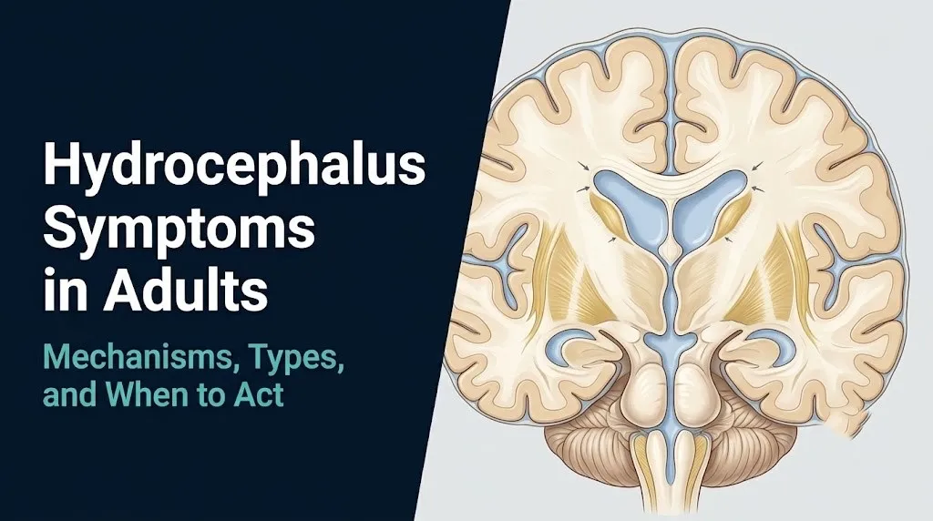
Hydrocephalus symptoms in adults are caused by the accumulation of excess cerebrospinal fluid (CSF) within the brain’s ventricular system, producing either elevated intracranial pressure or, in the case of normal pressure hydrocephalus, mechanical compression of periventricular brain structures at normal or near-normal pressure levels.
The symptom pattern varies by type. Adults with acute obstructive hydrocephalus develop severe headache, vomiting, and progressive loss of consciousness over hours. Adults with chronic communicating hydrocephalus develop more gradual symptoms: persistent headaches, cognitive slowing, and vision changes over weeks to months. Adults with normal pressure hydrocephalus develop the Hakim-Adams triad of gait disturbance, cognitive decline, and urinary incontinence over months to years, often without any significant headache.
| Hydrocephalus Type | Symptom Onset | Pressure Level | Primary Symptoms | Urgency |
|---|---|---|---|---|
| Acute obstructive | Hours to days | Markedly elevated | Severe headache, vomiting, decreased consciousness | Emergency |
| Chronic communicating | Weeks to months | Moderately elevated | Persistent headache, vision changes, cognitive slowing | Urgent referral |
| Normal pressure (NPH) | Months to years | Normal or near-normal | Gait disturbance, cognitive decline, urinary incontinence | Outpatient neurology |
| Post-traumatic | Days to months after injury | Variable | Depends on onset speed | Varies |
| Post-hemorrhagic | Days to weeks after bleed | Variable | Similar to acute or subacute | Urgent to emergency |
The most important practical principle from this overview: a person with any combination of progressive gait problems, cognitive decline, and urinary incontinence who is over 60 years old has NPH as a top differential diagnosis until a neurologist or neurosurgeon evaluates them. These three symptoms together are not “just aging.”
How CSF Circulation Works and Why Hydrocephalus Causes Symptoms
CSF production, circulation, and reabsorption form a continuous loop, and hydrocephalus develops when any part of this loop is disrupted, causing fluid to accumulate in the ventricular system and compress adjacent brain structures.
The choroid plexus, a specialized secretory epithelial structure lining the lateral ventricles and the third ventricle, produces approximately 500 milliliters of CSF per day. The total volume of CSF in the adult brain and spinal cord is approximately 150 milliliters, meaning the entire CSF volume turns over roughly three times every 24 hours. CSF produced in the lateral ventricles flows through the foramina of Monro into the third ventricle, then passes through the narrow cerebral aqueduct of Sylvius (also called the aqueduct of the midbrain) into the fourth ventricle, and exits into the subarachnoid space via the lateral foramina of Luschka and the medial foramen of Magendie.
From the subarachnoid space, CSF flows upward over the cerebral convexities and is reabsorbed into the venous blood at the arachnoid granulations, which project into the superior sagittal sinus along the top of the skull’s inner surface. The pressure gradient between the subarachnoid space and the venous sinus drives this passive reabsorption.
Think of the CSF system like the plumbing loop in a building with a pump (choroid plexus), pipes (ventricular pathways), and a drain (arachnoid granulations). Block the drain and water backs up into the pipes. Block a pipe and pressure builds upstream from the blockage. Either way, the backed-up fluid compresses whatever structures are nearby, which in the brain means critical white matter tracts, cranial nerve nuclei, and cortical regions.
Older adults with cerebral atrophy tolerate slowly developing ventricular enlargement longer than younger adults before symptoms become severe, because the atrophied brain provides additional space within the fixed skull. This is why NPH, which develops slowly, is far more likely to be misattributed to age-related cognitive decline in adults over 70 than in adults under 50.
Headache as a Symptom of Hydrocephalus in Adults
Headache in hydrocephalus is produced by elevated intracranial pressure stretching the pain-sensitive dura mater, compressing the intracranial venous sinuses, and distending the walls of the cerebral ventricles, producing a characteristic pressure-type or positional headache that differs in important ways from tension or migraine headache.
The dura mater, the outermost meningeal layer lining the inner skull, is richly innervated by branches of the trigeminal nerve and upper cervical nerve roots. When intracranial pressure rises, the dura is stretched over the expanding ventricles, and the trigeminal pain fibers transmit this as a deep, non-pulsatile, pressure-type headache that is typically bilateral and located at the forehead or behind the eyes. This is distinct from the throbbing, unilateral character of a typical migraine and from the bandlike pressure of a tension headache.
The classic hydrocephalus headache worsens with position changes that increase intracranial pressure, particularly lying flat (which reduces venous drainage from the head) and with activities that raise intra-abdominal and intracranial pressure together, such as coughing, sneezing, or straining. Headache that is worst in the morning after lying flat overnight, improves upon standing and moving around, and is accompanied by nausea or vomiting upon waking has a clinical profile consistent with elevated ICP.
According to the Mayo Clinic, headache is among the most frequently reported symptoms of hydrocephalus but is notably less prominent in normal pressure hydrocephalus than in obstructive or communicating hydrocephalus with frankly elevated pressure. Adults with NPH often report no significant headache at all, which is one of the clinical features that makes NPH harder to recognize than other hydrocephalus types.
Young adults with hydrocephalus from a specific obstructing lesion (such as a pineal region tumor blocking the cerebral aqueduct) develop headaches more acutely and more severely than older adults with gradual-onset communicating hydrocephalus, because the younger brain has less atrophic space to accommodate the rising pressure.
Key Takeaway: Hydrocephalus headache is classically worst in the morning and worsens with coughing, straining, or lying flat, but adults with normal pressure hydrocephalus often have no headache at all, making the other symptoms of the Hakim-Adams triad the more reliable recognition clues in this group.
Gait Disturbance and Balance Problems in Hydrocephalus
Gait disturbance in hydrocephalus, particularly in normal pressure hydrocephalus, is caused by transependymal CSF migration into the periventricular white matter compressing the corticospinal tract fibers that control lower limb movement, which are anatomically located closest to the ventricular walls and are therefore the first motor pathways affected by ventricular expansion.
The specific gait pattern of NPH is called magnetic gait or apraxic gait, clinically described as “marche à petits pas” (walking with small steps). A person with this gait pattern takes short, shuffling steps with their feet appearing to stick to the ground, has a wide-based stance, turns slowly and en bloc (the entire body turns together rather than the head and eyes leading), and has disproportionate difficulty initiating walking from a stopped position. The upper body often appears normal: arm swing may be preserved and the person does not necessarily appear weak or spastic when standing still.
The mechanistic reason for this specific pattern, which no competitor article explains, is anatomical. In the human corticospinal tract as it descends through the periventricular white matter toward the internal capsule, the fibers controlling the lower limbs are somatotopically arranged closest to the ventricular wall, while the fibers for the upper limbs and face are progressively further away. When ventricular expansion from NPH compresses the periventricular white matter, it preferentially disrupts the lower limb fibers first, producing leg weakness and gait disturbance before arm function or speech is affected.
The Hydrocephalus Association notes that gait disturbance is typically the earliest and most prominent component of NPH’s triad in most patients, often preceding cognitive changes by months or years. Patients frequently describe a progressive inability to walk on rough terrain, climb stairs, or rise from a low chair, even when their leg strength tests normally on a standard neurological examination.
Older adults with NPH are at substantially elevated fall risk from this gait pattern, because the magnetic gait produces instability on any uneven surface. A person over 60 with multiple unexplained falls, a shuffling gait, and no clear orthopedic explanation warrants neurological evaluation specifically considering NPH.
Cognitive Symptoms and Memory Problems in Adult Hydrocephalus
Cognitive symptoms in hydrocephalus result from compression of the frontal lobe white matter and frontal-subcortical circuits by ventricular enlargement, producing a characteristic pattern of slowed processing speed, impaired executive function, and retrieval-type memory difficulties that differs from the episodic memory failure pattern of Alzheimer’s disease.
The frontal lobes and their white matter connections to the basal ganglia, thalamus, and limbic system are particularly vulnerable to the periventricular compression of NPH. The resulting cognitive pattern is described as a subcortical dementia syndrome: slowed thinking speed (bradyphrenia), difficulty with planning and multitasking (executive dysfunction), mildly impaired attention, and trouble retrieving memories that are actually stored but difficult to access quickly. This is distinctly different from Alzheimer’s disease, in which the primary deficit is encoding new memories into the hippocampus, meaning new information is not stored at all rather than stored but hard to retrieve.
A practical clinical test of this distinction: a person with NPH-related cognitive changes can usually recall a word list if given enough time or a structured clue, because the memory trace was encoded. A person with early Alzheimer’s disease cannot recall the word list even with cues, because the hippocampal encoding mechanism is damaged.
According to research published in JAMA Neurology, the cognitive profile of NPH is most reliably assessed with neuropsychological testing that specifically evaluates processing speed, executive function, and cued recall versus free recall, because standard dementia screening tools like the Mini-Mental State Examination (MMSE) are not sufficiently sensitive to detect the frontal-subcortical pattern of NPH early in the course.
Women with hydrocephalus-related cognitive changes may have their symptoms attributed to perimenopausal or postmenopausal cognitive complaints before hydrocephalus is considered, because the symptom pattern of subjective cognitive slowing overlaps with the hormonal cognitive changes of menopause. Any woman with cognitive symptoms accompanied by gait changes or urinary urgency should have hydrocephalus included in her differential diagnosis regardless of menopausal status.
Urinary Incontinence as a Symptom of Hydrocephalus
Urinary incontinence in hydrocephalus is caused by compression of the frontal lobe micturition control centers and the periventricular white matter pathways connecting the medial frontal cortex to the pontine micturition center, disrupting the cortical inhibition of the urinary detrusor muscle and producing the specific pattern of urge incontinence rather than stress incontinence.
The micturition control pathway runs from the medial frontal cortex (specifically the anterior cingulate cortex and supplementary motor area) through the periventricular white matter to the pontine micturition center in the brainstem, and from there to the sacral spinal cord segments that control detrusor contraction. In health, the frontal cortex tonically inhibits the pontine micturition center, allowing voluntary bladder control by suppressing the urge to void until a socially appropriate time. When periventricular white matter is compressed by ventricular enlargement in NPH, this inhibitory pathway is disrupted and the detrusor becomes overactive.
The result is urge urinary incontinence, characterized by a sudden, compelling urge to urinate that cannot be delayed, followed by involuntary urination before reaching the bathroom. This is different from stress incontinence (leaking with coughing or physical exertion), which is produced by urethral sphincter weakness rather than detrusor overactivity. Distinguishing these two types matters clinically because urge incontinence in the context of gait disturbance and cognitive changes points strongly to a neurological cause, while isolated stress incontinence in a woman who has had multiple pregnancies points to pelvic floor weakness.
The NINDS notes that urinary incontinence is typically the last of the three NPH symptoms to appear in the classic presentation, after gait disturbance and cognitive decline have already been present for some time. Its appearance in the full triad context warrants prompt neurological evaluation.
Older men with NPH-related urge incontinence may initially be evaluated for benign prostatic hyperplasia (BPH) because both conditions produce urinary urgency and frequency. The co-occurrence of gait disturbance and cognitive slowing alongside urinary symptoms should prompt a clinician to consider NPH rather than attributing all symptoms to BPH alone.
Key Takeaway: Urinary incontinence in hydrocephalus is specifically urge incontinence caused by disrupted frontal inhibition of the detrusor muscle, not stress incontinence, and when it appears alongside gait disturbance and cognitive slowing in an adult over 60, this triad warrants neurological evaluation for NPH before assuming the cause is BPH or pelvic floor weakness.
Vision Problems Caused by Hydrocephalus in Adults
Vision problems in hydrocephalus are produced by elevated intracranial pressure transmitted through the optic nerve sheaths to the optic discs, causing papilledema, and by pressure on the sixth cranial nerve (abducens nerve) along its long intracranial course, producing diplopia (double vision) as a nonlocalizing sign of elevated ICP.
The optic nerve is surrounded by a sheath of dura mater that is continuous with the intracranial dura. When intracranial pressure rises, the elevated pressure is transmitted along this sheath to the optic nerve head at the back of the eye, producing swelling of the optic disc called papilledema. On fundoscopic examination, papilledema appears as blurring of the disc margins, disc elevation, and in severe cases, flame-shaped hemorrhages around the disc. Symptoms from papilledema include transient visual obscurations (brief episodes of graying or blacking out of vision, typically lasting seconds and triggered by postural changes), progressive blurring of peripheral vision, and in advanced cases, permanent visual field loss.
The sixth cranial nerve (abducens nerve) has the longest intracranial course of any cranial nerve, running along the base of the skull from the pons to the cavernous sinus and entering the orbit. This extended course makes it vulnerable to compression or traction from elevated ICP even when the site of elevated pressure is far from the sixth nerve itself, producing lateral gaze limitation and horizontal diplopia. Clinicians call this a “false localizing sign” because sixth nerve palsy from elevated ICP does not indicate pathology at the sixth nerve’s origin; it reflects globally elevated pressure.
In the specific context of obstructive hydrocephalus from a pineal region tumor or aqueductal stenosis, compression of the dorsal midbrain (the superior colliculi and pretectal area) can produce Parinaud’s syndrome: impaired upgaze, convergence-retraction nystagmus, and light-near dissociation of the pupils. This specific visual presentation points directly to pathology at the level of the midbrain or pineal region.
People over 50 being evaluated for vision changes should have fundoscopic examination to assess for papilledema before these changes are attributed to cataracts, glaucoma, or age-related macular degeneration, particularly when the vision changes are accompanied by headache.
Nausea, Vomiting, and Other Physical Symptoms of Hydrocephalus
Nausea and vomiting in hydrocephalus are produced by elevated intracranial pressure acting on the area postrema (the chemoreceptor trigger zone in the floor of the fourth ventricle) and by direct pressure on the brainstem structures controlling the vomiting reflex, producing the characteristic vomiting of elevated ICP that occurs explosively without preceding nausea in some acute cases.
The area postrema, located in the dorsal medulla at the floor of the fourth ventricle, is the primary brainstem structure mediating vomiting responses to elevated ICP. Because it sits at the floor of the fourth ventricle, it is particularly vulnerable to direct pressure from fourth ventricular dilation or from upward pressure transmitted through the ventricular system. Elevated ICP can also compress the dorsal vagal complex adjacent to the area postrema, further amplifying nausea and autonomic instability.
Projectile vomiting without nausea is the hallmark of acute elevated ICP in severe cases: the vomiting reflex is triggered so abruptly by brainstem stimulation that no prodromal nausea occurs. This pattern in an adult who is also developing headache and altered consciousness represents a neurological emergency.
Beyond nausea and vomiting, the broader physical symptom profile of hydrocephalus can include:
- Neck stiffness or pain from meningeal irritation (more prominent in post-meningitic or post-hemorrhagic hydrocephalus)
- Fatigue and generalized lethargy from globally elevated ICP reducing cerebral blood flow
- Tinnitus (ringing in the ears) from pressure effects on the cochlear aqueduct
- Photophobia (light sensitivity) from meningeal irritation
- Difficulty sleeping or changes in sleep-wake cycle from hypothalamic pressure in severe cases
According to the Cleveland Clinic, systemic physical symptoms including nausea and fatigue are more pronounced in acute or subacute hydrocephalus with significantly elevated pressure than in NPH, where physical symptoms other than gait disturbance are often minimal or absent.
Pregnant women with any pre-existing hydrocephalus condition or previously placed shunt who develop new headache with nausea in the third trimester should be evaluated by a neurosurgeon as well as their obstetrician, because expanding uterine size and increased blood volume during pregnancy can alter CSF dynamics and shunt function in previously compensated patients.
Normal Pressure Hydrocephalus Symptoms and the Classic Triad
Normal pressure hydrocephalus (NPH) is defined by the Hakim-Adams triad of gait disturbance, cognitive impairment, and urinary incontinence occurring in the setting of ventriculomegaly on brain imaging with normal or near-normal opening CSF pressure on lumbar puncture, and it is the most commonly missed treatable cause of dementia-like symptoms in adults over 60.
The three components of the triad do not always appear simultaneously. The natural history of NPH in most patients follows a predictable sequence: gait disturbance appears first and is most prominent, cognitive changes develop next over months to years, and urinary incontinence, typically urge incontinence, appears last. This ordering reflects the anatomical vulnerability of the periventricular motor fibers controlling the lower limbs before the more distant cortical and frontal-subcortical circuits are affected.
NPH was first described in 1965 by Hakim and Adams, who identified the characteristic triad in patients with enlarged ventricles but normal lumbar puncture opening pressures. The paradox of why NPH produces symptoms without globally elevated pressure is now explained by the transependymal CSF flow mechanism: even at normal CSF pressure, if the arachnoid granulation reabsorption capacity is reduced, CSF percolates through the ependymal lining into the periventricular white matter, where it acts as a mechanical compressive force on adjacent neural structures.
The Hydrocephalus Association reports that NPH affects an estimated 5% of people over age 80 and is misdiagnosed as Alzheimer’s disease, Parkinson’s disease, or vascular dementia in a substantial proportion of cases, partly because no single test is pathognomonic for NPH, and partly because the triad components can overlap with those conditions individually.
Adults with one or two components of the triad (most commonly gait disturbance plus cognitive changes without incontinence yet) should not wait for all three to appear before seeking neurological evaluation, because earlier treatment produces better outcomes in NPH than treatment initiated after all three components are fully established.
Key Takeaway: Normal pressure hydrocephalus produces its three symptoms in a specific order (gait first, then cognition, then urinary incontinence) because lower limb corticospinal fibers are anatomically closest to the ventricular wall and are compressed first, which means gait problems in an older adult should prompt NPH consideration even before the full triad appears.
Early Versus Late Stage Hydrocephalus Symptoms in Adults
Early stage hydrocephalus in adults produces symptoms that are subtle and often attributed to other conditions, while late stage hydrocephalus produces severe neurological deficits that may become irreversible without prompt treatment, making the ability to recognize early presentations clinically decisive.
In early communicating or NPH-type hydrocephalus, symptoms may include only mild morning headaches that resolve after standing, mild subjective cognitive slowing that a person notices in their work performance or daily tasks before formal testing detects it, mild unsteadiness on uneven surfaces that a person compensates for by avoiding those situations, and subtle urinary urgency without frank incontinence. These early symptoms are easily attributed to stress, aging, or other conditions, which is why the average delay from first symptoms to NPH diagnosis is commonly reported as multiple years in clinical literature.
As hydrocephalus progresses to a moderate or late stage:
- Headaches become persistent, present throughout the day, and less responsive to analgesics
- Cognitive deficits become measurable on formal neuropsychological testing, affecting occupational and social function
- Gait disturbance progresses to falls, inability to walk without assistance, and eventually wheelchair dependence in the most severe cases
- Urinary urgency progresses to frank incontinence requiring pads or protective garments
- In obstructive or communicating hydrocephalus with elevated pressure: papilledema, visual obscurations, and in the most severe cases, decreased level of consciousness
The critical inflection point is when consciousness begins to diminish. Any adult with known or suspected hydrocephalus who develops increasing drowsiness, confusion that is worsening over hours, or difficulty rousing warrants emergency evaluation the same day.
For individuals with previously placed ventriculoperitoneal (VP) shunts, a return of previously controlled symptoms, even subtly, can represent early shunt malfunction and warrants same-day evaluation by their neurosurgical team rather than waiting to see if symptoms worsen further.
Acute Obstructive Hydrocephalus Versus Chronic Hydrocephalus Symptoms
Acute obstructive hydrocephalus produces a rapid, severe symptom escalation over hours to days because a sudden blockage of CSF flow causes intracranial pressure to rise faster than the brain can compensate, while chronic hydrocephalus produces a gradual, progressive symptom pattern over weeks to years because the brain partially accommodates the slowly rising ventricular pressure.
Acute obstructive hydrocephalus most commonly results from sudden blockage of the cerebral aqueduct (by a tumor, hemorrhage, or acute inflammation), obstruction of the foramina of the fourth ventricle, or sudden hemorrhage into the ventricular system. The resulting rapid ICP elevation produces:
| Feature | Acute Obstructive Hydrocephalus | Chronic Communicating Hydrocephalus |
|---|---|---|
| Onset speed | Hours to 1 to 2 days | Weeks to months |
| Headache severity | Severe, constant, rapidly worsening | Mild to moderate, often positional |
| Vomiting | Projectile, may occur without nausea | Intermittent, usually with nausea |
| Level of consciousness | Rapidly declining | Preserved until late |
| Vision changes | Papilledema developing rapidly, diplopia | Papilledema developing slowly |
| Gait changes | May develop quickly with brainstem compression | Progressive over months |
| Urgency | Emergency | Urgent outpatient to urgent referral |
The mechanism of decompensation in acute obstructive hydrocephalus follows a predictable intracranial pressure cascade. When ICP approaches the mean arterial pressure, cerebral perfusion pressure (CPP) falls below the threshold needed to maintain neuronal oxygenation. The body’s compensatory response (the Cushing reflex) drives up systemic blood pressure while slowing heart rate, producing the Cushing’s triad of hypertension, bradycardia, and irregular respirations. This triad is a pre-terminal sign of imminent herniation.
Post-operative patients who have had recent neurosurgery, patients with known intracranial tumors, and patients with prior subarachnoid hemorrhage are at elevated risk for acute hydrocephalus. Any of these patients developing sudden severe headache or declining consciousness should be treated as a neurosurgical emergency.
Hydrocephalus Symptoms in Older Adults and After Age 60
Hydrocephalus in adults over 60 most commonly presents as normal pressure hydrocephalus with the Hakim-Adams triad, and its recognition is complicated by the fact that all three components of the triad (gait problems, cognitive decline, and urinary incontinence) individually occur commonly as part of multiple other age-related conditions, causing frequent diagnostic delay.
The specific challenge in older adults is that NPH symptoms overlap with Parkinson’s disease (which causes a shuffling gait), Alzheimer’s disease (which causes cognitive decline), and benign prostatic hyperplasia in men or pelvic floor dysfunction in women (which cause urinary urgency). When these three symptom domains are attributed to three separate age-related conditions rather than recognized as a single unifying neurological syndrome, the diagnosis of NPH is missed.
According to the NINDS, NPH is considered a potentially reversible cause of dementia-like symptoms, and surgical treatment with a ventriculoperitoneal shunt improves symptoms in a substantial proportion of carefully selected patients. The likelihood of improvement is generally better for gait disturbance than for cognitive symptoms, particularly when treatment is initiated before cognitive deficits become severe.
Older adults on anticoagulant medications (warfarin, direct oral anticoagulants) for atrial fibrillation or prior stroke are at elevated risk for intracranial hemorrhage causing post-hemorrhagic hydrocephalus. In this population, any new neurological symptom including unexplained worsening gait or new cognitive change warrants prompt evaluation with brain imaging rather than empiric attribution to their prior stroke.
The family member who first notices that an older adult “isn’t walking right” and “seems slower” in the same conversation often holds the first diagnostic clue to NPH. Any caregiver or family member observing this combination in an adult over 60 should raise it specifically with the patient’s primary care physician and request neurological referral.
Hydrocephalus After Brain Injury, Stroke, or Meningitis
Post-traumatic hydrocephalus, post-hemorrhagic hydrocephalus after stroke, and post-meningitic hydrocephalus each cause the condition through distinct mechanisms that produce CSF reabsorption failure at the arachnoid granulations, and each carries a distinct symptom timeline and presentation pattern.
Post-traumatic hydrocephalus develops in a subset of adults after moderate to severe traumatic brain injury (TBI). The mechanism involves subarachnoid blood products from the initial hemorrhage blocking the arachnoid granulations, combined with TBI-related inflammation promoting meningeal fibrosis that reduces long-term granulation function. Symptoms typically develop weeks to months after the initial TBI, and the presentation can be difficult to distinguish from post-concussive syndrome or TBI-related cognitive impairment. A progressive worsening of neurological function after an initial period of improvement following TBI is a clinical red flag for developing post-traumatic hydrocephalus.
Post-hemorrhagic hydrocephalus after subarachnoid hemorrhage (SAH) results from blood and fibrin degradation products clogging the arachnoid granulations in the immediate and subacute aftermath of the bleed. According to the Journal of Neurosurgery, communicating hydrocephalus develops in approximately 20 to 30% of patients following aneurysmal subarachnoid hemorrhage. Patients in the neurological intensive care unit after SAH are closely monitored with serial neurological examinations and CT head imaging for this complication.
Post-meningitic hydrocephalus occurs when bacterial meningitis induces arachnoid granulation fibrosis through TGF-beta-1 mediated scarring of the subarachnoid space, a process driven by the inflammatory response to the pathogen. Immunocompromised adults, including those with HIV/AIDS or receiving immunosuppressive therapy, are at elevated risk for both infectious meningitis and its complications including hydrocephalus.
Any adult with a history of TBI, subarachnoid hemorrhage, or bacterial meningitis who develops new progressive headache, cognitive changes, or gait disturbance should have CT or MRI brain imaging arranged by a primary care physician or neurologist within days, not weeks.
Key Takeaway: Post-traumatic, post-hemorrhagic, and post-meningitic hydrocephalus all develop through the same final pathway (arachnoid granulation dysfunction reducing CSF reabsorption) but on different timelines, and a progressive neurological decline weeks to months after a brain injury or bleed should immediately prompt consideration of developing hydrocephalus rather than attribution to the original injury alone.
Hydrocephalus Versus Dementia: How to Tell the Difference
Distinguishing hydrocephalus (particularly NPH) from Alzheimer’s disease, vascular dementia, and Parkinson’s disease requires recognizing that NPH produces a specific subcortical cognitive pattern, a specific gait pattern, and a specific urinary symptom that, taken together, create a clinical profile distinct from any of these conditions, even though individual symptom overlap exists.
The cognitive difference is the most clinically instructive. Alzheimer’s disease primarily disrupts episodic memory encoding in the hippocampus: the person cannot form new memories, forgets conversations that just occurred, and cannot recall even with cues. NPH disrupts retrieval speed and executive function: the person processes slowly, struggles to multitask, and has word-finding difficulties, but when given adequate time or a structured cue, can often recall information that was actually encoded. This is the difference between a failed memory trace (Alzheimer’s) and a slowed retrieval system (NPH).
The gait difference is equally distinctive. Parkinson’s disease produces a stooped, small-stepped gait with resting tremor, cogwheel rigidity, and masked facies. NPH produces a magnetic or apraxic gait with wide base, slow turns, and foot-floor adhesion, but typically without the resting tremor, rigidity, or facial changes of Parkinson’s. Asking a person to step over an imaginary line on the floor can reveal a striking improvement in step height in NPH (visual cueing temporarily unlocks the gait apraxia), while Parkinson’s gait does not improve with this visual cue in the same way.
Vascular dementia typically has a stepwise progression correlated with distinct vascular events (strokes), while NPH has a steadily progressive course without clear episodic worsening tied to vascular events.
A neurosurgeon or neurologist evaluating possible NPH will typically request brain MRI with FLAIR sequences, a formal lumbar puncture measuring opening CSF pressure, and a high-volume tap test (draining 30 to 50 mL of CSF and assessing for gait improvement in the subsequent 24 to 72 hours) before deciding on surgical intervention.
How Hydrocephalus Is Diagnosed in Adults
Hydrocephalus in adults is diagnosed through a combination of clinical history, physical and neurological examination, brain imaging (primarily MRI), lumbar puncture for opening pressure measurement, and in NPH cases, the high-volume tap test to assess whether CSF drainage produces temporary symptom improvement.
The diagnostic sequence typically progresses as follows when a neurologist or neurosurgeon evaluates a suspected hydrocephalus presentation:
- Clinical history: Duration and progression of symptoms, sequence of appearance (gait before cognition in NPH), prior brain injury, meningitis, hemorrhage, or neurosurgery, current medications, and any family history of neurological conditions.
- Neurological examination: Gait assessment (observing stance width, step length, turning pattern, and foot-floor clearance), cognitive screening (executive function, processing speed, memory with cued recall), fundoscopic examination for papilledema, cranial nerve testing for sixth nerve palsy.
- Brain MRI with FLAIR sequences: Evaluates ventricular size (using Evans’ ratio, the ratio of frontal horn width to maximum brain width; a ratio greater than 0.3 suggests ventriculomegaly), corpus callosum shape, periventricular FLAIR signal hyperintensities (indicating transependymal CSF flow), and any obstructing lesions.
- CT head: Faster and more available for acute presentations; excellent for identifying ventricular enlargement and obstructing hemorrhage; less detailed than MRI for soft tissue pathology.
- Lumbar puncture with opening pressure measurement: Confirms whether CSF pressure is elevated (above 25 cmH2O in communicating hydrocephalus) or normal/near-normal (8 to 18 cmH2O in NPH).
- High-volume tap test: Removal of 30 to 50 mL of CSF during lumbar puncture, followed by gait and cognitive assessment at 24 to 72 hours. Improvement in gait following the tap test has been shown in multiple studies to predict shunt responsiveness in NPH, though sensitivity is moderate and a negative tap test does not exclude NPH as the diagnosis.
- Intracranial pressure monitoring: In selected complex cases, a temporary ICP monitor is placed to measure pressure over 24 hours, looking for pathological B-waves that suggest intermittent ICP elevation even when mean pressure appears normal.
Adults being evaluated for possible hydrocephalus should bring a complete medication list (some medications affect CSF production), a timeline of symptom onset and progression written in advance, and any prior brain imaging on disc or digital format to the neurology or neurosurgery appointment.
Emergency Symptoms of Hydrocephalus: When to Call 911
Certain presentations of hydrocephalus in adults represent neurological emergencies. Do not wait to see if these resolve on their own.
Call 911 or go to the nearest emergency room immediately if you or someone you are with experiences:
- Sudden severe headache described as “the worst headache of my life,” particularly if accompanied by neck stiffness, photophobia, or vomiting (this can indicate subarachnoid hemorrhage causing acute hydrocephalus or a new intracranial hemorrhage requiring emergency neurosurgical evaluation)
- Rapidly declining level of consciousness, increasing confusion over hours, or difficulty rousing from sleep in a person with known hydrocephalus or a ventriculoperitoneal shunt (this indicates possible acute shunt malfunction with rising intracranial pressure approaching dangerous levels)
- Unequal pupils (one pupil larger than the other) combined with decreased consciousness and high blood pressure with slow heart rate (this is Cushing’s triad indicating impending transtentorial herniation, a neurosurgical emergency with minutes to act)
- A shunted patient who develops any return of symptoms that had previously been controlled by the shunt, including new headache, nausea, vomiting, or cognitive change, over hours to days (VP shunt malfunction can produce rapidly escalating ICP and requires neurosurgical evaluation the same day, not a scheduled appointment)
- Loss of consciousness, seizure, or sudden onset of focal neurological deficit (arm or leg weakness, speech difficulty, facial droop) in a person with known hydrocephalus
- Double vision with severe headache and altered mental status in any adult (sixth nerve palsy from acute ICP elevation combined with decreasing consciousness indicates a neurosurgical emergency)
These presentations can indicate acute hydrocephalus with impending transtentorial herniation, ventriculoperitoneal shunt malfunction with acute ICP crisis, or intracranial hemorrhage producing obstructive hydrocephalus, and all require emergency neurosurgical assessment, not an urgent care visit or a phone call scheduled for the following day.
Frequently Asked Questions About Hydrocephalus Symptoms in Adults
What are the first signs of hydrocephalus in adults?
The first signs of hydrocephalus in adults depend on the type: in normal pressure hydrocephalus, the earliest sign is usually a subtle gait disturbance with short, shuffling steps and difficulty on uneven surfaces, appearing months or years before cognitive or urinary symptoms develop.
In acute or subacute hydrocephalus with elevated pressure, the first signs are typically persistent morning headaches that worsen with lying flat, nausea, and mild cognitive slowing.
Any adult over 60 with unexplained progressive gait instability and subtle cognitive slowing should have NPH included in their neurological evaluation, even if incontinence is not yet present.
Can hydrocephalus symptoms come and go?
Hydrocephalus symptoms can fluctuate in early or compensated stages, where small changes in posture, hydration, or activity level temporarily alter intracranial pressure enough to worsen or improve symptoms.
A shunted patient may experience intermittent symptom fluctuation if the shunt valve pressure setting is suboptimal for their current condition.
Symptom fluctuation in a person with known or suspected hydrocephalus warrants reporting to a neurologist or neurosurgeon, not reassurance that the condition is improving, because fluctuation can precede a more sustained deterioration.
How do I know if my symptoms are NPH or dementia?
NPH produces a specific subcortical cognitive pattern of slow processing, executive dysfunction, and retrievable memories (recalled with cues), while Alzheimer’s disease produces episodic memory failure where new memories are not encoded and cannot be retrieved even with cues.
NPH’s gait disturbance (magnetic, wide-based, apraxic) is distinctly different from Parkinson’s disease (tremor-associated, with rigidity) and is not typically seen in early Alzheimer’s disease.
A neurologist or neurosurgeon can evaluate this distinction with brain MRI, lumbar puncture, and a high-volume tap test, which assesses whether CSF drainage temporarily improves gait, a response that supports NPH over a neurodegenerative diagnosis.
What does hydrocephalus headache feel like?
Hydrocephalus headache is typically a deep, non-pulsatile pressure sensation, often at the forehead or behind the eyes, that is worst in the morning after lying flat and worsens with coughing, straining, or bending forward.
It differs from a migraine in that it is not usually throbbing or unilateral, and it differs from a tension headache in that it worsens with lying flat rather than improving with rest.
Adults with normal pressure hydrocephalus often have no significant headache at all, which is a clinically useful distinguishing feature from the other types.
Can hydrocephalus go undetected for years in adults?
Normal pressure hydrocephalus frequently goes undetected for years in adults because its three symptoms, gait disturbance, cognitive decline, and urinary incontinence, are individually common in older adults and are often attributed to separate age-related conditions rather than recognized as a single neurological syndrome.
The Hydrocephalus Association reports that the average time from symptom onset to NPH diagnosis is often multiple years, during which treatable neurological compression continues.
Any adult over 60 with two or three components of the Hakim-Adams triad should be evaluated by a neurologist or neurosurgeon with brain MRI specifically assessing ventricular size before the diagnosis is dismissed as age-related changes.
When is hydrocephalus an emergency?
Hydrocephalus becomes an emergency when intracranial pressure rises fast enough to threaten cerebral perfusion and produce transtentorial herniation, specifically when a person with hydrocephalus develops decreasing consciousness, unequal pupils, or Cushing’s triad of high blood pressure, slow heart rate, and irregular breathing.
A patient with a ventriculoperitoneal shunt who develops any new or returning neurological symptoms, including new headache, vomiting, or confusion, over hours to days must be evaluated by a neurosurgeon the same day because shunt malfunction can produce rapidly rising ICP.
Call 911 immediately for any person with hydrocephalus who develops sudden severe headache, decreasing consciousness, seizure, or asymmetric pupils.
Recognizing What the Brain Is Telling You
The most important practical lesson from all of this is that no single hydrocephalus symptom in isolation confirms the diagnosis, but the combination and sequence of symptoms, particularly the gait-first, cognition-second, incontinence-third progression of NPH, is distinctive enough to warrant specific neurological investigation rather than symptomatic management of each complaint separately.
For anyone over 60 experiencing unexplained gait instability alongside cognitive slowing, ask a primary care physician specifically about normal pressure hydrocephalus and request a referral to a neurologist or neurosurgeon with brain MRI. For anyone with a known VP shunt, treat any return of previously controlled symptoms as a same-day neurosurgical question, not a wait-and-see situation.
The conditions on the other side of this diagnosis, Alzheimer’s disease, Parkinson’s disease, vascular dementia, are not surgically reversible. Hydrocephalus, in the right patient at the right stage, is. That distinction is worth the evaluation.