Silent Asthma Attack Symptoms: Signs, Stages & When to Call 911
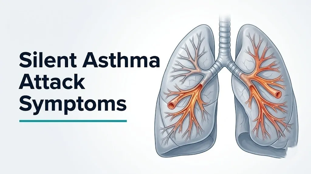
Silent asthma attack symptoms include chest tightness, progressive difficulty breathing, visible accessory muscle use in the neck and between the ribs, and in severe cases a complete absence of audible breath sounds, all of which occur without the audible wheeze that most people associate with asthma attacks. The silence is not reassuring. It is often a sign that the attack has become severe enough to reduce airflow to near zero.
According to the Global Initiative for Asthma (GINA), a silent chest during an acute asthma attack is classified as a life-threatening feature alongside SpO2 below 92 percent, a peak expiratory flow rate below 33 percent of personal best, altered mental status, and cyanosis. The Centers for Disease Control and Prevention reports that asthma causes approximately 3,500 deaths annually in the United States, many of which involve underestimated attack severity.
This article covers every recognized symptom of a silent asthma attack with the physiological mechanism behind each one, stages the presentation from early to near-fatal, addresses how the picture differs across age groups and health conditions, and gives you a clear framework for when to act and how fast.
What Is a Silent Asthma Attack
A silent asthma attack is an acute episode of severe airway obstruction in which the bronchospasm, mucosal edema, and mucus plugging are so extensive that insufficient airflow moves through the narrowed airways to produce the turbulent oscillations that create audible wheeze.
The term “silent” is clinically misleading in one important way. It does not mean the attack is mild. It means the attack may have progressed to the point where airflow is too restricted to generate the sound that normally signals an asthma attack to bystanders and clinicians. This is why the silent presentation is considered more dangerous, not less.
A person experiencing a silent asthma attack is typically working extremely hard to breathe. Their respiratory muscles are contracting forcefully. Their accessory muscles are recruited. Their chest may be visibly heaving. But the air moving through the severely narrowed bronchioles is so minimal that the stethoscope placed on their back finds near-silence rather than the expected wheeze.
The GINA 2023 guidelines identify “absent breath sounds” on auscultation during an acute asthma attack as one of the specific markers of a life-threatening attack requiring immediate emergency intervention, not outpatient management.
Individual variation note: People with long-standing poorly controlled asthma may have developed such a high subjective tolerance for dyspnea that they significantly underestimate the severity of even a near-fatal silent attack. Research published in the Journal of Allergy and Clinical Immunology has documented that poor perception of dyspnea is an independent risk factor for near-fatal and fatal asthma, making objective measurement (peak flow, pulse oximetry) more reliable than the patient’s own assessment of how bad the attack is.
Silent Asthma Attack Symptoms: The Complete Picture
The complete symptom picture of a silent asthma attack includes chest tightness or pressure, progressive breathlessness without audible wheeze, visible respiratory accessory muscle recruitment, reduced ability to speak in complete sentences, tachycardia, anxiety or agitation, and in advanced stages, cyanosis and altered mental status.
No single symptom is sufficient to recognize a silent attack. It is the combination of breathing difficulty without accompanying wheeze that should trigger concern. In a typical asthma attack, wheeze is the prominent sound that alerts both the patient and observers. In a silent attack, that auditory signal is absent, shifting the diagnostic burden entirely to visible physical signs and objective measurements.
| Symptom | Physiological Mechanism | Urgency Level |
|---|---|---|
| Chest tightness | Bronchial smooth muscle contraction, air trapping increasing lung compliance load | Early: use rescue inhaler and monitor |
| Breathlessness without wheeze | Severe airway obstruction reducing turbulent airflow below audible threshold | Moderate to severe: escalate care |
| Accessory muscle recruitment | Respiratory demand exceeds diaphragm capacity alone | Moderate to severe: seek immediate care |
| Inability to speak in full sentences | Respiratory effort required for ventilation leaves insufficient air for phonation | Severe: go to emergency room now |
| Tachycardia above 110 bpm | Sympathetic activation from hypoxemia and respiratory distress | Severe: emergency evaluation needed |
| Cyanosis (blue lips or fingertips) | SpO2 below approximately 85 percent; inadequate hemoglobin oxygen saturation | Life-threatening: Call 911 immediately |
| Altered mental status, confusion | Hypercapnia (rising CO2) from failing ventilatory effort; cerebral hypoxia | Life-threatening: Call 911 immediately |
Individual variation note: People with dark skin tones may not show visually apparent cyanosis on lip or fingertip examination even when SpO2 is critically low, because melanin pigmentation masks the color change. For these individuals, pulse oximetry is an even more important objective measurement, and the absence of visible cyanosis should never be interpreted as reassurance of adequate oxygen saturation.
Why Asthma Attacks Can Be Silent: The Mechanism Explained
A silent asthma attack occurs when the combination of bronchial smooth muscle contraction, bronchial mucosal edema, and intraluminal mucus plug formation reduces airflow through the small airways to a level insufficient to generate turbulent oscillations, eliminating the physical basis for audible wheeze.
Think of a garden hose with nearly pinched-off water flow. At partial constriction, the water rushing through the narrowed point creates noise. Pinch it completely shut, and silence returns, not because the pressure problem has resolved but because nothing is moving. The bronchiole in a severe asthma attack is that nearly pinched-off hose.
The inflammatory cascade driving this begins with IgE-mediated mast cell degranulation in the bronchial submucosa. Cross-linking of surface IgE by inhaled allergen (or other triggers in non-atopic asthma) releases pre-formed histamine and prostaglandin D2 (PGD2) from mast cell granules. These mediators produce immediate bronchospasm by acting on H1 receptors and prostanoid DP2 receptors on bronchial smooth muscle.
Simultaneously, de novo synthesis of leukotriene C4 and D4 (LTC4, LTD4) from arachidonic acid via 5-lipoxygenase produces sustained, prolonged bronchospasm more potent than histamine. The late-phase response, driven by IL-5 and IL-13 from Th2 lymphocytes and innate lymphoid cells type 2 (ILC2), recruits eosinophils that release major basic protein and eosinophil cationic protein, producing epithelial damage and further mucosal edema. IL-13 also drives goblet cell hyperplasia and mucus hypersecretion, producing the viscid, eosinophil-rich mucus plugs that can completely occlude small airways.
According to the American Journal of Respiratory and Critical Care Medicine, mucus plug formation in fatal asthma involves a distinctive composition of DNA, eosinophil granule proteins, and Charcot-Leyden crystals, confirming that mucus plugging rather than bronchospasm alone accounts for the most severe airflow obstruction in near-fatal presentations.
Individual variation note: Patients with aspirin-exacerbated respiratory disease (AERD), formerly called Samter’s triad (asthma, nasal polyps, and NSAID sensitivity), have constitutively overactive leukotriene synthesis pathways. Their attacks following NSAID ingestion can be particularly severe and rapid, producing silent chest within minutes through exaggerated LTC4 and LTD4 release.
Chest Tightness Without Wheezing in a Silent Asthma Attack
Chest tightness without wheezing is often the earliest and most consistently reported symptom of a silent asthma attack, produced by the combined sensation of bronchial smooth muscle contraction squeezing the airway walls and the increased resistive load against which the inspiratory and expiratory muscles must work.
Chest tightness in asthma is not pain in the cardiac sense. It is more accurately described as a constricting pressure or a sensation that the chest won’t fully expand. This feeling arises because the bronchial smooth muscle is maintained in sustained contraction, and the pleural pressure changes required to move air through the narrowed airways are substantially greater than during normal breathing, making each breath feel physically effortful.
What makes this symptom particularly important in the silent context is that many patients attribute chest tightness to anxiety, stress, or musculoskeletal causes rather than recognizing it as an asthma symptom. When wheeze is absent to reinforce the connection to asthma, chest tightness can be dismissed for hours before the attack is recognized.
The tightness typically worsens progressively during a silent attack. Early tightness may be mild and noticed only during exertion. As bronchospasm intensifies and mucus accumulates, the tightness becomes constant, present at rest, and increasingly severe with each attempted breath.
The National Asthma Education and Prevention Program Expert Panel Report 3 identifies chest tightness as one of the four cardinal symptoms of asthma alongside wheeze, cough, and shortness of breath, and notes specifically that some patients experience predominantly chest tightness rather than wheeze as their primary symptom, even during acute attacks.
Individual variation note: Older adults with coronary artery disease may find it genuinely difficult to distinguish asthma-related chest tightness from cardiac chest pressure. Both can occur without wheeze. Any chest tightness in an older adult with both cardiac and pulmonary disease that does not respond to two doses of a rescue bronchodilator inhaler warrants emergency evaluation to exclude myocardial ischemia as a concurrent or contributing cause.
Key Takeaway: Chest tightness that is worsening with each breath, present at rest, and not accompanied by wheeze is not necessarily a milder form of asthma attack; it may indicate that the attack is severe enough to have eliminated audible airflow.
Shortness of Breath and Silent Asthma Attacks
Shortness of breath in a silent asthma attack is produced by the combination of increased airway resistance requiring greater respiratory muscle effort, air trapping from incomplete expiration through obstructed airways producing lung hyperinflation, and worsening hypoxemia from ventilation-perfusion (V/Q) mismatch in the obstructed lung units.
The physiology of dyspnea in a silent attack differs from the dyspnea of a typical asthma attack in one important dimension: in a typical attack, the patient and any observer gets auditory confirmation that the breathing difficulty is asthma-related. In a silent attack, the breathlessness occurs without that auditory cue. The person may appear to be breathing, with visible chest movement, but the chest movement is producing far less airflow than the respiratory effort would suggest.
Air trapping is particularly important in severe silent attacks. Obstructed airways that allow air in during inspiration (when airways are slightly dilated by increasing lung volume) but resist expiration (when airways narrow during exhalation) cause progressive gas accumulation in the alveoli behind the obstructed segment. This dynamic hyperinflation flattens the diaphragm, placing it at a mechanical disadvantage for generating the force needed to move air. The patient must recruit increasingly forceful accessory muscle activity just to maintain an inadequate tidal volume.
The American Thoracic Society identifies the neural mechanisms of dyspnea as involving signals from the vagal airway receptors in the bronchial walls, the chest wall mechanoreceptors, and the central respiratory motor cortex, all of which send divergent signals when ventilatory effort is high but airflow is inadequate, producing the particularly distressing quality of breathlessness in severe asthma.
Individual variation note: Obese patients with asthma face compounded dyspnea during a silent attack because their elevated abdominal mass reduces the functional residual capacity of the lungs at baseline, meaning they begin any asthmatic episode with less respiratory reserve and develop air trapping more rapidly than normal-weight individuals with equivalent airway obstruction.
Accessory Muscle Use as a Sign of Silent Asthma Attack
Visible recruitment of accessory respiratory muscles, including the sternocleidomastoid muscle in the neck, the scalene muscles, and the intercostal muscles between the ribs, is one of the most reliable external signs of a severe silent asthma attack and indicates that the respiratory demand has exceeded the capacity of the diaphragm alone.
The diaphragm is the primary muscle of inspiration, generating approximately 70 percent of the inspiratory force during normal breathing. When airway resistance rises sharply during an asthma attack, the force required to move air through the narrowed airways increases proportionally. The diaphragm reaches its maximal force capacity, and the central nervous system recruits additional muscle groups to supplement inspiratory effort.
The sternocleidomastoid muscles, running from the mastoid process behind the ear to the sternum and clavicle, are the most visible accessory muscles. Their contraction during inspiration elevates the sternum and upper chest, and their prominent use produces a visually obvious neck muscle tensing that can be observed from several feet away. The scalene muscles elevate the first and second ribs. Both muscle groups are normally at rest during quiet breathing; their activation at rest is a clinical sign of severe respiratory distress.
Equally important is the visible intercostal retractions, the inward sucking of the skin between the ribs during inspiration, produced when the pressure inside the chest drops sharply below atmospheric pressure as the patient tries to inhale against severely obstructed airways. Suprasternal retractions (visible hollowing above the sternum) and supraclavicular retractions (hollowing above the collarbone) follow a similar mechanism and indicate even greater respiratory effort.
GINA 2023 specifically lists accessory muscle use and suprasternal retraction as features of a severe acute asthma attack, placing it in the same severity tier as inability to complete sentences in one breath and a peak flow of 33 to 50 percent of personal best.
Individual variation note: In infants and very young children, the chest wall is more compliant (more flexible) than in adults because the rib cage is still developing. This means retractions in young children may be more visually dramatic even with a moderate degree of airway obstruction, and conversely, a paradoxical inward movement of the lower chest during inspiration (see-saw breathing) can appear in severe infant bronchiolitis or asthma when the diaphragm contracts and the compliant chest wall collapses inward simultaneously.
Behavioral and Subtle Signs of Silent Asthma Attack
Behavioral changes, including unusual quietness, reduced physical activity, a preference for sitting upright or leaning forward, and visible anxiety without an obvious explanation, are the earliest observable signs of a silent asthma attack in patients who cannot or do not report their symptoms verbally.
The instinctive posture of a person in a severe silent asthma attack is the tripod position: sitting upright and leaning slightly forward with the hands resting on the knees. This position optimizes the mechanical advantage of the accessory muscles by fixing the shoulder girdle, allowing the pectoralis minor and serratus anterior muscles to assist with rib cage elevation during labored inspiration. A patient who spontaneously adopts this position during a breathing episode is communicating physiological distress through body language.
Anxiety and agitation in a silent asthma attack are not purely psychological. The hypoxemia-triggered chemoreceptor response in the carotid body (located at the bifurcation of the common carotid artery) generates powerful sympathetic activation as arterial oxygen tension falls. This produces the sense of air hunger, panic, and urgency that accompanies severe hypoxemia. When a person appears inexplicably anxious or panicked without a clear psychological trigger, and is also breathing hard, the anxiety may be physiologically driven by hypoxemia rather than situational.
Quick Tip:
- In children: watch for reluctance to walk or play that developed suddenly, unusual stillness, or preferring to sit upright in a child who normally runs around; these behavioral shifts can precede visible respiratory signs
- In adults: a person who has stopped talking mid-conversation to breathe, cannot finish sentences, or appears distracted by the effort of breathing is displaying a red flag behavioral sign
- In elderly patients: sudden onset of agitation or confusion in a person with known asthma, without an obvious neurological cause, should prompt immediate assessment of respiratory rate and oxygen saturation before attributing the change to dementia or psychiatric causes
Individual variation note: Adolescents with asthma may actively minimize their symptoms to peers or in social settings, appearing to control their breathing deliberately while suppressing visible signs of distress. Clinical observation in pediatric asthma studies published in Pediatrics has noted that adolescents are a high-risk group for underreporting attack severity and for delayed emergency presentations, in part due to social self-consciousness about appearing unwell.
Key Takeaway: A person sitting bolt upright, leaning forward, with tense neck muscles and the inability to complete a sentence is in a severe asthma attack regardless of whether you can hear any wheeze.
Peak Flow and Oxygen Saturation in a Silent Asthma Attack
Peak expiratory flow rate (PEFR) and oxygen saturation (SpO2) measured by pulse oximetry are the two objective measurements that provide reliable severity assessment when auscultatory wheeze is absent, and they are essential for determining whether a silent asthma attack is early-moderate or immediately life-threatening.
PEFR measures the fastest rate at which air can be expelled from the lungs during a forced maximal exhalation. In a severe silent asthma attack, the severely obstructed small airways substantially reduce this flow rate, even when the patient appears to be breathing with some effort. The critical thresholds from the NAEPP EPR-3 are:
- PEFR above 70 percent of personal best: mild obstruction; follow the action plan’s green zone guidance
- PEFR 40 to 69 percent of personal best: moderate obstruction; yellow zone; use rescue inhaler and reassess in 20 minutes
- PEFR below 40 percent of personal best: severe obstruction; red zone; initiate emergency plan
- PEFR below 33 percent of personal best: life-threatening; Call 911 immediately
SpO2 measured by pulse oximetry provides a non-invasive continuous estimate of arterial oxygen saturation. During a silent asthma attack, V/Q mismatch (obstructed airways that cannot be ventilated receiving continued blood flow) produces arterial hypoxemia.
The GINA 2023 guidelines define the following SpO2 thresholds during an acute asthma attack in adults:
- SpO2 above 95 percent: acceptable
- SpO2 90 to 95 percent: moderate concern; requires close monitoring and treatment escalation
- SpO2 below 90 percent: life-threatening; requires immediate oxygen supplementation and emergency intervention
Individual variation note: Children under 5 may not be able to perform a reliable peak flow maneuver because the technique requires a sustained maximal forced exhalation effort that young children cannot reproduce consistently. For this population, clinical assessment of respiratory rate, work of breathing (retractions, accessory muscle use), and ability to speak or cry becomes the primary severity measure rather than peak flow. A pediatrician or pediatric emergency medicine physician should assess any child under 5 with suspected severe asthma rather than relying on peak flow interpretation.
Silent Asthma Attack Stages: From Mild to Near-Fatal
A silent asthma attack progresses through clinically recognizable stages from early mild airway obstruction to a near-fatal presentation, and identifying the stage the patient is in determines whether the appropriate response is a rescue inhaler at home or an immediate 911 call.
| Stage | Peak Flow | SpO2 | Observable Signs | Action |
|---|---|---|---|---|
| Early (mild) | 70 to 100% of personal best | Above 95% | Chest tightness, minimal visible effort, no audible wheeze | Use rescue inhaler; monitor; contact doctor if no improvement |
| Moderate | 40 to 69% of personal best | 90 to 95% | Breathlessness at rest, early accessory muscle use, unable to complete some sentences | Use rescue inhaler; if no improvement in 20 min, go to ER |
| Severe | 33 to 40% of personal best | Below 92% | Obvious accessory muscle use, retractions, unable to speak in full sentences, tachycardia | Go to ER immediately; call for help; do not drive alone |
| Near-fatal | Below 33% of personal best | Below 90% | Silent chest, cyanosis, altered mental status, paradoxical breathing, bradycardia or exhaustion | Call 911 immediately |
Paradoxical breathing, where the abdomen moves inward during inspiration rather than outward, indicates that the diaphragm is fatiguing and may be about to fail. This is a pre-respiratory-arrest sign. A patient showing paradoxical breathing requires immediate emergency services regardless of any other assessment.
The NAEPP EPR-3 explicitly states that the absence of wheeze combined with severe dyspnea and accessory muscle use should be interpreted as indicating more severe obstruction rather than improving obstruction, and that clinical response should be urgently escalated in this scenario.
Individual variation note: Patients with a history of prior near-fatal asthma attack (previously requiring intubation or intensive care unit admission for asthma) have a substantially elevated risk of a subsequent fatal attack compared to patients without this history. This population should be managed more aggressively at an earlier stage of any acute attack, and their board-certified allergist-immunologist or pulmonologist should be aware of this history for treatment planning.
Silent Asthma Attack Symptoms in Children
Silent asthma attack symptoms in children include behavioral changes such as unusual quietness and reluctance to move, visible intercostal and suprasternal retractions, a respiratory rate above normal for age, nasal flaring, inability to speak or cry normally, and an oxygen saturation below 92 percent, all of which may be present without any audible wheeze.
Children, particularly those under 6, have airways that are proportionally smaller relative to their chest cavity than adult airways. This means that the same degree of mucosal edema and bronchospasm that produces moderate obstruction in an adult may produce severe obstruction in a child, because the absolute airway diameter reduction is more consequential in a smaller airway. A 1-millimeter reduction in airway radius reduces airflow by a factor of 16 (by Poiseuille’s law for laminar flow through a tube), which is why children can deteriorate from moderate to severe asthma faster than adults with an equivalent stimulus.
Observable signs in children that signal a severe silent attack:
- A child who has stopped playing and is sitting or lying very still, watching parents anxiously
- Skin pulling inward between the ribs (intercostal retractions) or above the collarbone (supraclavicular retractions) visible with each breath
- Nasal flaring: the nostrils widening with each inhalation as the child tries to reduce upper airway resistance
- A high-pitched “grunt” sound at the end of expiration (expiratory grunting): this is the child partially closing the glottis to maintain airway pressure and prevent complete alveolar collapse; it is a sign of severe respiratory distress
- Pallor or mottling of the skin
The American Academy of Pediatrics recommends that any child with suspected severe asthma, including silent presentation, receive emergency evaluation if two doses of a rescue bronchodilator administered 20 minutes apart produce no improvement in clinical appearance or respiratory effort.
Individual variation note: Infants under 12 months with their first episode of wheezing are more likely to have bronchiolitis than asthma, and the absence of wheeze in this age group can reflect the same severe-obstruction mechanism or can represent a different pathology entirely. A pediatric emergency medicine physician should evaluate any infant with significant respiratory distress without an established asthma diagnosis.
Silent Asthma in the Elderly: Why It Gets Missed
Silent asthma attacks in older adults are frequently misidentified as decompensated heart failure, COPD exacerbation, or deconditioning because wheeze is absent and the physiological signs overlap with these other common conditions in this age group.
Older adults with asthma face two compounding diagnostic challenges. First, many have coexisting cardiovascular or pulmonary disease that produces its own dyspnea, making it difficult to attribute new or worsening breathlessness specifically to asthma rather than to the comorbidity. Second, age-related changes in chest wall compliance and respiratory muscle strength mean that the work of breathing against obstructed airways may produce less visually dramatic accessory muscle use than in younger patients, reducing the external signs that normally signal distress.
The specific features that help distinguish a silent asthma attack from cardiac dyspnea in older adults:
- Asthma dyspnea typically responds to a short-acting beta-2 agonist inhaler (albuterol/salbutamol) within 15 to 20 minutes; cardiac dyspnea does not
- Asthma attacks often have a recognizable trigger (allergen, cold air, exercise, NSAID ingestion)
- Peak flow measurement will show reduced PEFR in an asthma exacerbation; in isolated cardiac dyspnea without concurrent airway disease, peak flow may be less severely reduced
- Cardiac dyspnea is typically associated with orthopnea (difficulty breathing when lying flat) and bilateral leg edema that are not primary features of asthma
Research published in the Journal of Allergy and Clinical Immunology has documented that asthma is significantly underdiagnosed in adults over 65, with many patients receiving only heart failure or COPD diagnoses despite having concurrent asthma that was never formally identified.
Individual variation note: Older adults taking non-cardioselective beta-blockers (such as propranolol or carvedilol) for cardiac indications face a specific risk: these drugs block the beta-2 adrenergic receptors on bronchial smooth muscle, reducing the bronchodilatory response to endogenous epinephrine during an asthma attack and potentially blunting the response to rescue inhaler use. A cardiologist and pulmonologist or allergist should collaborate on medication management for patients who have both asthma and heart disease requiring beta-blocker therapy.
Key Takeaway: In older adults and in children, objective measurements (peak flow and pulse oximetry) are more reliable than the presence or absence of wheeze for assessing silent asthma attack severity, because auscultatory findings are less trustworthy in both populations.
Exercise-Induced and Nocturnal Silent Asthma Symptoms
Exercise-induced bronchoconstriction (EIB) produces silent asthma attack symptoms predominantly in the minutes following exercise cessation rather than during peak exertion, with chest tightness, breathlessness, and performance decline occurring without audible wheeze in athletes and non-athletes alike.
EIB is triggered by the inhalation of large volumes of relatively dry, cold air during exercise, which dehydrates and cools the airway epithelium. The airway epithelium responds by producing osmotic stress in the periciliary fluid layer, triggering mast cell degranulation and release of histamine, LTC4, and LTD4. These mediators produce bronchospasm that typically peaks 5 to 10 minutes after exercise ceases, when the airways rewarm and the osmotic stress mediators reach maximal effect. Athletes often note that they feel fine during the run but cannot breathe normally in the cool-down period: this post-exercise timing is the key diagnostic clue.
Nocturnal asthma produces silent attacks during the early morning hours, typically between 2 and 4 AM, driven by the circadian nadir in endogenous cortisol production (which normally suppresses airway inflammation), the supine position increasing airway secretion pooling, reduced sympathetic tone during sleep increasing bronchoconstriction, and circadian changes in airway inflammatory cell activity with peak eosinophil activity in the early morning hours.
The American Thoracic Society official statement on exercise-induced bronchoconstriction notes that EIB affects an estimated 5 to 20 percent of the general population and up to 90 percent of people with pre-existing asthma, and that many cases are undiagnosed because the absence of wheeze leads athletes to attribute their symptoms to poor fitness rather than airway disease.
Individual variation note: Competitive athletes who experience breathlessness after exercise without wheeze are at risk of having undiagnosed EIB attributed to anxiety, deconditioning, or vocal cord dysfunction. An exercise challenge test performed in a pulmonology or sports medicine setting, with pre and post-exercise spirometry, is the appropriate diagnostic tool for athletes with suspected EIB who do not have typical wheeze.
Silent Asthma Attack During Pregnancy
A silent asthma attack during pregnancy requires a lower threshold for emergency intervention than in non-pregnant adults because the fetus depends entirely on maternal arterial oxygen saturation for its oxygen supply, and maternal SpO2 values that would be managed with outpatient bronchodilators in a non-pregnant patient can produce fetal hypoxia at the same values.
During normal pregnancy, maternal arterial oxygen saturation is slightly higher than in non-pregnant adults (typically 97 to 99 percent) because elevated progesterone levels increase respiratory drive and lower arterial CO2. When a pregnant woman with asthma develops a silent attack and her SpO2 begins to fall, the fetus experiences the oxygen debt through the uteroplacental circulation: the umbilical vein delivers oxygenated blood from the placenta to the fetus, and fetal oxygen delivery is directly dependent on maternal arterial oxygen tension.
The clinical standard for managing asthma in pregnancy, according to guidance from the American College of Obstetricians and Gynecologists, is that maternal SpO2 should be maintained above 95 percent (not the standard adult threshold of 90 percent) to ensure adequate fetal oxygenation. An asthmatic pregnant woman with an SpO2 of 92 percent would meet the “moderate” adult threshold but represents a potential fetal emergency.
Pregnant women with asthma should:
- Maintain a written asthma action plan specifically reviewed by their obstetrician and pulmonologist during pregnancy
- Keep rescue bronchodilator medication consistently accessible throughout pregnancy
- Report any acute asthma episode during pregnancy to their obstetric provider regardless of perceived severity
- Go to the emergency room immediately if SpO2 falls below 95 percent, if peak flow falls below 50 percent of personal best, or if rescue inhaler use does not produce rapid improvement
Individual variation note: Asthma severity commonly changes during pregnancy. Research from the Journal of Allergy and Clinical Immunology shows that approximately one-third of pregnant women with asthma experience worsening of their condition, one-third have improvement, and one-third remain unchanged. Women who were poorly controlled before conception are at highest risk for severe exacerbations during pregnancy.
Silent Asthma Attack vs Typical Asthma Attack
A silent asthma attack differs from a typical asthma attack primarily in the absence of audible wheeze, which paradoxically indicates greater rather than lesser airway obstruction, while sharing the same underlying inflammatory and bronchospastic mechanism as any acute asthma episode.
| Feature | Typical Asthma Attack | Silent Asthma Attack |
|---|---|---|
| Audible wheeze | Present; high-pitched expiratory or biphasic sound | Absent; breath sounds reduced or inaudible |
| Airway obstruction degree | Variable; moderate obstruction most commonly produces wheeze | Severe or near-complete obstruction; insufficient airflow for turbulence |
| Perceived urgency by patient and bystanders | Higher; wheeze acts as an audible alarm | Lower, paradoxically; absence of wheeze may be misread as mild |
| Actual clinical severity | Ranges from mild to moderate | Tends to represent moderate-to-severe or life-threatening obstruction |
| Peak flow at presentation | Variable | Typically below 50 percent of personal best |
| Diagnostic challenge | Lower; wheeze confirms asthma diagnosis | Higher; differential includes cardiac dyspnea, vocal cord dysfunction, pulmonary embolism |
| Response to rescue inhaler | Wheeze typically diminishes with improvement | Improvement may be assessed by return of wheeze (as airflow increases) and peak flow increase |
One counterintuitive clinical point deserves emphasis: in a person whose silent attack begins to respond to bronchodilator treatment, the return of audible wheeze during recovery can actually indicate improvement, because it means airflow has increased to a level sufficient to generate turbulence. The wheeze is now coming back because air is moving again.
The NAEPP EPR-3 addresses this directly, noting that a silent chest that remains silent after bronchodilator administration, rather than returning wheezing as expected with partial improvement, represents a continuing medical emergency requiring immediate escalation of care.
Individual variation note: Patients with vocal cord dysfunction (VCD), also called inducible laryngeal obstruction, can present with breathlessness without wheeze and without asthma. VCD produces inspiratory stridor and throat tightness rather than expiratory wheeze, and it is often misidentified as a silent asthma attack. A board-certified allergist or pulmonologist can distinguish VCD from asthma through laryngoscopy during symptoms and spirometry showing flattening of the inspiratory loop rather than the expiratory loop.
How Silent Asthma Attacks Are Diagnosed and Monitored
Silent asthma attacks are assessed using peak flow measurement, pulse oximetry, spirometry (when stable), clinical examination of respiratory effort and accessory muscle use, and in severe cases, arterial blood gas analysis, which provides direct measurement of arterial oxygen and carbon dioxide levels and identifies impending respiratory failure.
The diagnostic and monitoring process from first contact to confirmed assessment:
- Immediate clinical assessment: Assess respiratory rate (normal adult 12 to 20 breaths per minute; above 25 indicates significant distress), ability to complete sentences, visible accessory muscle use, skin color, and level of consciousness
- Pulse oximetry: Apply a pulse oximeter to the finger to obtain SpO2 immediately; this is the fastest objective measure available outside of a clinical setting
- Peak flow measurement: Ask the patient to perform a maximal forced exhalation into a peak flow meter; compare the result to the patient’s personal best or predicted value; if PEFR is below 40 percent, treat as severe
- Auscultation: Listen to all lung fields; the finding of reduced or absent breath sounds in a distressed patient is a sign of severe obstruction, not improvement
- Response to rescue bronchodilator: Administer albuterol (salbutamol) via a spacer and re-assess PEFR and SpO2 at 15 to 20 minutes; failure to improve peak flow by at least 10 percent, or persistence of severe symptoms, requires emergency escalation
- Arterial blood gas (ABG) in hospital setting: If SpO2 is below 92 percent or the patient is tiring, an ABG will directly measure PaO2, PaCO2, and pH; a rising PaCO2 (hypercapnia) in an asthmatic patient who is becoming “normalized” on blood gas represents a pre-arrest sign, because a severe asthmatic normally hyperventilates and maintains low PaCO2
According to GINA 2023, fractional exhaled nitric oxide (FeNO), which measures eosinophilic airway inflammation, is not a measurement used during acute attacks but is valuable for confirming Type 2 eosinophilic inflammation when the patient is stable and for guiding long-term anti-inflammatory therapy selection.
Individual variation note: Patients with chronic obstructive pulmonary disease (COPD) and asthma overlap (ACO) have reduced baseline peak flow values that make personal best comparisons difficult, because their “personal best” is itself lower than a healthy individual’s predicted value. A pulmonologist familiar with the patient’s baseline lung function is the most appropriate provider for interpreting peak flow in the ACO context.
What to Do During a Silent Asthma Attack
During a silent asthma attack, the correct response is determined by the objective severity: follow the patient’s written asthma action plan, administer rescue bronchodilator by inhaler with spacer or nebulizer, measure peak flow and SpO2, and escalate to emergency services without delay if severe thresholds are met.
To respond to a silent asthma attack step by step:
- Stop all physical activity immediately and help the person to a seated, upright position; the tripod position (leaning slightly forward, hands on knees) may ease breathing
- Administer rescue bronchodilator: give 2 to 4 puffs of a short-acting beta-2 agonist (albuterol/salbutamol) via a pressurized metered-dose inhaler with spacer, or via nebulizer if available; repeat every 20 minutes up to three times only if there is clear improvement
- Measure peak flow: if PEFR is below 40 percent of personal best, do not wait for a second bronchodilator dose; go to the emergency room or call 911 now
- Measure SpO2: if below 92 percent (or below 95 percent in pregnancy), call 911 immediately
- Do not give oral corticosteroids as a substitute for emergency care: they take hours to produce anti-inflammatory effect and will not relieve acute severe obstruction in the time window of an attack
- Do not leave the person alone: if they deteriorate to the point of inability to speak, altered mental status, or loss of consciousness, call 911 immediately and remain with them until help arrives
- Position for transport: if self-transport is necessary because 911 cannot reach in time, the person should sit upright in the vehicle, not reclined
Emergency Symptoms: When to Call 911 or Go to the ER
Certain symptoms associated with a silent asthma attack require immediate emergency evaluation. Do not wait to see if these resolve on their own.
Call 911 or go to the nearest emergency room immediately if you experience:
- Silent chest with absent or minimal breath sounds despite visible severe respiratory effort: this indicates near-complete airflow cessation and is a life-threatening emergency
- Oxygen saturation (SpO2) below 90 percent measured by pulse oximetry in a non-pregnant adult: indicates life-threatening hypoxemia requiring supplemental oxygen and emergency intervention
- Peak expiratory flow rate below 33 percent of personal best that does not improve after two rescue inhaler doses: indicates severe airway obstruction unresponsive to initial treatment
- Inability to speak more than one or two words between breaths: indicates that the entire respiratory effort is required for ventilation with no reserve for phonation
- Cyanosis: blue or gray coloration of the lips, fingertips, or around the mouth, indicating critically low arterial oxygen saturation
- Altered mental status, confusion, agitation, or drowsiness in a person known to have asthma who is also breathing with difficulty: indicates hypercapnia or severe hypoxemia with impending respiratory failure
- Paradoxical breathing (the abdomen moves inward during inspiration rather than outward): indicates diaphragmatic fatigue approaching respiratory muscle failure
- SpO2 below 95 percent during pregnancy: the fetal emergency threshold is higher than the standard adult threshold; do not apply adult SpO2 thresholds to pregnant women with asthma
- A child with visible retractions, inability to cry or speak normally, or reduced level of consciousness: in children, these signs require 911 immediately without waiting for failed rescue inhaler attempts
These presentations indicate near-fatal or life-threatening asthma and require emergency medical assessment at a hospital, not an urgent care center appointment or a phone call to a primary care office.
Frequently Asked Questions About Silent Asthma Attack Symptoms
What does a silent asthma attack feel like?
A silent asthma attack feels like a progressively tightening band around the chest combined with an inability to take a full breath, without the audible wheeze that typically accompanies asthma attacks.
Patients often describe the sensation as trying to breathe through a narrowing straw: the effort is intense, the air moved is minimal, and the chest tightness becomes more severe with each attempt.
Many people find the absence of wheeze confusing or even temporarily reassuring, which is why it is clinically important to know that the silence indicates severe obstruction rather than a mild episode.
Can you have an asthma attack without wheezing?
Yes, an asthma attack without wheezing is possible and is clinically referred to as a silent asthma attack or silent chest presentation.
The absence of wheeze occurs when airway obstruction is so severe that insufficient airflow moves through the narrowed bronchioles to generate the turbulent oscillations that produce audible sound.
According to GINA 2023 guidelines, absent breath sounds during an acute asthma attack is classified as a life-threatening feature, not a sign of mild disease.
How do you know if an asthma attack is serious if there is no wheeze?
Objective measurements are the most reliable way to assess severity when wheeze is absent: peak expiratory flow rate below 50 percent of personal best and SpO2 below 92 percent both indicate a serious attack requiring emergency care.
Visible signs including accessory muscle use in the neck and between the ribs, inability to complete sentences, and tachycardia above 110 beats per minute provide clinical confirmation of severity when objective measurement tools are unavailable.
Any silent asthma attack where two doses of rescue inhaler produce no measurable improvement should be treated as a medical emergency.
What are the signs of a silent asthma attack in a child?
Signs of a silent asthma attack in a child include unusual stillness or quietness, reluctance to walk or play, visible skin pulling inward between the ribs (intercostal retractions) and above the collarbone (supraclavicular retractions), nasal flaring, and inability to speak or cry in normal sentences.
Young children may also show an expiratory grunt, which is the sound of partial glottic closure used to maintain airway pressure, and is a sign of severe respiratory distress.
The American Academy of Pediatrics recommends emergency evaluation for any child whose silent asthma attack does not respond to two doses of rescue bronchodilator given 20 minutes apart.
When should I call 911 for a silent asthma attack?
Call 911 immediately if the person has absent or minimal breath sounds despite obvious severe respiratory effort, SpO2 below 90 percent (or below 95 percent in pregnancy), peak flow below 33 percent of personal best unresponsive to rescue inhaler, inability to speak more than one to two words, cyanosis, altered mental status, or paradoxical breathing.
Do not drive to the emergency room if the person is in this condition; they may stop breathing en route and require immediate resuscitation.
In children, visible retractions combined with inability to speak normally or reduced level of consciousness warrants 911 without waiting for further assessment.
How is a silent asthma attack different from a normal asthma attack?
A silent asthma attack differs from a typical asthma attack in that audible wheeze is absent, which occurs because airway obstruction has reduced airflow below the velocity required to generate turbulent sound rather than because the attack is mild.
Paradoxically, a silent chest during an acute attack indicates more severe obstruction than a typical wheezing attack, because the bronchi and bronchioles are so narrowed that near-complete airflow cessation has occurred.
The same inflammatory mechanism (bronchospasm, mucosal edema, and mucus plugging driven by mast cell and eosinophil activation) produces both types of attack, but the silent form requires more aggressive and urgent management.
Closing
The defining clinical fact about a silent asthma attack is this: the absence of wheeze does not mean the attack is mild. It may mean the attack is severe enough to have nearly stopped airflow entirely. That is the distinction that makes this condition specifically dangerous, and it is the one piece of information most general content on this topic fails to communicate clearly.
The next step depends on where you are in this picture. If you have asthma and you experience chest tightness or breathlessness without wheeze, measure your peak flow and SpO2 immediately and follow your written asthma action plan. If you do not have an action plan, make an appointment with a board-certified allergist-immunologist or pulmonologist to create one.
If someone around you is silently struggling to breathe, with visible neck and rib muscle effort and an inability to finish sentences, do not wait to hear wheeze before calling 911. That call cannot come too early. It absolutely can come too late.